Medial temporal lobe function and structure in mild cognitive impairment - PubMed (original) (raw)
Medial temporal lobe function and structure in mild cognitive impairment
Bradford C Dickerson et al. Ann Neurol. 2004 Jul.
Abstract
Functional magnetic resonance imaging (fMRI) was used to study memory-associated activation of medial temporal lobe (MTL) regions in 32 nondemented elderly individuals with mild cognitive impairment (MCI). Subjects performed a visual encoding task during fMRI scanning and were tested for recognition of stimuli afterward. MTL regions of interest were identified from each individual's structural MRI, and activation was quantified within each region. Greater extent of activation within the hippocampal formation and parahippocampal gyrus (PHG) was correlated with better memory performance. There was, however, a paradoxical relationship between extent of activation and clinical status at both baseline and follow-up evaluations. Subjects with greater clinical impairment, based on the Clinical Dementia Rating Sum of Boxes, recruited a larger extent of the right PHG during encoding, even after accounting for atrophy. Moreover, those who subsequently declined over the 2.5 years of clinical follow-up (44% of the subjects) activated a significantly greater extent of the right PHG during encoding, despite equivalent memory performance. We hypothesize that increased activation in MTL regions reflects a compensatory response to accumulating AD pathology and may serve as a marker for impending clinical decline.
Figures
Fig 1
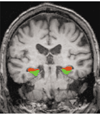
This coronal magnetic resonance image (MRI) displays the regions of interest (ROIs) for the hippocampal formation (red) and parahippocampal gyrus (green). ROIs were manually delineated from each individual subject’s structural MRI.
Fig 2
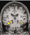
Example of a functional magnetic resonance image activation map for the novel versus repeated contrast (comparing the encoding of novel pictures vs repeated pictures; threshold p < 0.01).
Fig 3
Mean extent of right hippocampal (HF) and parahippocampal (PHG) activation for the subject group that remained stable after longitudinal clinical follow-up (stable) versus those with clinical decline (decliner); *p < 0.05. Error bars indicate 95% confidence intervals.
Comment in
- Doing more with less: the plight of the failing hippocampus.
Grafton ST. Grafton ST. Ann Neurol. 2004 Jul;56(1):7-9. doi: 10.1002/ana.20189. Ann Neurol. 2004. PMID: 15236396 No abstract available.
Similar articles
- Increased hippocampal activation in mild cognitive impairment compared to normal aging and AD.
Dickerson BC, Salat DH, Greve DN, Chua EF, Rand-Giovannetti E, Rentz DM, Bertram L, Mullin K, Tanzi RE, Blacker D, Albert MS, Sperling RA. Dickerson BC, et al. Neurology. 2005 Aug 9;65(3):404-11. doi: 10.1212/01.wnl.0000171450.97464.49. Neurology. 2005. PMID: 16087905 Free PMC article. - Medial temporal lobe atrophy and memory dysfunction as predictors for dementia in subjects with mild cognitive impairment.
Visser PJ, Scheltens P, Verhey FR, Schmand B, Launer LJ, Jolles J, Jonker C. Visser PJ, et al. J Neurol. 1999 Jun;246(6):477-85. doi: 10.1007/s004150050387. J Neurol. 1999. PMID: 10431775 - Discriminating accuracy of medial temporal lobe volumetry and fMRI in mild cognitive impairment.
Jauhiainen AM, Pihlajamäki M, Tervo S, Niskanen E, Tanila H, Hänninen T, Vanninen RL, Soininen H. Jauhiainen AM, et al. Hippocampus. 2009 Feb;19(2):166-75. doi: 10.1002/hipo.20494. Hippocampus. 2009. PMID: 18777563 - Functional abnormalities of the medial temporal lobe memory system in mild cognitive impairment and Alzheimer's disease: insights from functional MRI studies.
Dickerson BC, Sperling RA. Dickerson BC, et al. Neuropsychologia. 2008;46(6):1624-35. doi: 10.1016/j.neuropsychologia.2007.11.030. Epub 2007 Dec 8. Neuropsychologia. 2008. PMID: 18206188 Free PMC article. Review. - Functional alterations in memory networks in early Alzheimer's disease.
Sperling RA, Dickerson BC, Pihlajamaki M, Vannini P, LaViolette PS, Vitolo OV, Hedden T, Becker JA, Rentz DM, Selkoe DJ, Johnson KA. Sperling RA, et al. Neuromolecular Med. 2010 Mar;12(1):27-43. doi: 10.1007/s12017-009-8109-7. Neuromolecular Med. 2010. PMID: 20069392 Free PMC article. Review.
Cited by
- Effects of apolipoprotein E genotype on the off-line memory consolidation.
Wang DY, Han XJ, Li SF, Liu DQ, Yan CG, Zuo XN, Zhu CZ, He Y, Kiviniemi V, Zang YF. Wang DY, et al. PLoS One. 2012;7(12):e51617. doi: 10.1371/journal.pone.0051617. Epub 2012 Dec 12. PLoS One. 2012. PMID: 23251595 Free PMC article. - Perturbations of neural circuitry in aging, mild cognitive impairment, and Alzheimer's disease.
Leal SL, Yassa MA. Leal SL, et al. Ageing Res Rev. 2013 Jun;12(3):823-31. doi: 10.1016/j.arr.2013.01.006. Epub 2013 Feb 4. Ageing Res Rev. 2013. PMID: 23380151 Free PMC article. Review. - Anatomical and functional deficits in patients with amnestic mild cognitive impairment.
Han Y, Lui S, Kuang W, Lang Q, Zou L, Jia J. Han Y, et al. PLoS One. 2012;7(2):e28664. doi: 10.1371/journal.pone.0028664. Epub 2012 Feb 3. PLoS One. 2012. PMID: 22319555 Free PMC article. - Lateral entorhinal cortex dysfunction in amnestic mild cognitive impairment.
Tran TT, Speck CL, Gallagher M, Bakker A. Tran TT, et al. Neurobiol Aging. 2022 Apr;112:151-160. doi: 10.1016/j.neurobiolaging.2021.12.008. Epub 2021 Dec 30. Neurobiol Aging. 2022. PMID: 35182842 Free PMC article. - Cortical surface-based analysis reduces bias and variance in kinetic modeling of brain PET data.
Greve DN, Svarer C, Fisher PM, Feng L, Hansen AE, Baare W, Rosen B, Fischl B, Knudsen GM. Greve DN, et al. Neuroimage. 2014 May 15;92:225-36. doi: 10.1016/j.neuroimage.2013.12.021. Epub 2013 Dec 19. Neuroimage. 2014. PMID: 24361666 Free PMC article.
References
- Kordower JH, Chu Y, Stebbins GT, et al. Loss and atrophy of layer II entorhinal cortex neurons in elderly people with mild cognitive impairment. Ann Neurol. 2001;49:202–213. - PubMed
- de Toledo-Morrell L, Dickerson B, Sullivan MP, et al. Hemispheric differences in hippocampal volume predict verbal and spatial memory performance in patients with Alzheimer’s disease. Hippocampus. 2000;10:136–142. - PubMed
- Petersen RC, Jack CR, Jr, Xu YC, et al. Memory and MRI-based hippocampal volumes in aging and AD. Neurology. 2000;54:581–587. - PubMed
Publication types
MeSH terms
Grants and funding
- K23 NS 02189/NS/NINDS NIH HHS/United States
- P41 RR 14075/RR/NCRR NIH HHS/United States
- P41 RR014075/RR/NCRR NIH HHS/United States
- K23 AG 22509/AG/NIA NIH HHS/United States
- K23 NS002189/NS/NINDS NIH HHS/United States
- P01 AG 04953/AG/NIA NIH HHS/United States
- P01 AG004953/AG/NIA NIH HHS/United States
- K23 AG022509/AG/NIA NIH HHS/United States
LinkOut - more resources
Full Text Sources
Other Literature Sources
Medical


