Brain-derived neurotrophic factor mRNA and protein are targeted to discrete dendritic laminas by events that trigger epileptogenesis - PubMed (original) (raw)
Brain-derived neurotrophic factor mRNA and protein are targeted to discrete dendritic laminas by events that trigger epileptogenesis
Enrico Tongiorgi et al. J Neurosci. 2004.
Abstract
Dendritic targeting of mRNA and local protein synthesis are mechanisms that enable neurons to deliver proteins to specific postsynaptic sites. Here, we demonstrate that epileptogenic stimuli induce a dramatic accumulation of BDNF mRNA and protein in the dendrites of hippocampal neurons in vivo. BDNF mRNA and protein accumulate in dendrites in all hippocampal subfields after pilocarpine seizures and in selected subfields after other epileptogenic stimuli (kainate and kindling). BDNF accumulates selectively in discrete dendritic laminas, suggesting targeting to synapses that are active during seizures. Dendritic targeting of BDNF mRNA occurs during the time when the cellular changes that underlie epilepsy are occurring and is not seen after intense stimuli that are non-epileptogenic, including electroconvulsive seizures and high-frequency stimulation. MK801, an NMDA receptor antagonist that can prevent epileptogenesis but not acute seizures, prevents the dendritic accumulation of BDNF mRNA, indicating that dendritic targeting is mediated via NMDA receptor activation. Together, these results suggest that dendritic accumulation of BDNF mRNA and protein plays a critical role in the cellular changes leading to epilepsy.
Figures
Figure 1.
Pilocarpine seizures induce dendritic targeting of BDNF mRNA. Shown are representative coronal brain sections at the level of the dorsal hippocampus (plate 39) (Pellegrino et al., 1979), exhibiting total hybridization of the digoxigenin-labeled riboprobes. Nonspecific hybridization, estimated using the BDNF sense probe, is shown in the left column (sense). Under control conditions (control), BDNF mRNA is localized in the proximal dendrites of CA1 and CA3 pyramidal cells. Three hours after pilocarpine injection (300 mg/kg, i.p.), BDNF mRNA is found in the stratum radiatum of CA1, in the stratum lucidum, and in the radiatum of CA3 and in the proximal third of dentate gyrus (DG) granule cell dendrites. At 6 and 24 hr after injection, the dendritic staining appears to be less intense and more diffuse over the entire molecular layer in the DG and over the stratum radiatum only in CA1 and CA3. In chronic animals that did not experience seizures during the last 3 hr before being killed, dendritic staining was not different from control levels. These representative sections will not fully correlate with the mean changes in dendritic BDNF mRNA levels shown in Figure 2 because of slight differences among the four to six animals of each group. Scale bar: whole hippocampus, 750 μm; CA1, 75 μm; CA3, 150 μm; DG, 300 μm.
Figure 2.
Densitometric analysis of the effects of pilocarpine seizures. A, High-magnification of the CA1 region of a control animal (shown also in Fig. 1, control) showing proximal dendritic in situ staining for BDNF mRNA in pyramidal neurons and in interneurons within the stratum radiatum. B, CA1 region of a pilocarpine-treated rat (3 hr) showing dramatic enhancement of the BDNF mRNA in situ staining. Several strongly labeled dendrites are visible in the stratum radiatum but not in the stratum oriens. The white lines in A and B indicate the distance 0 from the cell soma used for the densitometric analysis. The left panels in _C_-E show the densitometric analysis of the dendritic labeling, expressed as pixel intensity (0-255 gray levels; 255 = white; 0 = black) as a function of the distance from the cell soma (in micrometers). Analysis was performed as described in Materials and Methods. Data are the means ± SE of four to six animals per group: control (thin line) and pilocarpine 3 hr (pilo 3h; thick line). Note that, under control conditions, the average gray level in CA1 and CA3 reaches background between 50 and 100 μm from the cell somas, whereas in the dentate gyrus it drops to background levels ∼40 μm from the cell bodies. *p < 0.05; ***p < 0.001 versus control values (□); ANOVA and post hoc Newman-Keuls test. lac-mol, Stratum lacunosum-moleculare; luc, stratum lucidum; ML, molecular layer; rad, stratum radiatum.
Figure 3.
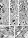
Postembedding in situ hybridization electron microscopy analysis of BDNF mRNA localization in CA1 apical dendrites. A, B, No staining was observed in proximal (A;<15 μm from cell soma) or distal (B;∼50 μm) dendrites with a BDNF sense probe. C, D, In control animals, a few gold grains for BDNF mRNA are associated with polyribosomes in proximal dendrites (C, arrows), whereas they are associated with microtubules in the distal dendrites (D, arrows). E, F, Three hours after pilocarpine administration, gold grains for BDNF mRNA are more abundant and larger both in proximal (E, arrows) and distal (F, arrows) dendrites and are mostly associated with microtubules and less frequently with polyribosomes. Under control conditions, gold grains for BDNF mRNA are also present at distal synapses (G, large black dots; ∼70 μm from the cell soma) close to the postsynaptic density and are accumulated at dendritic branchings (H; ∼45 μm from the cell soma), often in association with polyribosomes and in proximity to large dense core vesicles (arrowheads). Scale bars: (in F) A-F, 300 nm; G, 300 nm; H, 500 nm.
Figure 4.
Postembedding in situ hybridization electron microscopy in the dentate gyrus after pilocarpine seizures. BDNF mRNA is targeted to dendrites of dentate gyrus granule cells. A, Three hours after pilocarpine administration, gold grains for BDNF mRNA can be visualized in dentate gyrus dendrites associated with a large number of polyribosomes at dendritic hillock (A, B), proximal dendrite (A, C), and dendritic branchings at more distal dendritic domains (A, D). A few grains are also associated with microtubules (A, C, D). Scale bars: A, 2 μm; (in D) B-D, 1 μm.
Figure 5.
Pilocarpine seizure-induced dendritic BDNF mRNA is translated into protein. Shown are representative hematoxylin counterstained coronal brain sections at the level of the dorsal hippocampus (plate 39) (Pellegrino et al., 1979), exhibiting DAB-labeled BDNF-like immunoreactivity (LI). Omitting the primary antibody to estimate nonspecific signal yielded completely negative labeling (data not shown). Under control conditions (control), BDNF-LI is localized in the proximal dendrites. Peaking 6 hr after pilocarpine injection (pilo 6h; 300 mg/kg, i.p.), BDNF-LI is found in the stratum radiatum of CA1 pyramidal neurons, in the stratum lucidum, and in the radiatum of CA3 pyramidal neurons and in the proximal third of dentate gyrus granule cell dendrites. Increased dendritic BDNF-LI is also observed in spontaneously seizing animals killed 3-4 weeks after pilocapine administration. Scale bar: CA1 and DG, 75 μm; CA3, 120 μm.
Figure 6.
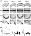
Kainate seizures induce dendritic targeting of BDNF mRNA in CA1 pyramidal neurons. A, Representative coronal brain sections at the level of the dorsal hippocampus, exhibiting total hybridization of the digoxigenin-labeled riboprobes. Six hours after kainate injection (10 mg/kg, i.p.), BDNF mRNA is found in the stratum radiatum of CA1. The serepresentative sections will not fully correlate with the mean changes in the dendritic BDNF mRNA levels shown in B because of slight differences in the three to seven animals of each group. DG, Dentate gyrus. Scale bar: whole hippocampus, 750 μm; CA1, 75 μm; CA3, 150 μm; DG, 300 μm. B, Densitometric analysis of the effects of kainate. The left panel shows the densitometric analysis of the dendritic labeling, expressed as pixel intensity (0-255 gray levels; 255 = white, 0 = black) as a function of the distance from the cell soma (in micrometers). Data are the mean of control (thin line; n = 5) and kainate 6 hr (KA 6h; thick line; n = 3). The other panels report the changes in BDNF mRNA levels in the different layers of CA1. No significant changes were observed in other hippocampal subfields. Analysis was performed as described in Materials and Methods. Data are the mean ± SE of three to seven animals per group. ***p < 0.001 versus control values (□); ANOVA and post hoc Newman-Keuls test. lac-mol, Stratum lacunosum-moleculare; rad, stratum radiatum.
Figure 7.
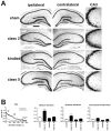
Class 2 kindled seizures induce dendritic targeting of BDNF mRNA in CA3 pyramidal neurons. A, Representative coronal brain sections at the level of the dorsal hippocampus, exhibiting total hybridization of the digoxigenin-labeled riboprobes. Three hours after class 2 kindled seizures, BDNF mRNA is found in the strata lucidum and radiatum of CA3. These representative sections will not fully correlate with the mean changes in the dendritic BDNF mRNA levels shown in B because of slight differences among the three to six animals of each group. B, Densitometric analysis of the effects of kindling. The left panel shows the densitometric analysis of the dendritic labeling, expressed as pixel intensity (0-255 gray levels; 255 = white; 0 = black) as a function of the distance from the cell soma (in micrometers). Data are the mean of sham-stimulated (control; thin line; n = 4) and class 2, 3 hr (class 2, 3h; thick line; n = 5). The other panels report the changes in BDNF mRNA levels in the different layers of CA3. No significant changes were observed in other hippocampal subfields nor at other stages of kindling development. Analysis was performed as described in Materials and Methods. Data are the mean ± SE of three to six animals per group. ***p < 0.001 versus control values (□); ANOVA and posthoc Newman-Keuls test.lac-mol, Stratum lacunosum-moleculare; luc, stratum lucidum; rad, stratum radiatum. Scale bar: whole hippocampus, 750 μm; CA3, 150 μm.
Figure 8.
Pilocarpine seizure-induced dendritic targeting of BDNF mRNA is glutamate receptor dependent. Animal pretreatment with the AMPA receptor antagonist NBQX [30 mg/kg, i.p.; administered 20 min before pilocarpine (pilo 3h + NBQX)] and with the NMDAR antagonist MK801 [1 mg/kg i.p.; administered 20 min before pilocarpine (pilo 3h + MK801)] prevented BDNF mRNA dendritic accumulation induced by pilocarpine (300 mg/kg, i.p.). Higher magnification of the CA1 subfield is shown in the bottom panels. Scale bar: whole hippocampus, 750 μm; CA1, 75 μm.
Similar articles
- Dendritic targeting of mRNAs for plasticity genes in experimental models of temporal lobe epilepsy.
Simonato M, Bregola G, Armellin M, Del Piccolo P, Rodi D, Zucchini S, Tongiorgi E. Simonato M, et al. Epilepsia. 2002;43 Suppl 5:153-8. doi: 10.1046/j.1528-1157.43.s.5.32.x. Epilepsia. 2002. PMID: 12121312 - Actions of brain-derived neurotrophic factor in slices from rats with spontaneous seizures and mossy fiber sprouting in the dentate gyrus.
Scharfman HE, Goodman JH, Sollas AL. Scharfman HE, et al. J Neurosci. 1999 Jul 1;19(13):5619-31. doi: 10.1523/JNEUROSCI.19-13-05619.1999. J Neurosci. 1999. PMID: 10377368 Free PMC article. - BDNF mRNA splice variants display activity-dependent targeting to distinct hippocampal laminae.
Chiaruttini C, Sonego M, Baj G, Simonato M, Tongiorgi E. Chiaruttini C, et al. Mol Cell Neurosci. 2008 Jan;37(1):11-9. doi: 10.1016/j.mcn.2007.08.011. Epub 2007 Aug 23. Mol Cell Neurosci. 2008. PMID: 17919921 - What is the biological significance of BDNF mRNA targeting in the dendrites? Clues from epilepsy and cortical development.
Tongiorgi E, Domenici L, Simonato M. Tongiorgi E, et al. Mol Neurobiol. 2006 Feb;33(1):17-32. doi: 10.1385/MN:33:1:017. Mol Neurobiol. 2006. PMID: 16388108 Review. - BDNF and epilepsy: too much of a good thing?
Binder DK, Croll SD, Gall CM, Scharfman HE. Binder DK, et al. Trends Neurosci. 2001 Jan;24(1):47-53. doi: 10.1016/s0166-2236(00)01682-9. Trends Neurosci. 2001. PMID: 11163887 Review.
Cited by
- The Contribution of Spatial and Temporal Molecular Networks in the Induction of Long-term Memory and Its Underlying Synaptic Plasticity.
Mirisis AA, Alexandrescu A, Carew TJ, Kopec AM. Mirisis AA, et al. AIMS Neurosci. 2016;3(3):356-384. doi: 10.3934/Neuroscience.2016.3.356. Epub 2016 Oct 22. AIMS Neurosci. 2016. PMID: 27819030 Free PMC article. - Centella asiatica L. Phytosome Improves Cognitive Performance by Promoting Bdnf Expression in Rat Prefrontal Cortex.
Sbrini G, Brivio P, Fumagalli M, Giavarini F, Caruso D, Racagni G, Dell'Agli M, Sangiovanni E, Calabrese F. Sbrini G, et al. Nutrients. 2020 Jan 29;12(2):355. doi: 10.3390/nu12020355. Nutrients. 2020. PMID: 32013132 Free PMC article. - Brain-derived neurotrophic factor and epilepsy--a missing link?
Scharfman HE. Scharfman HE. Epilepsy Curr. 2005 May-Jun;5(3):83-8. doi: 10.1111/j.1535-7511.2005.05312.x. Epilepsy Curr. 2005. PMID: 16145610 Free PMC article. - Group I mGluR-regulated translation of the neuronal glutamate transporter, excitatory amino acid carrier 1.
Ross JR, Ramakrishnan H, Porter BE, Robinson MB. Ross JR, et al. J Neurochem. 2011 Jun;117(5):812-23. doi: 10.1111/j.1471-4159.2011.07233.x. Epub 2011 Apr 11. J Neurochem. 2011. PMID: 21371038 Free PMC article. - Region-specific involvement of BDNF secretion and synthesis in conditioned taste aversion memory formation.
Ma L, Wang DD, Zhang TY, Yu H, Wang Y, Huang SH, Lee FS, Chen ZY. Ma L, et al. J Neurosci. 2011 Feb 9;31(6):2079-90. doi: 10.1523/JNEUROSCI.5348-10.2011. J Neurosci. 2011. PMID: 21307245 Free PMC article.
References
- Altar CA, Di Stefano PS (1998) Neurotrophin trafficking by anterograde transport. Trends Neurosci 21: 433-437. - PubMed
- Bengzon J, Kokaia Z, Ernfors P, Kokaia M, Leanza G, Nilsson OG, Persson H, Lindvall O (1993) Regulation of neurotrophin and trkA, trkB and trkC tyrosine kinase receptor messenger RNA expression in kindling. Neuroscience 53: 433-446. - PubMed
- Binder DK, Croll SD, Gall CM, Scharfman HE (2001) BDNF and epilepsy: too much of a good thing? Trends Neurosci 24: 47-53. - PubMed
- Brooks-Kayal AR, Shumate MD, Jin H, Rikhter TY, Coulter DA (1998) Selective changes in single cell GABA(A) receptor subunit expression and function in temporal lobe epilepsy. Nat Med 4: 1166-1172. - PubMed
Publication types
MeSH terms
Substances
LinkOut - more resources
Full Text Sources
Other Literature Sources
Medical


