A single in vivo exposure to cocaine abolishes endocannabinoid-mediated long-term depression in the nucleus accumbens - PubMed (original) (raw)
A single in vivo exposure to cocaine abolishes endocannabinoid-mediated long-term depression in the nucleus accumbens
Lawrence Fourgeaud et al. J Neurosci. 2004.
Abstract
In the nucleus accumbens (NAc), a key structure to the effects of all addictive drugs, presynaptic cannabinoid CB1 receptors (CB1Rs) and postsynaptic metabotropic glutamate 5 receptors (mGluR5s) are the principal effectors of endocannabinoid (eCB)-mediated retrograde long-term depression (LTD) (eCB-LTD) at the prefrontal cortex-NAc synapses. Both CB1R and mGluR5 are involved in cocaine-related behaviors; however, the impact of in vivo cocaine exposure on eCB-mediated retrograde synaptic plasticity remains unknown. Electrophysiological and biochemical approaches were used, and we report that a single in vivo cocaine administration abolishes eCB-LTD. This effect of cocaine was not present in D1 dopamine receptor (D1R) -/- mice and was prevented when cocaine was coadministered with the selective D1R antagonist 8-chloro-2,3,4,5-tetrahydro-3-5-1h-3-benzazepin-7-ol (0.5 mg/kg) or with the NMDA receptor (NMDAR) blocker (+)-5-methyl-10,11-dihydro-5H-dibenzo [a,d] cyclohepten-5,10-imine maleate (1 mg/kg), suggesting the involvement of D1R and NMDAR. We found that the cocaine-induced blockade of retrograde signaling was correlated with enhanced expression levels of Homer scaffolding proteins containing the coiled-coil domain and accompanied by a strong reduction of mGluR5 surface expression. The results suggest that cocaine-induced loss of eCB retrograde signaling is caused by a reduction in the ability of mGluR5 to translate anterograde glutamate transmission into retrograde eCB signaling.
Figures
Figure 1.
Single in vivo administration of cocaine abolishes eCB-LTD and increases CC-Homer protein levels. a, Top, Sample fEPSPs before (control) or after induction of eCB-LTD in mice injected in vivo with saline (sham) or cocaine (20 mg/kg) taken at the time indicated on graph below. Bottom, Average time courses of eCB-LTD. Single cocaine injection (n = 27) abolished eCB-LTD (compared with sham; n = 17; 50 min after tetanus; ***p < 0.05). The dashed line represents 100%. _b_, Representative Western blots of animals administered saline (sham) or cocaine _in vivo_. The blots show protein levels for CC-Homer using a pan-Homer antibody in the NAc 1 d after injection. Densitometry measurements are expressed as percentage of control ± SEM. Single cocaine exposure (_n_ = 7) markedly increased CC-Homer protein levels in the NAc, compared with saline (_n_ = 7; ***_p_ < 0.05). Immunoblots were quantified by scanning densitometry. _c_, Top, Densitometry measurements expressed as percentage of control ± SEM and representative Western blots of animals administered saline (sham) or cocaine _in vivo_. The blots show protein levels for CC-Homer using a pan-Homer antibody in the NAc 1 week after single cocaine injection. One week after a single exposure to cocaine, CC-Homer protein levels were back to normal (_n_ = 5) compared with saline (_n_ = 5; _p_ > 0.05). Bottom, eCB-LTD expressed as percentage of baseline 50 min after LTD induction: eCB-LTD was identical 1 week after a single exposure to cocaine (n = 7) and in sham mice (n = 17; p > 0.05). The dashed line represents 100%.
Figure 2.
Role of D1R and NMDAR in the cocaine-induced blockade of eCB-LTD. a, Cocaine-induced inhibition of eCB-LTD was prevented by a D1R antagonist. Animals that had received SCH23390 (0.5 mg/kg) displayed eCB-LTD when coinjected with cocaine (n = 9; ***p < 0.05 compared with cocaine alone). D1R -/- mice display normal eCB-LTD (_n_ = 7) but were insensitive to cocaine administration compared with wild type (_n_ = 9; ***_p_ < 0.05). The dashed line represents 100%. _b_, Representative Western blots of animals administered saline (sham), SCH23390 (SCH; 0.5 mg/kg) alone, or SCH23390 plus cocaine (SCH + Coc) _in vivo_. The blots show protein levels for CC-Homer using a pan-Homer antibody in the NAc 1 d after injection. Densitometry measurements are expressed as percentage of control ± SEM. In SCH23390-injected animals, single cocaine exposure (_n_ = 6) did not alter CC-Homer protein levels in the NAc, compared with saline (_n_ = 5; _p_ > 0.05) and SCH23390 alone (n = 6; p > 0.05). c, Cocaine-induced inhibition of eCB-LTD is prevented by an NMDAR antagonist. Animals receiving MK801 display normal eCB-LTD when coinjected with cocaine (n = 13; ***p < 0.05 compared with cocaine alone). The dashed line represents 100%. _d_, Representative Western blots of animals administered saline (sham), MK801 (1 mg/kg) alone, or MK801 plus cocaine (MK801 + Coc) _in vivo_. The blots show protein levels for CC-Homer using a pan-Homer antibody in the NAc 1 d after injection. Densitometry measurements are expressed as percentage of control ± SEM. In MK801-injected animals, single cocaine exposure (_n_ = 8) did not alter CC-Homer protein levels in the NAc, compared with saline (_n_ = 8; _p_ > 0.05) and MK801 alone (n = 8; p > 0.05).
Figure 3.
CB1R presynaptic functions and protein levels are not altered by single in vivo cocaine exposure. a, CB1R-mediated fEPSP inhibition. Single cocaine injection (n = 5) did not alter the CB1R-mediated fEPSP inhibition [compared with sham; n = 4; 20 min after application of the CB1R agonist CP55,940 (10 μ
m
); p > 0.05]. b, Densitometry measurements expressed as percentage of control ± SEM. Single cocaine exposure (n = 6) did not alter CB1R protein levels in the NAc, compared with saline (n = 5; p > 0.05).
Figure 4.
A single in vivo cocaine exposure does not induce a generalized alteration of prefrontal cortex-NAc synapses. a, Single cocaine injection (n = 7) did not alter mGluR2/3-LTD [compared with sham; n = 10; 60 min after application of the selective mGluR2/3 agonist LY354740 (200 n
m
) for 10 min; p > 0.05]. The dashed line represents 100%. b, Single cocaine exposure did not change NAc protein levels of mGluR2/3. Densitometry measurements are expressed as percentage of control ± SEM. Single cocaine exposure (n = 4) did not alter mGluR2/3 protein levels in the NAc, compared with saline (n = 3; p > 0.05). c, Input-output curves for sham-treated mice and mice treated with a single injection of cocaine were similar. d, Left, Paired-pulse ratios were augmented after eCB-LTD induction in sham animals (1.21 ± 0.0910 min before tetanus compared with 1.36 ± 0.09 50 min after tetanus; n = 12; ***p < 0.05 but not in cocaine-treated animals (1.17 ± 0.08 10 min before tetanus compared with 1.12 ± 0.09 50 min after tetanus; _n_ = 20; _p_ > 0.05). Right, Baseline paired-pulse ratios were identical in vehicle- and cocaine-treated animals [1.21 ± 0.09 (n = 12) in sham animals compared with 1.17 ± 0.08 (n = 20) in cocaine-treated animals; p > 0.05], suggesting that basal probability of transmitter release was unchanged after cocaine injection. tet, Tetanus.
Figure 5.
Cocaine administration causes a loss of surface mGluR5. a, Single cocaine exposure did not change total NAc protein levels of mGluR5. Representative Western blots of animals administered saline (sham) or cocaine in vivo are shown. The blots demonstrate protein levels for mGluR5 using an mGluR5 antibody in the NAc 1 d after injection. Densitometry measurements are expressed as percentage of control ± SEM. Single cocaine exposure (n = 6) did not alter total mGluR5 protein levels in the NAc, compared with saline (n = 5; p > 0.05). b, Representative Western blots of animals administered in vivo with saline (sham) or cocaine showing biotinylated mGluR5 in the NAc 1 d after injection. Densitometry measurements are expressed as percentage of control ± SEM. Single cocaine exposure (n = 9) decreased biotinylated mGluR5 protein levels in the NAc, compared with saline (n = 8; ***p < 0.05). c, Average time courses of mGluR5-mediated LTD. Single cocaine injection (n = 11) abolished mGluR5-LTD [compared with sham; n = 13; 40 min after application of the selective mGluR5 agonist (S)-DHPG (100 μ
m
) for 10 min; p < 0.05]. Coc, Cocaine.
Figure 6.
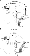
Schematic illustrating the interactions between the eCB-mediated retrograde signaling and the CC-Homer family of scaffolding proteins at excitatory NAc synapses. a, In naive or sham animals, retrograde eCB-LTD is normally induced after activation of postsynaptic mGluR5 and production of eCB. b, One day after cocaine exposure, heightened protein levels of CC-Homer caused the intracellular retention of postsynaptic mGluR5. The associated loss of postsynaptic mGluR5 surface expression prevents the induction of eCB-dependent retrograde signaling. The effects of cocaine on eCB-dependent LTD require D1R and NMDAR. RyR, Ryanodine receptor; G, G-protein; iGluRs, ionotropic GluRs.
Similar articles
- Neuroadaptations in the cellular and postsynaptic group 1 metabotropic glutamate receptor mGluR5 and Homer proteins following extinction of cocaine self-administration.
Ghasemzadeh MB, Vasudevan P, Mueller C, Seubert C, Mantsch JR. Ghasemzadeh MB, et al. Neurosci Lett. 2009 Mar 13;452(2):167-71. doi: 10.1016/j.neulet.2008.12.028. Epub 2008 Dec 24. Neurosci Lett. 2009. PMID: 19118598 Free PMC article. - Exogenous and endogenous cannabinoids control synaptic transmission in mice nucleus accumbens.
Robbe D, Alonso G, Manzoni OJ. Robbe D, et al. Ann N Y Acad Sci. 2003 Nov;1003:212-25. doi: 10.1196/annals.1300.013. Ann N Y Acad Sci. 2003. PMID: 14684448 - Activation of NMDA receptors reduces metabotropic glutamate receptor-induced long-term depression in the nucleus accumbens via a CaMKII-dependent mechanism.
Huang CC, Hsu KS. Huang CC, et al. Neuropharmacology. 2012 Dec;63(8):1298-307. doi: 10.1016/j.neuropharm.2012.08.008. Epub 2012 Aug 28. Neuropharmacology. 2012. PMID: 22947307 - Endocannabinoid-mediated synaptic plasticity in the CNS.
Chevaleyre V, Takahashi KA, Castillo PE. Chevaleyre V, et al. Annu Rev Neurosci. 2006;29:37-76. doi: 10.1146/annurev.neuro.29.051605.112834. Annu Rev Neurosci. 2006. PMID: 16776579 Review. - Endocannabinoid signaling and synaptic plasticity in the brain.
Zhu PJ. Zhu PJ. Crit Rev Neurobiol. 2006;18(1-2):113-24. doi: 10.1615/critrevneurobiol.v18.i1-2.120. Crit Rev Neurobiol. 2006. PMID: 17725514 Review.
Cited by
- Group I mGluRs and long-term depression: potential roles in addiction?
Grueter BA, McElligott ZA, Winder DG. Grueter BA, et al. Mol Neurobiol. 2007 Dec;36(3):232-44. doi: 10.1007/s12035-007-0037-7. Epub 2007 Jul 27. Mol Neurobiol. 2007. PMID: 17955198 - Repeated cocaine administration impairs group II metabotropic glutamate receptor-mediated long-term depression in rat medial prefrontal cortex.
Huang CC, Yang PC, Lin HJ, Hsu KS. Huang CC, et al. J Neurosci. 2007 Mar 14;27(11):2958-68. doi: 10.1523/JNEUROSCI.4247-06.2007. J Neurosci. 2007. PMID: 17360919 Free PMC article. - mGluR5 antagonism inhibits cocaine reinforcement and relapse by elevation of extracellular glutamate in the nucleus accumbens via a CB1 receptor mechanism.
Li X, Peng XQ, Jordan CJ, Li J, Bi GH, He Y, Yang HJ, Zhang HY, Gardner EL, Xi ZX. Li X, et al. Sci Rep. 2018 Feb 27;8(1):3686. doi: 10.1038/s41598-018-22087-1. Sci Rep. 2018. PMID: 29487381 Free PMC article. - Long-Term Plasticity of Neurotransmitter Release: Emerging Mechanisms and Contributions to Brain Function and Disease.
Monday HR, Younts TJ, Castillo PE. Monday HR, et al. Annu Rev Neurosci. 2018 Jul 8;41:299-322. doi: 10.1146/annurev-neuro-080317-062155. Epub 2018 Apr 25. Annu Rev Neurosci. 2018. PMID: 29709205 Free PMC article. Review. - Long-Term Plasticity in Amygdala Circuits: Implication of CB1-Dependent LTD in Stress.
Li B, Ge T, Cui R. Li B, et al. Mol Neurobiol. 2018 May;55(5):4107-4114. doi: 10.1007/s12035-017-0643-y. Epub 2017 Jun 7. Mol Neurobiol. 2018. PMID: 28593436
References
- Brady AM, Glick SD, O'Donnell P (2003) Changes in electrophysiological properties of nucleus accumbens neurons depend on the extent of behavioral sensitization to chronic methamphetamine. Ann NY Acad Sci 1003: 358-363. - PubMed
- Brakeman PR, Lanahan AA, O'Brien R, Roche K, Barnes CA, Huganir RL, Worley PF (1997) Homer: a protein that selectively binds metabotropic glutamate receptors. Nature 386: 284-288. - PubMed
- Castane A, Valjent E, Ledent C, Parmentier M, Maldonado R, Valverde O (2002) Lack of CB1 cannabinoid receptors modifies nicotine behavioural responses, but not nicotine abstinence. Neuropharmacology 43: 857-867. - PubMed
- Chiamulera C, Epping-Jordan MP, Zocchi A, Marcon C, Cottiny C, Tacconi S, Corsi M, Orzi F, Conquet F (2001) Reinforcing and locomotor stimulant effects of cocaine are absent in mGluR5 null mutant mice. Nat Neurosci 4: 873-874. - PubMed
Publication types
MeSH terms
Substances
LinkOut - more resources
Full Text Sources
Medical


