Cryo-electron microscopy of vitreous sections - PubMed (original) (raw)
Review
Cryo-electron microscopy of vitreous sections
Ashraf Al-Amoudi et al. EMBO J. 2004.
Abstract
Since the beginning of the 1980s, cryo-electron microscopy of a thin film of vitrified aqueous suspension has made it possible to observe biological particles in their native state, in the absence of the usual artefacts of dehydration and staining. Combined with 3-d reconstruction, it has become an important tool for structural molecular biology. Larger objects such as cells and tissues cannot generally be squeezed in a thin enough film. Cryo-electron microscopy of vitreous sections (CEMOVIS) provides then a solution. It requires vitrification of a sizable piece of biological material and cutting it into ultrathin sections, which are observed in the vitrified state. Each of these operations raises serious difficulties that have now been overcome. In general, the native state seen with CEMOVIS is very different from what has been seen before and it is seen in more detail. CEMOVIS will give its full potential when combined with computerized electron tomography for 3-d reconstruction.
Figures
Figure 1
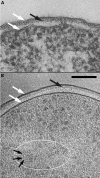
Gram-negative bacteria. (A) Escherichia coli K12 embedded in conventional resin (from Beveridge, 1999, Figure 1, with permission). (B) Pseudomonas aeruginosa PAO1 prepared by CEMOVIS (Matias et al, 2003). The cells in exponential growth were put in 20% (wt/vol) dextran (42 kDa) and vitrified in a Leica EMPACT (Vienna, Austria) high-pressure freezer. The sections were cut at −160°C in a Leica UCS/FCS cryomicrotome with a 45° Diatome diamond cryo-knife (Biel, Switzerland). Observation was made at −180°C in a Philips CM12 EM (Eindhoven, The Netherlands) operating at 80 kV and equipped with a Gatan 626 cryo-specimen holder (Warrendale, PA) and a Gatan 600CW Multiscan CCD camera. The total electron dose did not exceed 800 e/nm2. Technical details are described in previous publications (Dubochet et al, 1988; Al-Amoudi et al, 2002) or are in preparation. White arrows: cell membrane and outer membrane; black arrows: peptidoglycan layer; the white ellipse marks a region of the nucleoid; small black arrows: 2 nm high-contrast dots identified as portions of DNA filaments seen along their axis. Scale bar: 100 nm.
Figure 2
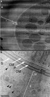
Cyanobacterium L. majuscula prepared by CEMOVIS. (A) Low-magnification view. I, II and III are different zones of the extracellular matrix; white asterisk: bulk medium with crevasses. Scale bar: 1 μm. (B) High-magnification view of the envelope. CM: cell membrane; P: peptidoglycan; OM: outer membrane; S: putative S-layer; black arrows: additional layers; II and III: zones of the extracellular matrix; black arrowheads: filaments in zone II. Scale bar: 100 nm.
Figure 3
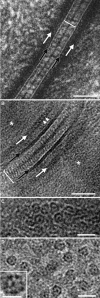
Epidermal desmosome and IFs. (A) Desmosome from neonatal mouse epidermis prepared by freeze substitution (from He et al, 2003, from Figure 1B, with permission). (B) Human desmosome at midportion of the viable part of forearm epidermis prepared by CEMOVIS. A ca. 200 μm thick layer was sliced with a razor blade and immediately cooled in a Baltec HPM 010 (Balzers, Liechtenstein) high-pressure freezing apparatus. The rest of the preparation was as for Figure 2B. See text for more explanations. (C) Same region as in (B) but with IFs oriented along the viewing direction. (D) As in (C) but in stratum corneum. The fine structure of the IFs is best resolved in the thinnest sections (inset). (C, D) Modified from Norlén and Al-Amoudi (2004), with permission. Section thickness: 70 nm (B, C); less than 50 nm (D). Scale bar: 50 nm (A, B); 20 nm (C, D); 10 nm (inset, D).
Figure 4
Stereo-pair of an 895 bp supercoiled double-stranded DNA molecule. Preparation by vitrification of a thin layer of 200 μg/ml DNA solution. Tilt angle: ±15°. The first exposure is on the right. Electron dose for each micrograph: ca. 2000 e/nm2. Scale bar: 50 nm. Courtesy Dr Jan Bednar.
Similar articles
- Cryo-electron microscopy of vitreous sections of native biological cells and tissues.
Al-Amoudi A, Norlen LP, Dubochet J. Al-Amoudi A, et al. J Struct Biol. 2004 Oct;148(1):131-5. doi: 10.1016/j.jsb.2004.03.010. J Struct Biol. 2004. PMID: 15363793 - Exploring skin structure using cryo-electron microscopy and tomography.
Norlen L. Norlen L. Eur J Dermatol. 2008 May-Jun;18(3):279-84. doi: 10.1684/ejd.2008.0387. Epub 2008 May 13. Eur J Dermatol. 2008. PMID: 18474455 Review. - Comparison of the envelope architecture of E. coli using two methods: CEMOVIS and cryo-electron tomography.
Kishimoto-Okada A, Murakami S, Ito Y, Horii N, Furukawa H, Takagi J, Iwasaki K. Kishimoto-Okada A, et al. J Electron Microsc (Tokyo). 2010;59(5):419-26. doi: 10.1093/jmicro/dfq056. Epub 2010 Jul 13. J Electron Microsc (Tokyo). 2010. PMID: 20630858 - Cryo-electron microscopy of vitreous sections.
Chlanda P, Sachse M. Chlanda P, et al. Methods Mol Biol. 2014;1117:193-214. doi: 10.1007/978-1-62703-776-1_10. Methods Mol Biol. 2014. PMID: 24357365 - Closer to the native state. Critical evaluation of cryo-techniques for Transmission Electron Microscopy: preparation of biological samples.
Mielanczyk L, Matysiak N, Michalski M, Buldak R, Wojnicz R. Mielanczyk L, et al. Folia Histochem Cytobiol. 2014;52(1):1-17. doi: 10.5603/FHC.2014.0001. Folia Histochem Cytobiol. 2014. PMID: 24802956 Review.
Cited by
- In situ fate of Chikungunya virus replication organelles.
Girard J, Le Bihan O, Lai-Kee-Him J, Girleanu M, Bernard E, Castellarin C, Chee M, Neyret A, Spehner D, Holy X, Favier A-L, Briant L, Bron P. Girard J, et al. J Virol. 2024 Jul 23;98(7):e0036824. doi: 10.1128/jvi.00368-24. Epub 2024 Jun 28. J Virol. 2024. PMID: 38940586 Free PMC article. - Electron microscopy for imaging organelles in plants and algae.
Weiner E, Pinskey JM, Nicastro D, Otegui MS. Weiner E, et al. Plant Physiol. 2022 Feb 4;188(2):713-725. doi: 10.1093/plphys/kiab449. Plant Physiol. 2022. PMID: 35235662 Free PMC article. - Cytomegalovirus primary envelopment occurs at large infoldings of the inner nuclear membrane.
Buser C, Walther P, Mertens T, Michel D. Buser C, et al. J Virol. 2007 Mar;81(6):3042-8. doi: 10.1128/JVI.01564-06. Epub 2006 Dec 27. J Virol. 2007. PMID: 17192309 Free PMC article. - Neurons as a model system for cryo-electron tomography.
Zuber B, Lučić V. Zuber B, et al. J Struct Biol X. 2022 Mar 9;6:100067. doi: 10.1016/j.yjsbx.2022.100067. eCollection 2022. J Struct Biol X. 2022. PMID: 35310407 Free PMC article. - Lamellar body ultrastructure revisited: high-pressure freezing and cryo-electron microscopy of vitreous sections.
Vanhecke D, Herrmann G, Graber W, Hillmann-Marti T, Mühlfeld C, Studer D, Ochs M. Vanhecke D, et al. Histochem Cell Biol. 2010 Oct;134(4):319-26. doi: 10.1007/s00418-010-0736-4. Epub 2010 Sep 1. Histochem Cell Biol. 2010. PMID: 20809233
References
- Abbott A (2002) The society of proteins. Nature 417: 894–896 - PubMed
- Adrian M, Dubochet J, Lepault J, McDowall AW (1984) Cryo-electron microscopy of viruses. Nature 308: 32–36 - PubMed
- Al-Amoudi A, Dubochet J, Gnaegi H, Lüthi W, Studer D (2003) An oscillating cryo-knife reduces cutting-induced deformation of vitreous ultrathin sections. J Microsc 212: 26–33 - PubMed
- Al-Amoudi A, Dubochet J, Studer D (2002) Amorphous solid water produced by cryosectioning of crystalline ice at 113 K. J Microsc 207: 146–153 - PubMed
- Al-Amoudi A, Norlén LPO, Dubochet J (2004) Cryo-electron microscopy of vitreous sections of native biological cells and tissues. J Struct Biol (in press) - PubMed
Publication types
MeSH terms
Substances
LinkOut - more resources
Full Text Sources
Other Literature Sources


