Hippocampal plasticity across multiple days of exposure to novel environments - PubMed (original) (raw)
Comparative Study
Hippocampal plasticity across multiple days of exposure to novel environments
Loren M Frank et al. J Neurosci. 2004.
Abstract
The hippocampus is essential for learning complex spatial relationships, but little is known about how hippocampal neural activity changes as animals learn about a novel environment. We studied the formation of new place representations in rats by examining the changes in place-specific firing of neurons in the CA1 region of the hippocampus and the relationship between these changes and behavioral change across multiple days of exposure to novel places. We found that many neurons showed very rapid changes on the first day of exposure to the novel place, including many cases in which a previously silent neuron developed a place field over the course of a single pass through the environment. Across the population, the largest changes in neural activity occurred on day 2 of exposure to a novel place, but only if the animal had little experience (<4 min) in that location on day 1. Longer exposures on day 1 were associated with smaller changes on day 2, suggesting that hippocampal neurons required 5-6 min of experience to form a stable spatial representation. Even after the representation stabilized, the animals' behavior remained different in the novel places, suggesting that other brain regions continued to distinguish novel from familiar locations. These results show that the hippocampus can form new spatial representations quickly but that stable hippocampal representations are not sufficient for a place to be treated as familiar.
Figures
Figure 1.
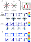
A, The familiar and novel configurations of the T-maze task. In the familiar configuration, the animals ran between arms 1 and 3 and 1 and 7, whereas in the novel configuration, either arm 3 or arm 7 was replaced with a novel arm that had not been visited previously (e.g., arm 6). B, Correlations of place field structure from run 1 to run 2 across the 3 d of exposure to the novel arm. The green bar represents the average correlation between the time-averaged place fields from the novel arm in run 2 and the adjacent familiar arm in run 1, whereas the red bar represents the correlations for the familiar outside arm that was visited in both runs 1 and 2. The asterisks denote significant differences (p < 0.00001). C, The place-related activity of a neuron that was active in the familiar configuration and maintained the same place field in the novel configuration. The top row shows the color-coded firing rate on the four trajectories through the familiar environment (depicted below each plot). The firing rate color map is shown to the right of the plots. The bottom row shows the firing rate in the novel environment. D, The place-related activity of a neuron recorded simultaneously from the same tetrode that was not active in the familiar configuration but developed a place field in the novel arm. The trajectories are the same as in C.
Figure 2.
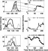
Place field plasticity on the first day of exposure to the novel arm. Each row represents the activity of a single neuron in one direction on the track. A, C, E, Examples of the spatial intensity (place field) at four different times. B, D, F, The integrated spatial intensity plotted as a function of the total experience in the novel arm. The four gray circles represent the times for the place fields shown in A, C, and E. The tick marks on the top _x_-axis represent the times at which the animal completed a full pass through the novel arm. These three neurons illustrate the variety of changes seen during the novel experience, including the sudden development of new fields (A, B), changes in location of fields (C, D), and decreases in activity of neurons with fields that were apparent from the beginning of the experience (E, F).
Figure 3.
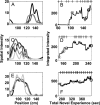
Examples of rapid place field formation in three neurons during the first day of exposure to the novel arm. Each row represents the activity of a single neuron in one direction on the track. As in Figure 2, A, D, and G represent the place field at four times corresponding to the gray circles in B, E, and H, and the tick marks on the top _x_-axis represent times when the animal completed a full pass through the novel arm. C, F, I, The spike rasters showing the activity of the neuron on each pass through the field. These neurons showed little or no activity for the first 1-2 min of experience in the novel arm, followed by the rapid emergence of place-specific firing. Once place-specific activity was visible, it evolved over the course of the session with adeclining rate. These plots show that the animal traversed the entire arm three to six times before the place field became visible and that the place fields developed rapidly over the course of one to two passes. These raster plots must be interpreted cautiously because they do not include information about the amount of time the animal spent in each location.
Figure 4.
Place field plasticity during the second day of experience. As in Figure 2, A, C, and E represent the place fields of three neurons at four times corresponding to the gray circles in B, D,and F, and the tick marks on the top _x_-axis represent at which the animal completed a full pass through the novel arm. The integrated intensity is plotted as a function of the total novel experience, computed as the length of experience on day 1 plus the length on day 2. In most cases in which the animal had little experience during the first day, cells showed large, fast changes on day 2 that were qualitatively similar to the fastest changes seen during day 1. In contrast, as the amount of experience increased, the amount of place field plasticity decreased (C-F).
Figure 5.
Place field plasticity over days. A, The cumulative distribution of the derivatives of the place field area as a function of the total experience in the novel arm on day 2. Each color corresponds to a particular number of minutes of experience (see legend). B, The cumulative distributions of derivatives on day 2 as a function of the total number of complete passes through the place. C, The plasticity indices as a function of total experience for each minute on each day. The index was calculated as the mean absolute value of the derivatives from each minute and measures the overall tendency to show changes. Bars represent mean ± SEM. D, The plasticity indices as a function of experience within each day. The missing bars in C and D represent minutes of experience that did not contain sufficient data sets or numbers of neurons to be included in the analyses.
Figure 6.
Place field properties in the novel and familiar environments. A, Place field directionality in the novel and familiar arms across the 3 d of experience. The asterisks represent a significant difference within the day. B, The plasticity indices for place fields in the familiar arm as a function of total experience in the familiar arm for each minute on days 1 and 2. There were insufficient data to perform the same analysis on day 3. The increase in plasticity seen in the novel arms was not present in the familiar arms. C, The plasticity indices as a function of experience within 1 d for the familiar arm days 1-3 and for the familiar arms in the run 1 familiar configuration. _D_-F, Histograms of the proportional change of place fields recorded from neurons active in the novel arm on day 1 (A), the familiar arm on day 1 (B), or in one of the familiar arms from the run 1 in the familiar configuration (C). The proportional change was, on average, larger in the novel arms.
Figure 7.
Changes in the activity of putative inhibitory interneurons in the novel and familiar arms. Each plot represents the average, normalized change in firing rate of all putative inhibitory neurons in the novel or familiar arm(s). A-C, The change in firing rate in the novel arm on days 1-3. _D_-F, The change in firing rate in the familiar arm on days 1-3. G, The change in firing rate in the familiar arms from the run 1 familiar configuration. The pattern of change in the novel arm did not resemble that seen in the familiar arms until day 3.
Figure 8.
The velocity of the animal in the novel and familiar arms on each day. Each plot shows the mean ± SEM of the velocity during each minute of experience on each day. _A_-C, The velocity on days 1-3. There were insufficient data to reliably calculate the velocity for the third minute of experience on day 3. D, The velocity in the familiar arms of the run 1 familiar configuration. The velocity remained different between the novel and familiar arms through day 3.
Similar articles
- Hippocampal and cortical place cell plasticity: implications for episodic memory.
Frank LM, Brown EN, Stanley GB. Frank LM, et al. Hippocampus. 2006;16(9):775-84. doi: 10.1002/hipo.20200. Hippocampus. 2006. PMID: 16921502 Review. - Rats with hippocampal lesion show impaired learning and memory in the ziggurat task: a new task to evaluate spatial behavior.
Faraji J, Lehmann H, Metz GA, Sutherland RJ. Faraji J, et al. Behav Brain Res. 2008 May 16;189(1):17-31. doi: 10.1016/j.bbr.2007.12.002. Epub 2007 Dec 15. Behav Brain Res. 2008. PMID: 18192033 - Cognitive aging and the hippocampus: how old rats represent new environments.
Wilson IA, Ikonen S, Gureviciene I, McMahan RW, Gallagher M, Eichenbaum H, Tanila H. Wilson IA, et al. J Neurosci. 2004 Apr 14;24(15):3870-8. doi: 10.1523/JNEUROSCI.5205-03.2004. J Neurosci. 2004. PMID: 15084668 Free PMC article. - Ventral Midline Thalamus Is Necessary for Hippocampal Place Field Stability and Cell Firing Modulation.
Cholvin T, Hok V, Giorgi L, Chaillan FA, Poucet B. Cholvin T, et al. J Neurosci. 2018 Jan 3;38(1):158-172. doi: 10.1523/JNEUROSCI.2039-17.2017. Epub 2017 Nov 13. J Neurosci. 2018. PMID: 29133436 Free PMC article. - Parallel processing across neural systems: implications for a multiple memory system hypothesis.
Mizumori SJ, Yeshenko O, Gill KM, Davis DM. Mizumori SJ, et al. Neurobiol Learn Mem. 2004 Nov;82(3):278-98. doi: 10.1016/j.nlm.2004.07.007. Neurobiol Learn Mem. 2004. PMID: 15464410 Review.
Cited by
- Dissociation between the experience-dependent development of hippocampal theta sequences and single-trial phase precession.
Feng T, Silva D, Foster DJ. Feng T, et al. J Neurosci. 2015 Mar 25;35(12):4890-902. doi: 10.1523/JNEUROSCI.2614-14.2015. J Neurosci. 2015. PMID: 25810520 Free PMC article. - Hippocampal lesions impair rapid learning of a continuous spatial alternation task.
Kim SM, Frank LM. Kim SM, et al. PLoS One. 2009;4(5):e5494. doi: 10.1371/journal.pone.0005494. Epub 2009 May 8. PLoS One. 2009. PMID: 19424438 Free PMC article. - Developmental Designs and Adult Functions of Cortical Maps in Multiple Modalities: Perception, Attention, Navigation, Numbers, Streaming, Speech, and Cognition.
Grossberg S. Grossberg S. Front Neuroinform. 2020 Feb 6;14:4. doi: 10.3389/fninf.2020.00004. eCollection 2020. Front Neuroinform. 2020. PMID: 32116628 Free PMC article. Review. - On the Integration of Space, Time, and Memory.
Eichenbaum H. Eichenbaum H. Neuron. 2017 Aug 30;95(5):1007-1018. doi: 10.1016/j.neuron.2017.06.036. Neuron. 2017. PMID: 28858612 Free PMC article. Review. - A Learned Map for Places and Concepts in the Human Medial Temporal Lobe.
Herweg NA, Kunz L, Schonhaut D, Brandt A, Wanda PA, Sharan AD, Sperling MR, Schulze-Bonhage A, Kahana MJ. Herweg NA, et al. J Neurosci. 2023 May 10;43(19):3538-3547. doi: 10.1523/JNEUROSCI.0181-22.2023. Epub 2023 Mar 31. J Neurosci. 2023. PMID: 37001991 Free PMC article.
References
- Abbott LF, Blum KI (1996) Functional significance of long-term potentiation for sequence learning and prediction. Cereb Cortex 6: 406-416. - PubMed
- Aggleton JP, Hunt PR, Rawlins JN (1986) The effects of hippocampal lesions upon spatial and non-spatial tests of working memory. Behav Brain Res 19: 133-146. - PubMed
- Amaral DG, Witter MP (1995) Hippocampal formation. In: The rat nervous system (Paxinos G, ed), pp 443-493. San Diego: Academic.
- Barnett SA (1975) The rat. Chicago: University of Chicago.
- Best PJ, White AM, Minai A (2001) Spatial processing in the brain: the activity of hippocampal place cells. Annu Rev Neurosci 24: 459-486. - PubMed
Publication types
MeSH terms
Grants and funding
- MH61673/MH/NIMH NIH HHS/United States
- R01 MH059733/MH/NIMH NIH HHS/United States
- MH65108/MH/NIMH NIH HHS/United States
- R01 CA094143/CA/NCI NIH HHS/United States
- R01 DA015644/DA/NIDA NIH HHS/United States
- DA015644/DA/NIDA NIH HHS/United States
- F32 MH065108/MH/NIMH NIH HHS/United States
- MH59733/MH/NIMH NIH HHS/United States
- K02 MH061637/MH/NIMH NIH HHS/United States
LinkOut - more resources
Full Text Sources
Other Literature Sources
Miscellaneous


