A Francisella tularensis pathogenicity island required for intramacrophage growth - PubMed (original) (raw)
A Francisella tularensis pathogenicity island required for intramacrophage growth
Francis E Nano et al. J Bacteriol. 2004 Oct.
Abstract
Francisella tularensis is a gram-negative, facultative intracellular pathogen that causes the highly infectious zoonotic disease tularemia. We have discovered a ca. 30-kb pathogenicity island of F. tularensis (FPI) that includes four large open reading frames (ORFs) of 2.5 to 3.9 kb and 13 ORFs of 1.5 kb or smaller. Previously, two small genes located near the center of the FPI were shown to be needed for intramacrophage growth. In this work we show that two of the large ORFs, located toward the ends of the FPI, are needed for virulence. Although most genes in the FPI encode proteins with amino acid sequences that are highly conserved between high- and low-virulence strains, one of the FPI genes is present in highly virulent type A F. tularensis, absent in moderately virulent type B F. tularensis, and altered in F. tularensis subsp. novicida, which is highly virulent for mice but avirulent for humans. The G+C content of a 17.7-kb stretch of the FPI is 26.6%, which is 6.6% below the average G+C content of the F. tularensis genome. This extremely low G+C content suggests that the DNA was imported from a microbe with a very low G+C-containing chromosome.
Figures
FIG. 1.
Gene organization and G+C content of the FPI. Top graph, fractional G+C content of the 300-kb region of the F. tularensis subsp. tularensis (strain Schu4) chromosome that encompasses the FPI (
http://artedi.ebc.uu.se/Projects/Francisella/
); bottom graph, G+C content of the FPI. The ORFs (arrows) from pdpD through pdpA are derived from the DNA sequence of F. tularensis subsp. novicida strain U112, determined in this work (GenBank accession no. AY293579) and the remaining ORFs and sequence data are derived from the F. tularensis subsp. tularensis Schu4 sequence. At the left end of the FPI are ORFs tnpAB, which show exact identity to genes encoding presumed transposases previously found in F. tularensis (GenBank accession no. AAL06399 and AAL064100); at the right end is tnpA only. The small opposing arrows indicate the approximate positions of 16-bp inverted repeats that have previously been shown to be associated with tnpA (21). The ORF labeled pmcA shows 44% identity to conserved domains found in putative molecular chaperones, the most closely related of which is the vdcC gene of Bacillus anthracis (NP_656320). The short nature of seven of the eight ORFs between pdpB and pdpC suggests that the area has many sequencing errors or is composed of nonfunctional vestigial genes. However, the DNA sequence is highly conserved between F. tularensis subsp. tularensis and novicida, suggesting that these ORFs represent functioning genes. Arrows labeled 62 and 116, locations of the transposon insertions that originally indicated a cluster of virulence-associated genes; arrow labeled 304-2, location of the insertion that gave rise to the mutant described in this work, codon 401 of pdpA (821 codons). Hatched line, region of the allelic replacement in the JL12 mutant; solid thick lines, extent of the F. tularensis DNA insert in the complementing plasmid pVIC314 and the noncomplementing plasmid pVIC311. rrsH and rrlH, 16S and 23S rRNA genes, respectively; fdn, A subunit of formate dehydrogenase. The smaller ORFs are not drawn to scale; the numbers of ORFs cited in the text are based on the F. tularensis subsp. novicida DNA sequence.
FIG. 2.
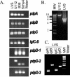
PCR amplification of FPI segments. (A) Results when primers were used to amplify internal fragments of pdpA, pdpB, pdpC, pdpD, and the junction between iglC and iglD. The resulting PCR products are shown; they provide evidence that the amplified regions in pdpA, pdpB, pdpC, and iglC-D in the F. tularensis strains that were tested are very similar. The three primer pairs used to amplify the region encompassing codons 123 to 356 (pdpD-1), codons 468 to 560 (in U112; codons 468 to 512 in Schu4; pdpD-2), and codons 906 to 1131 for pdpD-3 demonstrate that the pdpD gene is missing or is substantially different in sequence from the form found in strains U112, Schu4, and B38. The different sizes of the PCR products with the pdpD-2 set of primers with U112 DNA as the template relative to the sizes for the other template products confirm the difference in the DNA sequences. (B) Long-range PCR encompassing pdpD to iglD. Primers were used to amplify a region of approximately 10 kb in strain U112. The product in the LVS reaction is approximately 5.5 kb. Lane MW, molecular weight markers. (C) Left to right, PCR products obtained by using primers for iglA, iglB, iglC, and iglD. Molecular weight markers are as in panel B. Lane water, PCR done with primers for iglA but with no template. Similar reactions were done with the three other sets of primers with identical results (data not shown).
FIG. 3.
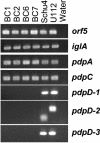
PCR analysis of clinical isolates of F. tularensis. The chromosomes of four type B isolates were amplified with primers corresponding to three regions of the pdpD gene as well as surrounding areas of the chromosome. The orf5 locus is 4 kb to the left of pdpD as shown in Fig. 1. These results show that pdpD is missing or substantially different in type B clinical isolates compared to the form found in type A strains or the F. tularensis subsp. novicida biotype. Lane water is as defined for Fig. 2.
FIG. 4.
Properties of the pdpD genomic region and phenotypes of pdpD mutant. (A) Relative sizes of the pdpD genes in F. tularensis strain Schu4, F. tularensis subsp. novicida, and the LVS. (B) Recombinant constructs used to make the pdpD::Emr allelic replacement mutant JL12, as described in the Materials and Methods. (C) Immunoblot of _E. coli_-produced, isolated recombinant IglA (lane 1) and IglA expressed in the pdpD mutant, JL12 (lane 2), and wild-type U112 (lane 3). When normalized to the amount of total protein loaded per lane, the intensity of the IglA band is sixfold higher in the U112 lane than in the JL12 lane. (D) Relative growth of wild-type F. tularensis subsp. novicida U112 and JL12 in bone marrow-derived macrophages after 72 h. Representative data from one of three repetitions are shown.
FIG. 5.
Growth of F. tularensis pdpA mutant and control strains in mouse macrophages. Bone marrow-derived macrophages were infected, and samples were lysed every 24 h to determine the number of viable bacteria (CFU) on agar medium. Squares, growth of parent strain F. tularensis (U112); inverted triangles, control strain R4 with a random transposon insertion not affecting intracellular growth; triangles, mutant 304-2; diamonds, mutant 304-2 with complementing plasmid pVIC314; circles, mutant 304-2 with plasmid pVIC311, which should not complement the genetic defect. Representative data from one of three repetitions are shown.
FIG. 6.
Infection of mice with F. tularensis pdpA mutant and control strains. Mice were infected intradermally with doses ranging from 2 × 10⁵ to 2 × 10⁷ CFU of the indicated bacteria; actual infection doses were confirmed by retrospective plate count. Squares, survival of mice following infection with 2 × 10⁵ CFU of the parent strain of F. tularensis (U112); inverted triangles, infection with 2 × 10⁵ CFU of the control strain R4, which has a random transposon insertion not affecting intracellular growth; triangles, infection with 2 × 10⁷ CFU of mutant 304-2; diamonds, infection with 2 × 10⁷ CFU of mutant 304-2 with complementing plasmid pVIC314; circles, infection with 2 × 10⁷ CFU of mutant 304-2 with plasmid pVIC311, which should not complement the genetic defect. Although lines in this graph are offset slightly for clarity, all mice survived (100% survival) infection with mutant 304-2 and mutant 304-2 with the noncomplementing plasmid pVIC311, and similarly all mice died (0% survival) following infection with the wild type and mutant 304-2 with complementing plasmid pVIC314. Representative data from one of four repetitions are shown.
Similar articles
- Molecular and genetic basis of pathogenesis in Francisella tularensis.
Barker JR, Klose KE. Barker JR, et al. Ann N Y Acad Sci. 2007 Jun;1105:138-59. doi: 10.1196/annals.1409.010. Epub 2007 Mar 29. Ann N Y Acad Sci. 2007. PMID: 17395737 Review. - Proteomics analysis of the Francisella tularensis LVS response to iron restriction: induction of the F. tularensis pathogenicity island proteins IglABC.
Lenco J, Hubálek M, Larsson P, Fucíková A, Brychta M, Macela A, Stulík J. Lenco J, et al. FEMS Microbiol Lett. 2007 Apr;269(1):11-21. doi: 10.1111/j.1574-6968.2006.00595.x. Epub 2007 Jan 15. FEMS Microbiol Lett. 2007. PMID: 17227466 - A Francisella tularensis pathogenicity island protein essential for bacterial proliferation within the host cell cytosol.
Santic M, Molmeret M, Barker JR, Klose KE, Dekanic A, Doric M, Abu Kwaik Y. Santic M, et al. Cell Microbiol. 2007 Oct;9(10):2391-403. doi: 10.1111/j.1462-5822.2007.00968.x. Epub 2007 May 21. Cell Microbiol. 2007. PMID: 17517064 - The Francisella pathogenicity island.
Nano FE, Schmerk C. Nano FE, et al. Ann N Y Acad Sci. 2007 Jun;1105:122-37. doi: 10.1196/annals.1409.000. Epub 2007 Mar 29. Ann N Y Acad Sci. 2007. PMID: 17395722 Review.
Cited by
- Pathogenicity and virulence of Francisella tularensis.
Degabriel M, Valeva S, Boisset S, Henry T. Degabriel M, et al. Virulence. 2023 Dec;14(1):2274638. doi: 10.1080/21505594.2023.2274638. Epub 2023 Nov 8. Virulence. 2023. PMID: 37941380 Free PMC article. Review. - Disruption of Francisella tularensis Schu S4 iglI, iglJ, and pdpC genes results in attenuation for growth in human macrophages and in vivo virulence in mice and reveals a unique phenotype for pdpC.
Long ME, Lindemann SR, Rasmussen JA, Jones BD, Allen LA. Long ME, et al. Infect Immun. 2013 Mar;81(3):850-61. doi: 10.1128/IAI.00822-12. Epub 2012 Dec 28. Infect Immun. 2013. PMID: 23275090 Free PMC article. - Current status of vaccine development for tularemia preparedness.
Hong KJ, Park PG, Seo SH, Rhie GE, Hwang KJ. Hong KJ, et al. Clin Exp Vaccine Res. 2013 Jan;2(1):34-9. doi: 10.7774/cevr.2013.2.1.34. Epub 2013 Jan 15. Clin Exp Vaccine Res. 2013. PMID: 23596588 Free PMC article. - Expression cloning and periplasmic orientation of the Francisella novicida lipid A 4'-phosphatase LpxF.
Wang X, McGrath SC, Cotter RJ, Raetz CR. Wang X, et al. J Biol Chem. 2006 Apr 7;281(14):9321-30. doi: 10.1074/jbc.M600435200. Epub 2006 Feb 8. J Biol Chem. 2006. PMID: 16467300 Free PMC article. - A New Species of the γ-Proteobacterium Francisella, F. adeliensis Sp. Nov., Endocytobiont in an Antarctic Marine Ciliate and Potential Evolutionary Forerunner of Pathogenic Species.
Vallesi A, Sjödin A, Petrelli D, Luporini P, Taddei AR, Thelaus J, Öhrman C, Nilsson E, Di Giuseppe G, Gutiérrez G, Villalobo E. Vallesi A, et al. Microb Ecol. 2019 Apr;77(3):587-596. doi: 10.1007/s00248-018-1256-3. Epub 2018 Sep 5. Microb Ecol. 2019. PMID: 30187088
References
- Amer, A. O., and M. S. Swanson. 2002. A phagosome of one's own: a microbial guide to life in the macrophage. Curr. Opin. Microbiol. 5:56-61. - PubMed
- Anthony, L. S. D., M. Z. Gu, S. C. Cowley, W. W. Leung, and F. E. Nano. 1991. Transformation and allelic replacement in Francisella spp. J. Gen. Microbiol. 137:2697-2703. - PubMed
- Baron, G. S., and F. E. Nano. 1998. MglA and MglB are required for the intramacrophage growth of Francisella novicida. Mol. Microbiol. 29:247-259. - PubMed
Publication types
MeSH terms
LinkOut - more resources
Full Text Sources
Other Literature Sources


