Bbs2-null mice have neurosensory deficits, a defect in social dominance, and retinopathy associated with mislocalization of rhodopsin - PubMed (original) (raw)
. 2004 Nov 23;101(47):16588-93.
doi: 10.1073/pnas.0405496101. Epub 2004 Nov 11.
Melissa Fath, Robert F Mullins, Charles Searby, Michael Andrews, Roger Davis, Jeaneen L Andorf, Kirk Mykytyn, Ruth E Swiderski, Baoli Yang, Rivka Carmi, Edwin M Stone, Val C Sheffield
Affiliations
- PMID: 15539463
- PMCID: PMC534519
- DOI: 10.1073/pnas.0405496101
Bbs2-null mice have neurosensory deficits, a defect in social dominance, and retinopathy associated with mislocalization of rhodopsin
Darryl Y Nishimura et al. Proc Natl Acad Sci U S A. 2004.
Abstract
Bardet-Biedl syndrome (BBS) is a heterogeneous, pleiotropic human disorder characterized by obesity, retinopathy, polydactyly, renal and cardiac malformations, learning disabilities, hypogenitalism, and an increased incidence of diabetes and hypertension. No information is available regarding the specific function of BBS2. We show that mice lacking Bbs2 gene expression have major components of the human phenotype, including obesity and retinopathy. In addition, these mice have phenotypes associated with cilia dysfunction, including retinopathy, renal cysts, male infertility, and a deficit in olfaction. With the exception of male infertility, these phenotypes are not caused by a complete absence of cilia. We demonstrate that BBS2 retinopathy involves normal retina development followed by apoptotic death of photoreceptors, the primary ciliated cells of the retina. Photoreceptor cell death is preceded by mislocalization of rhodopsin, indicating a defect in transport. We also demonstrate that Bbs2(-/-) mice and a second BBS mouse model, Bbs4(-/-), have a defect in social function. The evaluation of Bbs2(-/-) mice indicates additional phenotypes that should be evaluated in human patients, including deficits in social interaction and infertility.
Figures
Fig. 1.
Bbs2 gene targeting. (A) Strategy for the targeted deletion of Bbs2. Upon homologous recombination, exons 5–14 are replaced with a neomycin cassette. (B) (Upper) Northern blot analysis of Bbs2 expression in kidney total cellular RNA from WT (+/+), heterozygous (+/–), and homozygous (–/–) animals. The probe is a Bbs2 partial 3′ cDNA. (Lower) The same blot probed with β-actin as a loading control. (C) PCR genotyping of heterozygous (+/–), WT (+/+), and homozygous (–/–) mice. The top and middle amplimers correspond to 5′ and 3′ regions of the neomycin resistance gene, respectively. The bottom band is derived from amplification with Bbs2 internal primers.
Fig. 2.
_Bbs_2 gene expression. (A) Northern blot analysis of mouse Bbs2 RNA (20 μg) isolated from embryos 4.5 embryonic days postconception (E4.5) through E18.5 demonstrates early and widespread Bbs2 expression. The blot was sequentially hybridized with 32P-labeled Bbs2 and β-actin probes. (B) Northern blot analysis of Bbs2 gene expression in adult mouse tissues. Poly(A) mRNA (2 μg) was sequentially hybridized with 32P-labeled Bbs2 and β-actin probes. (C) Northern blot analysis of Bbs2 gene expression in discrete regions of the adult mouse brain. Total RNA (10 μg) was sequentially hybridized with 32P-labeled Bbs2 and β-actin probes.
Fig. 3.
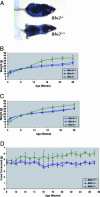
Weight gain and food consumption of Bbs2+/+, Bbs2+/–, and _Bbs2_–/– mice. (A) Photograph depicting typical obesity phenotype of _Bbs2_–/– mice. (B) Bbs2 female weight versus age comparison (minimum of five animals per group). (C) Weight versus age for all animals (males and females combined with a minimum of 10 mice per group). _Bbs2_–/– mice are statistically different from Bbs2+/+ and Bbs2+/– mice. (D) Bbs2 average food consumed (males and females combined with a minimum of six animals per group). Food consumption was averaged every 7 days.
Fig. 4.
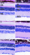
Hematoxylin/eosin staining of age-matched Bbs2+/+ (A, C, and E) and _Bbs2_–/– (B, D, and F) mouse retinas. Mice were examined at 7 weeks (A and B), 5 months (C and D), and 10 months (E and F). Some degenerative changes in the ONL are apparent in 7-week-old mice, although inner and outer segments are clearly distinguishable. By 5 months of age (C and D), the distinction between inner and outer segments is less apparent and the ONL is significantly thinner than in Bbs2+/+ eyes. At 10 months of age (E and F), the ONL is largely degenerated and no inner or outer segments are present. RPE, retinal pigment epithelium; IS, inner segments; OS, outer segments; INL, inner nuclear layer; GCL, ganglion cell layer. (Magnification: ×160.)
Fig. 5.
Localization of rhodopsin in 7-week Bbs2+/+ (A), 7-week _Bbs2_–/– (B), and 5-month _Bbs2_–/– (C) mice. (A) In Bbs2+/+ retinas, rhodopsin (green) is largely confined to the outer segments (OS), with little labeling of inner segments (IS). (B) _Bbs2_–/– mice at 7 weeks of age also predominantly localize rhodopsin to the OS, although some cell bodies in the ONL also appear to be labeled (arrows). (C) At 5 months of age _Bbs2_–/– mice do not exhibit partitioning of rhodopsin between the IS and OS. Labeling of ONL cell bodies is readily apparent. Photoreceptor degeneration in the _Bbs2_–/– mouse is caused by apoptosis as revealed by TUNEL labeling of the ONL (D, red). Ultrastructural analysis of _Bbs2_–/– mouse retina at 5 months of age reveals membranous whorls in the subretinal space (F) as compared with the orderly arrangement of membranous disks in Bbs2+/– (E) and WT mice. (Magnification: ×380, A_–_C; ×110, D; and ×3,000, E and F.)
Fig. 6.
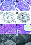
Hematoxylin/eosin-stained sections of the testes of Bbs2+/+ (A) and _Bbs2_–/– (B) mice show that, whereas WT mice possess numerous flagella in the seminiferous tubules (*), the _Bbs2_–/– mice have no flagella. Some condensed chromatin, indicating spermatogenesis, is detectable. Transmission electron micrographs through the tracheal cilia of Bbs2+/+ (C) and _Bbs2_–/– (D) mice reveal a normal 9-plus -2 pattern of microtubules. In some cases, vesicles were observed in association with these microtubules in _Bbs2_–/– mice (C, arrow). Kidneys from Bbs2+/+ (E) and _Bbs2_–/– (F) mice are shown. _Bbs2_–/– mice have numerous cysts, some of which involve the glomerulus. Scanning electron microscopic views of renal tubules from Bbs2+/+ (G) and _Bbs2_–/– (H) mice showing primary cilia. Some cells from _Bbs2_–/– mice show abnormally tapered cilia. (Magnification: ×100, A and B; ×90,000, C and D; ×90, E and F; and ×2,500 G and H.)
Similar articles
- Bardet-Biedl syndrome type 4 (BBS4)-null mice implicate Bbs4 in flagella formation but not global cilia assembly.
Mykytyn K, Mullins RF, Andrews M, Chiang AP, Swiderski RE, Yang B, Braun T, Casavant T, Stone EM, Sheffield VC. Mykytyn K, et al. Proc Natl Acad Sci U S A. 2004 Jun 8;101(23):8664-9. doi: 10.1073/pnas.0402354101. Epub 2004 Jun 1. Proc Natl Acad Sci U S A. 2004. PMID: 15173597 Free PMC article. - Mkks-null mice have a phenotype resembling Bardet-Biedl syndrome.
Fath MA, Mullins RF, Searby C, Nishimura DY, Wei J, Rahmouni K, Davis RE, Tayeh MK, Andrews M, Yang B, Sigmund CD, Stone EM, Sheffield VC. Fath MA, et al. Hum Mol Genet. 2005 May 1;14(9):1109-18. doi: 10.1093/hmg/ddi123. Epub 2005 Mar 16. Hum Mol Genet. 2005. PMID: 15772095 - Ectopic expression of human BBS4 can rescue Bardet-Biedl syndrome phenotypes in Bbs4 null mice.
Chamling X, Seo S, Bugge K, Searby C, Guo DF, Drack AV, Rahmouni K, Sheffield VC. Chamling X, et al. PLoS One. 2013;8(3):e59101. doi: 10.1371/journal.pone.0059101. Epub 2013 Mar 15. PLoS One. 2013. PMID: 23554981 Free PMC article. - [Current status and implication of research on Bardet-Biedl syndrome].
Shen T, Yan XM, Xiao CJ. Shen T, et al. Zhonghua Yi Xue Yi Chuan Xue Za Zhi. 2013 Oct;30(5):570-3. doi: 10.3760/cma.j.issn.1003-9406.2013.05.013. Zhonghua Yi Xue Yi Chuan Xue Za Zhi. 2013. PMID: 24078572 Review. Chinese. - Use of isolated populations in the study of a human obesity syndrome, the Bardet-Biedl syndrome.
Sheffield VC. Sheffield VC. Pediatr Res. 2004 Jun;55(6):908-11. doi: 10.1203/01.pdr.0000127013.14444.9c. Pediatr Res. 2004. PMID: 15155861 Review.
Cited by
- BBSome Component BBS5 Is Required for Cone Photoreceptor Protein Trafficking and Outer Segment Maintenance.
Bales KL, Bentley MR, Croyle MJ, Kesterson RA, Yoder BK, Gross AK. Bales KL, et al. Invest Ophthalmol Vis Sci. 2020 Aug 3;61(10):17. doi: 10.1167/iovs.61.10.17. Invest Ophthalmol Vis Sci. 2020. PMID: 32776140 Free PMC article. - Smelling the roses and seeing the light: gene therapy for ciliopathies.
McIntyre JC, Williams CL, Martens JR. McIntyre JC, et al. Trends Biotechnol. 2013 Jun;31(6):355-63. doi: 10.1016/j.tibtech.2013.03.005. Epub 2013 Apr 17. Trends Biotechnol. 2013. PMID: 23601268 Free PMC article. Review. - Loss of Bardet Biedl syndrome proteins causes defects in peripheral sensory innervation and function.
Tan PL, Barr T, Inglis PN, Mitsuma N, Huang SM, Garcia-Gonzalez MA, Bradley BA, Coforio S, Albrecht PJ, Watnick T, Germino GG, Beales PL, Caterina MJ, Leroux MR, Rice FL, Katsanis N. Tan PL, et al. Proc Natl Acad Sci U S A. 2007 Oct 30;104(44):17524-9. doi: 10.1073/pnas.0706618104. Epub 2007 Oct 24. Proc Natl Acad Sci U S A. 2007. PMID: 17959775 Free PMC article. - BBS6, BBS10, and BBS12 form a complex with CCT/TRiC family chaperonins and mediate BBSome assembly.
Seo S, Baye LM, Schulz NP, Beck JS, Zhang Q, Slusarski DC, Sheffield VC. Seo S, et al. Proc Natl Acad Sci U S A. 2010 Jan 26;107(4):1488-93. doi: 10.1073/pnas.0910268107. Epub 2010 Jan 4. Proc Natl Acad Sci U S A. 2010. PMID: 20080638 Free PMC article. - Mutations in a guanylate cyclase GCY-35/GCY-36 modify Bardet-Biedl syndrome-associated phenotypes in Caenorhabditis elegans.
Mok CA, Healey MP, Shekhar T, Leroux MR, Héon E, Zhen M. Mok CA, et al. PLoS Genet. 2011 Oct;7(10):e1002335. doi: 10.1371/journal.pgen.1002335. Epub 2011 Oct 13. PLoS Genet. 2011. PMID: 22022287 Free PMC article.
References
- Green, J. S., Parfrey, P. S., Harnett, J. D., Farid, N. R., Cramer, B. C., Johnson, G., Heath, O., McManamon, P. J., O'Leary, E. & Pryse-Phillips, W. (1989) N. Engl. J. Med. 321, 1002–1009. - PubMed
- Biedl, A. (1922) Dtsch. Med. Wochenschr. 48, 1630.
- Bardet, G. (1920) Ph.D thesis (University of Paris, Paris).
- Elbedour, K., Zucker, N., Zalzstein, E., Barki, Y. & Carmi, R. (1994) Am. J. Med. Genet. 52, 164–169. - PubMed
- Harnett, J. D., Green, J. S., Cramer, B. C., Johnson, G., Chafe, L., McManamon, P., Farid, N. R., Pryse-Phillips, W. & Parfrey, P. S. (1988) N. Engl. J. Med. 319, 615–618. - PubMed
Publication types
MeSH terms
Substances
LinkOut - more resources
Full Text Sources
Other Literature Sources
Molecular Biology Databases


