Co-activation of ERK, NF-kappaB, and GADD45beta in response to ionizing radiation - PubMed (original) (raw)
Co-activation of ERK, NF-kappaB, and GADD45beta in response to ionizing radiation
Tieli Wang et al. J Biol Chem. 2005.
Abstract
NF-kappaB has been well documented to play a critical role in signaling cell stress reactions. The extracellular signal-regulated kinase (ERK) regulates cell proliferation and survival. GADD45beta is a primary cell cycle element responsive to NF-kappaB activation in anti-apoptotic responses. The present study provides evidence demonstrating that NK-kappaB, ERK and GADD45beta are co-activated by ionizing radiation (IR) in a pattern of mutually dependence to increase cell survival. Stress conditions generated in human breast cancer MCF-7 cells by the administration of a single exposure of 5 Gy IR resulted in the activation of ERK but not p38 or JNK, along with an enhancement of the NF-kappaB transactivation and GADD45beta expression. Overexpression of dominant negative Erk (DN-Erk) or pre-exposure to ERK inhibitor PD98059 inhibited NF-kappaB. Transfection of dominant negative mutant IkappaB that blocks NF-kappaB nuclear translocation, inhibited ERK activity and GADD45beta expression and increased cell radiosensitivity. Interaction of p65 and ERK was visualized in living MCF-7 cells by bimolecular fluorescence complementation analysis. Antisense inhibition of GADD45beta strikingly blocked IR-induced NF-kappaB and ERK but not p38 and JNK. Overall, these results demonstrate a possibility that NF-kappaB, ERK, and GADD45beta are able to coordinate in a loop-like signaling network to defend cells against the cytotoxicity induced by ionizing radiation.
Figures
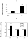
Fig. 1. NF-_κ_B and ERK but not p38 or JNK was activated by IR
A, NF-_κ_B activation by 5 Gy IR. MCF-7 cells were co-transfected with NF-_κ_B luciferase and _β_-galactosidase reporters for 6 h, and luciferase activity was measured 24 h after exposure to sham-IR (−) or a single dose of 5 Gy IR (+). Luciferase reporter activity was normalized to _β_-galactosidase (mean ± S.E., n = 3). B, p-ERK but not p-JNK or p-p38 was induced by IR. MCF-7 cells treated with sham-IR (0 Gy) and IR (5 Gy) were collected 24 h after irradiation. p-ERK, p-JNK, and p-p38 antibodies were incubated with 20 _μ_g of whole cell lysates, and the immunocomplex was labeled with streptavidin-phycoerythrin and detected by the Bio-Plex (Bio-Rad laboratory) protein array system as described under “Experimental Procedures” (mean ± S.E., n = 3; **, p < 0.01).
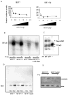
Fig. 2. Overexpression of mI_κ_B inhibited NF-_κ_B activity and Elk-1 phosphorylation
A, basal and radiation-induced NF-_κ_B activity was dose-dependently inhibited by transfection of dominate negative mutant I_κ_B (mI_κ_B). NF-_κ_B luciferase reporters were co-transfected with the indicated amounts of mI_κ_B plasmids into MCF-7 cells, and luciferase activity was measured 24 h after treatment with or without 5 Gy IR. B, IR-induced NF-_κ_B DNA binding activity was inhibited in stable transfectants of mI_κ_B. Wild type MFC-7 and stable transfectants of mI_κ_B (MCF-7/mI_κ_B) or empty vector (MCF-7/V) were treated with or without 5 Gy IR. NF-_κ_B DNA binding activity was assayed with DNA retardation gel analysis. MCF-7/V+cold oligonucleotide was included as a negative control with the presence of unlabeled NF-_κ_B nucleotides. C, NF-_κ_B subunits p65 and p50 were detected in the NF-_κ_B·DNA complexes induced by IR. Nuclear extracts from 5 Gy IR-treated MCF-7 cells were assayed with DNA retardation gel with or without antibody to NF-κ_B subunit p65 and p50 as indicated. NF-κ_B and the antibody super-shifted bands are indicated with arrowheads. D, expression of mI_κ_B inhibited IR-induced ERK kinase activity. Wild type MFC-7, empty vector control transfectants (MCF-7/V) and mI_κ_B MCF-7 transfectants (MCF-7/mI_κ_B) were irradiated with 0 (−) or 5 Gy (+) IR. Phos-pho-Elk-1 (sc-355; Santa Cruz Biotechnology) was detected by Western blotting as a downstream ERK kinase substrate. E, expression of mI_κ_B inhibited IR-induced GADD45_β. Wild type MFC-7, empty vector control transfectants (MCF-7/V), and mI_3_B MCF-7 transfectants (MCF-7/mI_κ_B) were irradiated with 0 (−) or 5 Gy (+) IR. GADD45_β was detected by Western blot (antibody sc-8775; Santa Cruz Biotechnology).
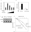
Fig. 3. Expression of dominant negative Erk, DN-Erk, or mI_κ_B in MCF-7 cells blocked NF-_κ_B activity and increased cell sensitivity to radiation
A, NF-_κ_B activity was dose-dependently inhibited by transfection of DN-Erk. NF-_κ_B luciferase reporters were co-transfected with the indicated amounts of DN_-_Erk into MCF-7 cells, and luciferase activity was measured 24 h after treatment with or without 5 Gy IR (mean ± S.E., n = 3, p < 0.01) compared with the control of MCF-7 (without IR). B, basal and IR-induced NF-κ_B activities were inhibited in MCF-7/DN-Erk transfectants. NF-κ_B luciferase reporters were transfected into parental MCF-7 and DN-Erk transfectants (MCF-7/DNErk), and luciferase activity was measured 24 h after exposure to 5 Gy IR. C, IR-induced GADD45_β was inhibited in MCF-7/DN-Erk transfectants. GADD45_β was measured with Western blotting in MCF-7/V and MCF-7/DN-Erk transfectants 24 h after exposure to 5 Gy IR. D, radiosensitivity was increased in MCF-7/DN-Erk and MCF-7/mI_κ_B transfectants. Wild type MCF-7, vector control MCF-7/V, MCF-7/mI_κ_B, and MCF-7/DN-Erk transfectants were irradiated with a range of IR doses (0–12 Gy), and clonogenic survival was determined 18 days after IR. Colonies with more than 50 cells were counted, and survival fractions of each cell line were normalized to the plating efficiency.
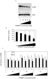
Fig. 4. PD98059 inhibited p-ERK and NF-_κ_B activity and increased MCF-7 cell radiosensitivity
A, ERK inhibitor PD98059 dose-dependently reduced IR-induced p-ERK. MCF-7 cells treated with the indicated concentrations of PD98059 for 2 h before exposure to 5 Gy IR and p-ERK activity was determined by Western blotting with antibody of p-ERK (sc-7383; sc-93 for ERK). B, PD98059 dose-dependently inhibited NF-_κ_B luciferase reporter activity. MCF-7 cells transfected with NF-_κ_B luciferase reporters were exposed to the indicated concentrations of PD98059 for 2 h, and luciferase activity was determined 24 h after IR. C, PD98059-increased IR-induced cytotoxicity. MCF-7 cells cultured in 6-well plates were treated with PD98059 by the indicated concentrations of PD98059 for 2 h before 5 Gy IR. Cell proliferation was determined by cell numbers counted 24, 48, and 72 h after radiation (mean ± S.E., n = 5; **, p < 0.01).
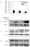
Fig. 5. Transfection of antisense Gadd45β reduced IR-induced p-ERK but not p-p38 and p-JNK
A, MCF-7 cells were incubated with antisense (AS) Gadd45β oligonucleotides 0.2 _μ_M for 24 h, and p-ERK, p-JNK, and p-p38 kinase activities were measured 24 h after 5 Gy IR with a Bio-Plex kit (**, p < 0.01, n = 3). B–D, antisense Gadd45β inhibited p-ERK. 40 _μ_g of whole cell lysate was analyzed by Western blotting with antibodies of p-ERK (sc-7383; sc-93 for ERK as control; B), p-p38 (sc-7975-R, sc-728 for p-38 as control; C), and p-JNK (sc-6254; sc-4061 for JNK1 as control; D); each blot was re-hybridized with the control antibody; results are representative of three blots.
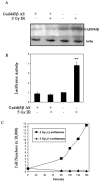
Fig. 6. Antisense blocking GADD45_β_ inhibited NF-_κ_B causing increased radiosensitivity
A, MCF-7 cells were incubated with 5 ml of transfection mixture containing 0.2 _μ_M antisense (AS) Gadd45β oligonucleotides for 6 h and further transfected with 0.1 μ_M antisense Gadd45β oligonucleotides for 24 h before IR with 5 Gy. Cell lysate was prepared 24 h after IR, and Western blotting was performed to confirm GADD45_β inhibition. B, antisense Gadd45β inhibited IR-induced NF-_κ_B. MCF-7 cells transfected with NF-_κ_B luciferase reporters were treated with or without antisense transfection and exposed to IR. Lu-ciferase activity was determined 24 h after IR (**, p < 0.01, n = 3). C, MCF-7 cells in multiple-well plates were treated with (+) or without (−) antisense Gadd45β oligonucleotides for 24 h followed by 2 Gy IR. Cell proliferation was calculated at different time intervals after IR (data are the combined mean values of three experiments).
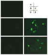
Fig. 7. Visualization of p65/ERK interactions in living MCF-7 cells
Using bimolecular fluorescence complementation analysis, fluorescence images of MCF-7 cells expressing the fusion proteins indicated in each panel were acquired 14–16 h after co-transfection. A, p65-YC156 alone; B, Erk1-YN173 alone; C, p65-YC156 with Erk1-YN173; D, Erk2-YN173 alone; E, p65-YC156 with Erk2-YN173.
Similar articles
- Apoptosis induced by trimethyltin chloride in human neuroblastoma cells SY5Y is regulated by a balance and cross-talk between NF-κB and MAPKs signaling pathways.
Qing Y, Liang Y, Du Q, Fan P, Xu H, Xu Y, Shi N. Qing Y, et al. Arch Toxicol. 2013 Jul;87(7):1273-85. doi: 10.1007/s00204-013-1021-9. Epub 2013 Feb 20. Arch Toxicol. 2013. PMID: 23423712 - The cellular response to oxidative stress: influences of mitogen-activated protein kinase signalling pathways on cell survival.
Wang X, Martindale JL, Liu Y, Holbrook NJ. Wang X, et al. Biochem J. 1998 Jul 15;333 ( Pt 2)(Pt 2):291-300. doi: 10.1042/bj3330291. Biochem J. 1998. PMID: 9657968 Free PMC article. - Akt stimulates the transactivation potential of the RelA/p65 Subunit of NF-kappa B through utilization of the Ikappa B kinase and activation of the mitogen-activated protein kinase p38.
Madrid LV, Mayo MW, Reuther JY, Baldwin AS Jr. Madrid LV, et al. J Biol Chem. 2001 Jun 1;276(22):18934-40. doi: 10.1074/jbc.M101103200. Epub 2001 Mar 20. J Biol Chem. 2001. PMID: 11259436 - Gadd45 proteins as critical signal transducers linking NF-kappaB to MAPK cascades.
Yang Z, Song L, Huang C. Yang Z, et al. Curr Cancer Drug Targets. 2009 Dec;9(8):915-30. doi: 10.2174/156800909790192383. Curr Cancer Drug Targets. 2009. PMID: 20025601 Free PMC article. Review. - Gadd45 in the liver: signal transduction and transcriptional mechanisms.
Tian J, Locker J. Tian J, et al. Adv Exp Med Biol. 2013;793:69-80. doi: 10.1007/978-1-4614-8289-5_5. Adv Exp Med Biol. 2013. PMID: 24104474 Review.
Cited by
- Dual targeting of IGF-1R and PDGFR inhibits proliferation in high-grade gliomas cells and induces radiosensitivity in JNK-1 expressing cells.
Carapancea M, Cosaceanu D, Budiu R, Kwiecinska A, Tataranu L, Ciubotaru V, Alexandru O, Banita M, Pisoschi C, Bäcklund ML, Lewensohn R, Dricu A. Carapancea M, et al. J Neurooncol. 2007 Dec;85(3):245-54. doi: 10.1007/s11060-007-9417-0. Epub 2007 Jun 14. J Neurooncol. 2007. PMID: 17568996 - Shigatoxin-induced endothelin-1 expression in cultured podocytes autocrinally mediates actin remodeling.
Morigi M, Buelli S, Zanchi C, Longaretti L, Macconi D, Benigni A, Moioli D, Remuzzi G, Zoja C. Morigi M, et al. Am J Pathol. 2006 Dec;169(6):1965-75. doi: 10.2353/ajpath.2006.051331. Am J Pathol. 2006. PMID: 17148661 Free PMC article. - NF-kB as a key player in regulation of cellular radiation responses and identification of radiation countermeasures.
Singh V, Gupta D, Arora R. Singh V, et al. Discoveries (Craiova). 2015 Mar 31;3(1):e35. doi: 10.15190/d.2015.27. Discoveries (Craiova). 2015. PMID: 32309561 Free PMC article. Review. - Grifola frondosa Extract Containing Bioactive Components Blocks Skin Fibroblastic Inflammation and Cytotoxicity Caused by Endocrine Disrupting Chemical, Bisphenol A.
Kim JH, Lim SR, Jung DH, Kim EJ, Sung J, Kim SC, Choi CH, Kang JW, Lee SJ. Kim JH, et al. Nutrients. 2022 Sep 15;14(18):3812. doi: 10.3390/nu14183812. Nutrients. 2022. PMID: 36145189 Free PMC article. - Phosphoproteomics profiling of human skin fibroblast cells reveals pathways and proteins affected by low doses of ionizing radiation.
Yang F, Waters KM, Miller JH, Gritsenko MA, Zhao R, Du X, Livesay EA, Purvine SO, Monroe ME, Wang Y, Camp DG 2nd, Smith RD, Stenoien DL. Yang F, et al. PLoS One. 2010 Nov 30;5(11):e14152. doi: 10.1371/journal.pone.0014152. PLoS One. 2010. PMID: 21152398 Free PMC article.
References
- Hartwell LH, Kastan MB. Science. 1994;266:1821–1828. - PubMed
- Bebien M, Salinas S, Becamel C, Richard V, Linares L, Hipskind RA. Oncogene. 2003;22:1836–1847. - PubMed
- Amundson SA, Bittner M, Chen Y, Trent J, Meltzer P, Fornace AJ., Jr Oncogene. 1999;18:3666–3672. - PubMed
- Li Z, Xia L, Lee ML, Khaletskiy A, Wang J, Wong JYC, Li JJ. Radiat Res. 2001;155:543–553. - PubMed
Publication types
MeSH terms
Substances
LinkOut - more resources
Full Text Sources
Research Materials
Miscellaneous
