Oral mucosal endotoxin tolerance induction in chronic periodontitis - PubMed (original) (raw)
Oral mucosal endotoxin tolerance induction in chronic periodontitis
Manoj Muthukuru et al. Infect Immun. 2005 Feb.
Abstract
The oral mucosa is exposed to a high density and diversity of gram-positive and gram-negative bacteria, but very little is known about how immune homeostasis is maintained in this environment, particularly in the inflammatory disease chronic periodontitis (CP). The cells of the innate immune response recognize bacterial structures via the Toll-like receptors (TLR). This activates intracellular signaling and transcription of proteins essential for the induction of an adaptive immune response; however, if unregulated, it can lead to destructive inflammatory responses. Using single-immunoenzyme labeling, we show that the human oral mucosa (gingiva) is infiltrated by large numbers of TLR2(+) and TLR4(+) cells and that their numbers increase significantly in CP, relative to health (P < 0.05, Student's t test). We also show that the numbers of TLR2(+) but not TLR4(+) cells increase linearly with inflammation (r(2) = 0.33, P < 0.05). Double-immunofluorescence analysis confirms that TLR2 is coexpressed by monocytes (MC)/macrophages (mphi) in situ. Further analysis of gingival tissues by quantitative real-time PCR, however, indicates that despite a threefold increase in the expression of interleukin-1beta (IL-1beta) mRNA during CP, there is significant (30-fold) downregulation of TLR2 mRNA (P < 0.05, Student's t test). Also showing similar trends are the levels of TLR4 (ninefold reduction), TLR5 (twofold reduction), and MD-2 (sevenfold reduction) mRNA in CP patients compared to healthy persons, while the level of CD14 was unchanged. In vitro studies with human MC indicate that MC respond to an initial stimulus of lipopolysaccharide (LPS) from Porphyromonas gingivalis (PgLPS) or Escherichia coli (EcLPS) by upregulation of TLR2 and TLR4 mRNA and protein; moreover, IL-1beta mRNA is induced and tumor necrosis factor alpha (TNF-alpha), IL-10, IL-6, and IL-8 proteins are secreted. However, restimulation of MC with either PgLPS or EcLPS downregulates TLR2 and TLR4 mRNA and protein and IL-1beta mRNA and induces a ca. 10-fold reduction in TNF-alpha secretion, suggesting the induction of endotoxin tolerance by either LPS. Less susceptible to tolerance than TNF-alpha were IL-6, IL-10, and IL-8. These studies suggest that certain components of the innate oral mucosal immune response, most notably TLRs and inflammatory cytokines, may become tolerized during sustained exposure to bacterial structures such as LPS and that this may be one mechanism used in the oral mucosa to attempt to regulate local immune responses.
Figures
FIG. 1.
Oral mucosa infiltrated with TLR2+ and TLR4+ cells. Representative serial sections of interproximal gingival tissue from patients with CP (A and C) persons in relative health (B and D) were single-immunoenzyme stained as described in Materials and Methods. Images of TLR2+ cells (A and B) and TLR4+ cells (C and D) were obtained with a 20× objective (panels A1, A2, B1, and B2), a 40× objective (panels B3, B4, C3, and D2), a 10× objective (panels C1, C2, and D1), 100× objective (panels A3, C4, D3, and D4) and captured using image-enhanced light microscopy. The specificity of the antibodies was confirmed by negative staining with the respective isotype controls (e.g., panels A4 and B4). The sections were counterstained with hematoxylin.
FIG. 2.
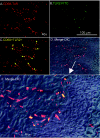
TLR2+ cells in the oral mucosa include MC/mφ. A representative serial section of tissue from a person with CP was stained with monoclonal anti-CD68-Texas Red (CD68-TxR) followed by monoclonal anti-TLR2-FITC (TLR2-FITC) antibodies, as indicated in Materials and Methods. Conjugated mouse immunoglobulins of the same isotypes were used as controls (not shown). (A to C) Shown are image-enhanced double-fluorescence images, sharpened digitally with two-dimensional-deconvolution software. Evident are CD68+ MC/mφ (red) (A), TLR2+ cells (green) (B), and TLR2+ MC/mφ (merge, yellow arrows) (C). (D) A digital merge of a differential interference contrast image with panel C. (E) Final digital enlargement with a magnification of approximately ×1,500.
FIG. 3.
Increased infiltration of gingiva with TLR2- and TLR4-positive cells in persons with CP. Interproximal gingival tissues from subjects with gingival health and those with disease (CP) were single-immunoenzyme labeled as described in Materials and Methods and analyzed by image-enhanced histomorphometry. (A) Shown are the mean number of immunoreactive cells/20× field; error bars indicate standard errors. Asterisks indicate a statistically significant increase in diseased versus healthy tissue by Student's t test (P < 0.05). (B and C) Linear-regression analysis of the number of TLR2+ (B) and TLR4+ (C) cells with the number of H&E-positive inflammatory cells.
FIG. 4.
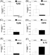
Negative regulation of TLR mRNA in human gingiva from subjects with CP. Eight of the tissues used for immunohistochemistry were randomly selected and bisected for total RNA extraction as described in Materials and Methods, along with four additional samples form healthy and diseased subjects. The total RNA was normalized among all samples, and real-time RT-PCR was used to quantitate TLR2 (A), TLR4 (B), TLR5 (C), CD14 (D), and MD2 (E) mRNA expression levels, along with IL-1β as an inflammatory marker (F). For each transcript, conventional PCR-amplified products (cleaned, with concentrations determined at OD260) were used as standards (from 0.1 to 0.00001 ng) to generate a standard curve for absolute real-time quantitation. The absolute mRNA levels of all the genes were then normalized to β-actin levels of individual tissue samples. All quantitations were performed in triplicate. The normalized initial concentration of each transcript in every sample was converted to the initial copy number (see Materials and Methods). Error bars indicate standard error. The level of mRNA for TLR2 in diseased samples was statistically different from that in samples from healthy controls (P < 0.05, Student's t test).
FIG. 5.
Positive and negative regulation of TLR mRNA in MC by stimulus or challenge with PgLPS or EcLPS. (A and B) MC were stimulated at 37°C for 24 h with 1,000 ng of PgLPS (Pg), EcLPS (Ec), or no LPS (---), challenged with PgLPS (PgPg) or EcLPS (EcEc) for an additional 1 h, pelleted, washed, and processed for total RNA and for analysis of (A) TLR2 mRNA and (B) TLR4 mRNA by quantitative real-time PCR as described in Materials and Methods. The assay was performed three separate times, and the means and standard errors of three separate analyses are shown. (C and D) Fluorescence-activated all sorter analysis of human MC stimulated as in panels (A and B), except that the challenge occurred for 24 h to enable the proteins to be transcribed. A detailed account of the method is given in Materials and Methods. Histograms show mean fluorescence intensity, while numbers in parentheses indicate the geometric means of MC stimulated with the indicated regimen of stimulus and challenge. Results are representative of five separate experiments. FLT-Height, fluorescence intensity of TLRs.
FIG. 6.
Negative regulation of cytokine response by TLR-mediated endotoxin tolerance. (A to D) Supernatants from MC treated with LPS as described in Materials and Methods were analyzed for TNF-α (A), IL-8 (B), IL-6 (C), and IL-10 (D) by flow cytometry using a CBA kit. The assay was performed in triplicate. Based on the standard curve for each cytokine, the CBA software calculates levels in picograms per milliliter. (E) Mean levels of IL-1β mRNA from triplicate samples analyzed by real-time PCR, as described in the legend to Fig. 3 and in Materials and Methods. Error bars indicate standard deviation.
Similar articles
- Intracellular signaling and cytokine induction upon interactions of Porphyromonas gingivalis fimbriae with pattern-recognition receptors.
Hajishengallis G, Sojar H, Genco RJ, DeNardin E. Hajishengallis G, et al. Immunol Invest. 2004 May;33(2):157-72. doi: 10.1081/imm-120030917. Immunol Invest. 2004. PMID: 15195695 - Porphyromonas gingivalis lipopolysaccharide signaling in gingival fibroblasts-CD14 and Toll-like receptors.
Wang PL, Ohura K. Wang PL, et al. Crit Rev Oral Biol Med. 2002;13(2):132-42. doi: 10.1177/154411130201300204. Crit Rev Oral Biol Med. 2002. PMID: 12097356 Review. - Interactions of oral pathogens with toll-like receptors: possible role in atherosclerosis.
Hajishengallis G, Sharma A, Russell MW, Genco RJ. Hajishengallis G, et al. Ann Periodontol. 2002 Dec;7(1):72-8. doi: 10.1902/annals.2002.7.1.72. Ann Periodontol. 2002. PMID: 16013219 Review.
Cited by
- Microbial carriage state of peripheral blood dendritic cells (DCs) in chronic periodontitis influences DC differentiation, atherogenic potential.
Carrion J, Scisci E, Miles B, Sabino GJ, Zeituni AE, Gu Y, Bear A, Genco CA, Brown DL, Cutler CW. Carrion J, et al. J Immunol. 2012 Sep 15;189(6):3178-87. doi: 10.4049/jimmunol.1201053. Epub 2012 Aug 13. J Immunol. 2012. PMID: 22891282 Free PMC article. - Human squamous cell carcinomas evade the immune response by down-regulation of vascular E-selectin and recruitment of regulatory T cells.
Clark RA, Huang SJ, Murphy GF, Mollet IG, Hijnen D, Muthukuru M, Schanbacher CF, Edwards V, Miller DM, Kim JE, Lambert J, Kupper TS. Clark RA, et al. J Exp Med. 2008 Sep 29;205(10):2221-34. doi: 10.1084/jem.20071190. Epub 2008 Sep 15. J Exp Med. 2008. PMID: 18794336 Free PMC article. - AAV2/1-TNFR:Fc gene delivery prevents periodontal disease progression.
Cirelli JA, Park CH, MacKool K, Taba M Jr, Lustig KH, Burstein H, Giannobile WV. Cirelli JA, et al. Gene Ther. 2009 Mar;16(3):426-36. doi: 10.1038/gt.2008.174. Epub 2008 Dec 11. Gene Ther. 2009. PMID: 19078994 Free PMC article. - Exposure of periodontal ligament progenitor cells to lipopolysaccharide from Escherichia coli changes osteoblast differentiation pattern.
Albiero ML, Amorim BR, Martins L, Casati MZ, Sallum EA, Nociti FH Jr, Silvério KG. Albiero ML, et al. J Appl Oral Sci. 2015 Mar-Apr;23(2):145-52. doi: 10.1590/1678-775720140334. J Appl Oral Sci. 2015. PMID: 26018305 Free PMC article. - Macrophage-specific TLR2 signaling mediates pathogen-induced TNF-dependent inflammatory oral bone loss.
Papadopoulos G, Weinberg EO, Massari P, Gibson FC 3rd, Wetzler LM, Morgan EF, Genco CA. Papadopoulos G, et al. J Immunol. 2013 Feb 1;190(3):1148-57. doi: 10.4049/jimmunol.1202511. Epub 2012 Dec 21. J Immunol. 2013. PMID: 23264656 Free PMC article.
References
- Abreu, M. T., P. Vora, E. Faure, L. S. Thomas, E. T. Arnold, and M. Arditi. 2001. Decreased expression of Toll-like receptor-4 and MD-2 correlates with intestinal epithelial cell protection against dysregulated proinflammatory gene expression in response to bacterial lipopolysaccharide. J. Immunol. 167:1609-1616. - PubMed
- Beutler, B., K. Hoebe, X. Du, and R. J. Ulevitch. 2003. How we detect microbes and respond to them: the Toll-like receptors and their transducers. J. Leukoc. Biol. 74:479-485. - PubMed
- Cristofaro, P., S. M. Opal. 2003. The Toll-like receptors and their role in septic shock. Expert Opin. Ther. Targets 7:603-612. - PubMed
- Cohen, N., J. Morisset, and D. Emilie. 2004. Induction of tolerance by Porphyromonas gingivalis on APCs: a mechanism implicated in periodontal infection. J. Dent. Res. 83:429-433. - PubMed
Publication types
MeSH terms
Substances
LinkOut - more resources
Full Text Sources
Other Literature Sources
Research Materials
Miscellaneous


