Avian brains and a new understanding of vertebrate brain evolution - PubMed (original) (raw)
Review
doi: 10.1038/nrn1606.
Onur Güntürkün, Laura Bruce, András Csillag, Harvey Karten, Wayne Kuenzel, Loreta Medina, George Paxinos, David J Perkel, Toru Shimizu, Georg Striedter, J Martin Wild, Gregory F Ball, Jennifer Dugas-Ford, Sarah E Durand, Gerald E Hough, Scott Husband, Lubica Kubikova, Diane W Lee, Claudio V Mello, Alice Powers, Connie Siang, Tom V Smulders, Kazuhiro Wada, Stephanie A White, Keiko Yamamoto, Jing Yu, Anton Reiner, Ann B Butler; Avian Brain Nomenclature Consortium
Affiliations
- PMID: 15685220
- PMCID: PMC2507884
- DOI: 10.1038/nrn1606
Review
Avian brains and a new understanding of vertebrate brain evolution
Erich D Jarvis et al. Nat Rev Neurosci. 2005 Feb.
Abstract
We believe that names have a powerful influence on the experiments we do and the way in which we think. For this reason, and in the light of new evidence about the function and evolution of the vertebrate brain, an international consortium of neuroscientists has reconsidered the traditional, 100-year-old terminology that is used to describe the avian cerebrum. Our current understanding of the avian brain - in particular the neocortex-like cognitive functions of the avian pallium - requires a new terminology that better reflects these functions and the homologies between avian and mammalian brains.
Figures
Figure 1. Avian and mammalian brain relationships
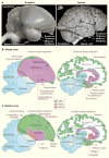
a | Side view of a songbird (zebra finch) and human brain to represent avian and mammalian species. In this view, the songbird cerebrum covers the thalamus; the human cerebrum covers the thalamus and midbrain. Inset (left) next to the human brain is the zebra finch brain to the same scale. Human brain image reproduced, with permission, courtesy of John W. Sundsten, Digital Anatomist Project. b | Classic view of avian and mammalian brain relationships. Although past authors had different opinions about which brain regions are pallium versus subpallium, we have coloured individual brain regions according to the meaning of the names given to those brain regions. Ac, accumbens; B, nucleus basalis; Cd, caudate nucleus; CDL, dorsal lateral corticoid area; E, ectostriatum; GP, globus pallidus (i, internal segment; e, external segment); HA, hyperstriatum accessorium; HV, hyperstriatum ventrale; IHA, interstitial hyperstriatum accessorium; L2, field L2; LPO, lobus parolfactorius; OB, olfactory bulb; Pt, putamen; TuO, olfactory tubercle. c | Modern consensus view of avian and mammalian brain relationships according to the conclusions of the Avian Brain Nomenclature Forum. Solid white lines are lamina (cell-sparse zones separating brain subdivisions). Large white areas in the human cerebrum are axon pathways called white matter. Dashed grey lines divide regions that differ by cell density or cell size; dashed white lines separate primary sensory neuron populations from adjacent regions. Abbreviations where different from b: E, entopallium; B, basorostralis; HA, hyperpallium apicale; Hp, hippocampus; IHA, interstitial hyperpallium apicale; MV, mesopallium ventrale.
Figure 2. Simplified modern view of vertebrate evolution
The diagram begins with the fish group that contains the most recent ancestors of land vertebrates. This differs from the classic view in that instead of giving rise to reptiles, ancestral amphibians are thought to have given rise to stem amniotes. Stem amniotes then split into at least two groups: the sauropsids, which gave rise to all modern reptiles as we know them today; and the therapsids, which, through a series of now-extinct intermediate forms, evolved into mammals. Many sauropsids (reptiles) are currently living. Solid horizontal lines indicate temporal fossil evidence. Dashed lines indicate proposed ancestral links based on other types of data. MYA, million years ago Based on REFS ,.
Figure 3. Auditory and vocal pathways of the songbird brain within the context of the new consensus view of avian brain organization
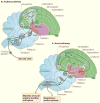
Only the most prominent and/or most studied connections are indicated. a | The auditory pathway. Most of the hindbrain connectivity is extrapolated from non-songbird species. For clarity, reciprocal connections in the pallial auditory areas are not indicated. b | The vocal pathways. Black arrows show connections of the posterior vocal pathway (or vocal motor pathway), white arrows indicate the anterior vocal pathway (or pallial–basal ganglia–thalamic–pallial loop) and dashed lines show connections between the two pathways. Av, avalanche; B, basorostralis; CLM, caudal lateral mesopallium; CMM, caudal medial mesopallium; CN, cochlear nucleus; CSt, caudal striatum; DLM, dorsal lateral nucleus of the medial thalamus; DM, dorsal medial nucleus; E, entopallium; HVC (a letter-based name); L1, L2, L3, fields L1, L2 and L3; LAreaX, lateral AreaX of the striatum; LLD, lateral lemniscus, dorsal nucleus; LLI, lateral lemniscus, intermediate nucleus; LLV, lateral lemniscus, ventral nucleus; LMAN, lateral magnocellular nucleus of the anterior nidopallium; LMO, lateral oval nucleus of the mesopallium; MLd, dorsal lateral nucleus of the mesencephalon; NCM, caudal medial nidopallium; NIf, interfacial nucleus of the nidopallium; nXIIts, nucleus XII, tracheosyringeal part; OB, olfactory bulb; Ov, ovoidalis; PAm, para-ambiguus; RA, robust nucleus of the arcopallium; RAm, retroambiguus; SO, superior olive; Uva, nucleus uvaeformis.
Similar articles
- Revised nomenclature for avian telencephalon and some related brainstem nuclei.
Reiner A, Perkel DJ, Bruce LL, Butler AB, Csillag A, Kuenzel W, Medina L, Paxinos G, Shimizu T, Striedter G, Wild M, Ball GF, Durand S, Güntürkün O, Lee DW, Mello CV, Powers A, White SA, Hough G, Kubikova L, Smulders TV, Wada K, Dugas-Ford J, Husband S, Yamamoto K, Yu J, Siang C, Jarvis ED; Avian Brain Nomenclature Forum. Reiner A, et al. J Comp Neurol. 2004 May 31;473(3):377-414. doi: 10.1002/cne.20118. J Comp Neurol. 2004. PMID: 15116397 Free PMC article. - The Avian Brain Nomenclature Forum: Terminology for a New Century in Comparative Neuroanatomy.
Reiner A, Perkel DJ, Bruce LL, Butler AB, Csillag A, Kuenzel W, Medina L, Paxinos G, Shimizu T, Striedter G, Wild M, Ball GF, Durand S, Gütürkün O, Lee DW, Mello CV, Powers A, White SA, Hough G, Kubikova L, Smulders TV, Wada K, Dugas-Ford J, Husband S, Yamamoto K, Yu J, Siang C, Jarvis ED. Reiner A, et al. J Comp Neurol. 2004;473:E1-E6. doi: 10.1002/cne.20119. J Comp Neurol. 2004. PMID: 19626136 Free PMC article. - A new avian brain nomenclature: why, how and what.
Reiner A. Reiner A. Brain Res Bull. 2005 Sep 15;66(4-6):317-31. doi: 10.1016/j.brainresbull.2005.05.007. Brain Res Bull. 2005. PMID: 16144608 Review. - Songbirds and the revised avian brain nomenclature.
Reiner A, Perkel DJ, Mello CV, Jarvis ED. Reiner A, et al. Ann N Y Acad Sci. 2004 Jun;1016:77-108. doi: 10.1196/annals.1298.013. Ann N Y Acad Sci. 2004. PMID: 15313771 Free PMC article. Review. - Parallel evolution in mammalian and avian brains: comparative cytoarchitectonic and cytochemical analysis.
Rehkämper G, Zilles K. Rehkämper G, et al. Cell Tissue Res. 1991 Jan;263(1):3-28. doi: 10.1007/BF00318396. Cell Tissue Res. 1991. PMID: 2009552 Review.
Cited by
- Learning-related neuronal activation in the zebra finch song system nucleus HVC in response to the bird's own song.
Bolhuis JJ, Gobes SM, Terpstra NJ, den Boer-Visser AM, Zandbergen MA. Bolhuis JJ, et al. PLoS One. 2012;7(7):e41556. doi: 10.1371/journal.pone.0041556. Epub 2012 Jul 25. PLoS One. 2012. PMID: 22848527 Free PMC article. - Morphology, biochemistry and connectivity of Cluster N and the hippocampal formation in a migratory bird.
Heyers D, Musielak I, Haase K, Herold C, Bolte P, Güntürkün O, Mouritsen H. Heyers D, et al. Brain Struct Funct. 2022 Nov;227(8):2731-2749. doi: 10.1007/s00429-022-02566-y. Epub 2022 Sep 17. Brain Struct Funct. 2022. PMID: 36114860 Free PMC article. - Morphology of axonal projections from the high vocal center to vocal motor cortex in songbirds.
Yip ZC, Miller-Sims VC, Bottjer SW. Yip ZC, et al. J Comp Neurol. 2012 Aug 15;520(12):2742-56. doi: 10.1002/cne.23084. J Comp Neurol. 2012. PMID: 22684940 Free PMC article. - Characterization of respiratory neurons in the rostral ventrolateral medulla, an area critical for vocal production in songbirds.
McLean J, Bricault S, Schmidt MF. McLean J, et al. J Neurophysiol. 2013 Feb;109(4):948-57. doi: 10.1152/jn.00595.2012. Epub 2012 Nov 21. J Neurophysiol. 2013. PMID: 23175802 Free PMC article. - The Development of Object Recognition Requires Experience with the Surface Features of Objects.
Wood JN, Wood SMW. Wood JN, et al. Animals (Basel). 2024 Jan 17;14(2):284. doi: 10.3390/ani14020284. Animals (Basel). 2024. PMID: 38254453 Free PMC article.
References
- Edinger L. Investigations on the Comparative Anatomy of the Brain. 1–5. Moritz Diesterweg; Frankfurt/Main: 1888–1903. (Translation from German)
- Darwin C. The Origin of Species. Murray: 1859.
- Edinger L. The Anatomy of the Central Nervous System of Man and of Vertebrates in General. 5. F. A. Davis Company; Philadelphia: 1896.
- Edinger L. The relations of comparative anatomy to comparative psychology. Comp Neurol Psychol. 1908;18:437–457.
- Northcutt RG. Changing views of brain evolution. Brain Res Bull. 2001;55:663–674. - PubMed


