Effects of intravenous dimethyl sulfoxide on ischemia evolution in a rat permanent occlusion model - PubMed (original) (raw)
Effects of intravenous dimethyl sulfoxide on ischemia evolution in a rat permanent occlusion model
Juergen Bardutzky et al. J Cereb Blood Flow Metab. 2005 Aug.
Abstract
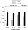
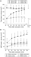
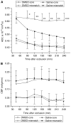
Dimethyl sulfoxide (DMSO) has a variety of biological actions that suggest efficacy as a neuroprotectant. We (1) tested the neuroprotective potential of DMSO at different time windows on infarct size using 2,3,5-triphenyltetrazolium staining and (2) investigated the effects of DMSO on ischemia evolution using quantitative diffusion and perfusion imaging in a permanent middle cerebral artery occlusion (MCAO) model in rats. In experiment 1, DMSO treatment (1.5 g/kg intravenously over 3 h) reduced infarct volume 24 h after MCAO by 65% (P<0.00001) when initiated 20 h before MCAO, by 44% (P=0.0006) when initiated 1 h after MCAO, and by 17% (P=0.11) when started 2 h after MCAO. Significant infarct reduction was also observed after a 3-day survival in animals treated 1 h after MCAO (P=0.005). In experiment 2, treatment was initiated 1 h after MCAO and maps for cerebral blood flow (CBF) and apparent diffusion coefficient (ADC) were acquired before treatment and then every 30 mins up to 4 h. Cerebral blood flow characteristics and CBF-derived lesion volumes did not differ between treated and untreated animals, whereas the ADC-derived lesion volume essentially stopped progressing during DMSO treatment, resulting in a persistent diffusion/perfusion mismatch. This effect was mainly observed in the cortex. Our data suggest that DMSO represents an interesting candidate for acute stroke treatment.
Figures
Figure 1
2,3,5-Triphenyltetrazolium chloride (TTC) -defined corrected infarct volume 24 or 72 h after permanent middle cerebral artery occlusion (MCAO). Data are expressed as mean ±s.d. *P<0.01; **P<0.001; ***P<0.00001.
Figure 2
Representative apparent diffusion coefficient (ADC) and cerebral blood flow (CBF) maps from one control and one dimethyl sulfoxide (DMSO)-treated animal subjected to permanent middle cerebral artery occlusion (MCAO). Three of eight maps are shown at 45 mins (baseline), 120, 180, and 240 mins after occlusion for ADC maps, and at 45 mins for CBF maps. Dimethyl sulfoxide treatment was initiated 1 h after occlusion.
Figure 3
(A) Temporal evolution of total apparent diffusion coefficient (ADC)- and cerebral blood flow (CBF)-derived lesion volumes (mean±s.d.) by using previously validated viability thresholds in permanently occluded rats. Dimethyl sulfoxide (DMSO) or saline infusion was initiated 1 h after occlusion. Saline group: CBF-lesion=closed diamonds, ADC-lesion= closed squares, 2,3,5-triphenyltetrazolium chloride (TTC)-lesion=gray square; DMSO group: CBF-lesion=open triangles, ADC-lesion=open circles, TTC-lesion=gray circle. (B) Temporal evolution of cortical and subcortical ADC lesion volumes (mean±s.d.). Saline lesion: cortex=closed circles, subcortex =closed squares; DMSO lesion: cortex=open circles, subcortex=open squares. *P<0.05 for differences in ADC lesion volumes at same time points. **_P_=0.002 for difference in TTC lesion volume at 24 h after occlusion.
Figure 4
Temporal evolution of apparent diffusion coefficient (ADC) (A) and cerebral blood flow (CBF) (B) in the core and mismatch regions defined before treatment (45 mins) in permanently occluded rats. Dimethyl sulfoxide (DMSO) or saline was administered 1 h after occlusion. Dimethyl sulfoxide group: core=open triangle, mismatch=open circles. Saline group: core=closed triangles, mismatch=closed circles. *P<0.05; **P<0.01; ***P<0.001.
Similar articles
- Differences in ischemic lesion evolution in different rat strains using diffusion and perfusion imaging.
Bardutzky J, Shen Q, Henninger N, Bouley J, Duong TQ, Fisher M. Bardutzky J, et al. Stroke. 2005 Sep;36(9):2000-5. doi: 10.1161/01.STR.0000177486.85508.4d. Epub 2005 Jul 21. Stroke. 2005. PMID: 16040589 Free PMC article. - Normobaric hyperoxia delays perfusion/diffusion mismatch evolution, reduces infarct volume, and differentially affects neuronal cell death pathways after suture middle cerebral artery occlusion in rats.
Henninger N, Bouley J, Nelligan JM, Sicard KM, Fisher M. Henninger N, et al. J Cereb Blood Flow Metab. 2007 Sep;27(9):1632-42. doi: 10.1038/sj.jcbfm.9600463. Epub 2007 Feb 21. J Cereb Blood Flow Metab. 2007. PMID: 17311078 - Comparison between coated vs. uncoated suture middle cerebral artery occlusion in the rat as assessed by perfusion/diffusion weighted imaging.
Bouley J, Fisher M, Henninger N. Bouley J, et al. Neurosci Lett. 2007 Feb 2;412(3):185-90. doi: 10.1016/j.neulet.2006.11.003. Epub 2006 Nov 22. Neurosci Lett. 2007. PMID: 17123725 - Role of dimethyl sulfoxide in prostaglandin-thromboxane and platelet systems after cerebral ischemia.
de la Torre JC. de la Torre JC. Ann N Y Acad Sci. 1983;411:293-308. doi: 10.1111/j.1749-6632.1983.tb47311.x. Ann N Y Acad Sci. 1983. PMID: 6349494 Review. - Elevated body swing test after focal cerebral ischemia in rodents: methodological considerations.
Ingberg E, Gudjonsdottir J, Theodorsson E, Theodorsson A, Ström JO. Ingberg E, et al. BMC Neurosci. 2015 Aug 5;16:50. doi: 10.1186/s12868-015-0189-8. BMC Neurosci. 2015. PMID: 26242584 Free PMC article. Review.
Cited by
- Neuroprotective Action of the CB1/2 Receptor Agonist, WIN 55,212-2, against DMSO but Not Phenobarbital-Induced Neurotoxicity in Immature Rats.
Huizenga MN, Forcelli PA. Huizenga MN, et al. Neurotox Res. 2019 Jan;35(1):173-182. doi: 10.1007/s12640-018-9944-9. Epub 2018 Aug 24. Neurotox Res. 2019. PMID: 30141144 Free PMC article. - Mitochondrial functional state impacts spontaneous neocortical activity and resting state FMRI.
Sanganahalli BG, Herman P, Hyder F, Kannurpatti SS. Sanganahalli BG, et al. PLoS One. 2013 May 1;8(5):e63317. doi: 10.1371/journal.pone.0063317. Print 2013. PLoS One. 2013. PMID: 23650561 Free PMC article. - Multi-site laser Doppler flowmetry for assessing collateral flow in experimental ischemic stroke: Validation of outcome prediction with acute MRI.
Cuccione E, Versace A, Cho TH, Carone D, Berner LP, Ong E, Rousseau D, Cai R, Monza L, Ferrarese C, Sganzerla EP, Berthezène Y, Nighoghossian N, Wiart M, Beretta S, Chauveau F. Cuccione E, et al. J Cereb Blood Flow Metab. 2017 Jun;37(6):2159-2170. doi: 10.1177/0271678X16661567. Epub 2016 Jan 1. J Cereb Blood Flow Metab. 2017. PMID: 27466372 Free PMC article. - Cerebroprotective Effects of the TLR4-Binding DNA Aptamer ApTOLL in a Rat Model of Ischemic Stroke and Thrombectomy Recanalization.
Aliena-Valero A, Hernández-Jiménez M, López-Morales MA, Tamayo-Torres E, Castelló-Ruiz M, Piñeiro D, Ribó M, Salom JB. Aliena-Valero A, et al. Pharmaceutics. 2024 May 30;16(6):741. doi: 10.3390/pharmaceutics16060741. Pharmaceutics. 2024. PMID: 38931862 Free PMC article. - Neuroprotective activity of (1S,2E,4R,6R,-7E,11E)-2,7,11-cembratriene-4,6-diol (4R) in vitro and in vivo in rodent models of brain ischemia.
Martins AH, Hu J, Xu Z, Mu C, Alvarez P, Ford BD, El Sayed K, Eterovic VA, Ferchmin PA, Hao J. Martins AH, et al. Neuroscience. 2015 Apr 16;291:250-259. doi: 10.1016/j.neuroscience.2015.02.001. Epub 2015 Feb 10. Neuroscience. 2015. PMID: 25677097 Free PMC article.
References
- Back T, Zhao W, Ginsberg MD. Three-dimensional image analysis of brain glucose metabolism–blood flow uncoupling and its electrophysiological correlates in the acute ischemic penumbra following middle cerebral artery occlusion. J Cereb Blood Flow Metab. 1995;15:566–77. - PubMed
- Belayev L, Zhao W, Busto R, Ginsberg MD. Transient middle cerebral artery occlusion by intraluminal suture: I. Three-dimensional autoradiographic image-analysis of local cerebral glucose metabolism-blood flow interrelationships during ischemia and early recirculation. J Cereb Blood Flow Metab. 1997;17:1266–80. - PubMed
- Camp PE, James HE, Werner R. Acute dimethyl sulfoxide therapy in experimental brain edema: Part I. Effects on intracranial pressure, blood pressure, central venous pressure, and brain water and electrolyte content. Neurosurgery. 1981;9:28–33. - PubMed
- Chalela JA, Kang DW, Luby M, Ezzeddine M, Latour LL, Todd JW, Dunn B, Warach S. Early magnetic resonance imaging findings in patients receiving tissue plasminogen activator predict outcome: Insights into the pathophysiology of acute stroke in the thrombolysis era. Ann Neurol. 2004;55:105–12. - PubMed
- Christou I, Alexandrov AV, Burgin WS, Wojner AW, Felberg RA, Malkoff M, Grotta JC. Timing of recanalization after tissue plasminogen activator therapy determined by transcranial doppler correlates with clinical recovery from ischemic stroke. Stroke. 2000;31:1812–6. - PubMed
MeSH terms
Substances
Grants and funding
- R01 NS045879-04/NS/NINDS NIH HHS/United States
- R01 NS045879-03/NS/NINDS NIH HHS/United States
- R01 NS045879-02/NS/NINDS NIH HHS/United States
- R01 NS045879-01A1/NS/NINDS NIH HHS/United States
- R01 NS045879-06/NS/NINDS NIH HHS/United States
- R01 NS045879-05/NS/NINDS NIH HHS/United States
- R01 NS045879/NS/NINDS NIH HHS/United States
LinkOut - more resources
Full Text Sources
Other Literature Sources


