Decisions under uncertainty: probabilistic context influences activation of prefrontal and parietal cortices - PubMed (original) (raw)
Comparative Study
Decisions under uncertainty: probabilistic context influences activation of prefrontal and parietal cortices
Scott A Huettel et al. J Neurosci. 2005.
Abstract
Many decisions are made under uncertainty; that is, with limited information about their potential consequences. Previous neuroimaging studies of decision making have implicated regions of the medial frontal lobe in processes related to the resolution of uncertainty. However, a different set of regions in dorsal prefrontal and posterior parietal cortices has been reported to be critical for selection of actions to unexpected or unpredicted stimuli within a sequence. In the current study, we induced uncertainty using a novel task that required subjects to base their decisions on a binary sequence of eight stimuli so that uncertainty changed dynamically over time (from 20 to 50%), depending on which stimuli were presented. Activation within prefrontal, parietal, and insular cortices increased with increasing uncertainty. In contrast, within medial frontal regions, as well as motor and visual cortices, activation did not increase with increasing uncertainty. We conclude that the brain response to uncertainty depends on the demands of the experimental task. When uncertainty depends on learned associations between stimuli and responses, as in previous studies, it modulates activation in the medial frontal lobes. However, when uncertainty develops over short time scales as information is accumulated toward a decision, dorsal prefrontal and posterior parietal contributions are critical for its resolution. The distinction between neural mechanisms subserving different forms of uncertainty resolution provides an important constraint for neuroeconomic models of decision making.
Figures
Figure 1.
Experimental design. A, In each trial, subjects viewed a series of eight context stimuli that provided information about which decision to make in that trial. Each context stimulus was a circle or triangle presented rapidly (duration, 400 ms; interval, 350 ms) at fixation. After a variable delay interval, subjects indicated their decision by pressing one of four buttons on a response box: high-confidence circle, low-confidence circle, low-confidence triangle, and high-confidence triangle. Subsequently, after a second variable delay, a feedback stimulus was revealed that was consistent or inconsistent with the decision. B, Uncertainty varied systematically with the number of stimuli of each type, as shown for these example sequences. If all stimuli were of one type, there was only 20% uncertainty; that is, the feedback stimulus was 80% likely to be of the presented type. However, as the proportion of stimuli grew more even, the uncertainty associated with selecting the more probable stimulus increased.
Figure 2.
Behavioral results. For each subject, we counted the number of each possible response as a function of the numbers of stimuli of each type that were presented, from 0 triangles/8 circles to 8 triangles/0 circles (_x_-axis). We weighted high-confidence judgments as ± 2 and low-confidence judgments as ± 1 and plotted the resulting mean judgment on the _y_-axis. All subjects' decisions (colored lines) showed sensitivity to the number of stimuli presented, indicating that our stimulus manipulation had the expected effects on decision uncertainty.
Figure 3.
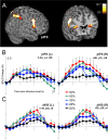
Regions of frontal and insular cortices for which task-related activation was significantly modulated by uncertainty. A, Lateral and cross-sectional views of frontal cortex, with the red-yellow color map (corrected p range, 0.05-0.000001; uncorrected range, 10-7 to 10-12) indicating regions that exhibited significant task-related activation for which amplitude across conditions depended on stimulus uncertainty. B, Within regions of dlPFC, notably the posterior inferior frontal sulcus (pIFS), significant effects of uncertainty on fMRI signal were observed. In these regions, fMRI activation increased as the proportion of uncertainty in the decision increased. L, Left; R, right. C, Similar uncertainty effects were observed within the anterior insula. All of the time courses on this and subsequent figures present amplitude of fMRI BOLD signal (_y_-axis) as a function of the time since the onset of the context-phase stimuli (_x_-axis).
Figure 4.
Regions of parietal cortex for which task-related activation was significantly modulated by uncertainty. A, Cross-sectional view of parietal and occipital cortices, with the red-yellow color map (corrected p range, 0.05-0.000001; uncorrected range, 10-7 to 10-12) indicating regions that exhibited significant task-related activation for which amplitude across conditions depended on stimulus uncertainty. B, Within posterior parietal cortex, fMRI BOLD activation systematically scaled with decision uncertainty, as plotted here for regions along the intraparietal sulcus, bilaterally. IPS, Intraparietal sulcus; L, left; R, right.
Figure 5.
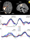
Regions with a significant intercept in the regression analyses, indicating significant activation in the absence of uncertainty. A, In the displayed superior and sagittal views, significant uncertainty-independent activation was observed primarily within visual and motor regions (corrected p range, 0.05-0.000001; uncorrected range, 10-7 to 10-12). B, Activation in visual regions [e.g., middle occipital gyrus (MOG)] peaked early in the trial, reflecting the presentation of the context stimuli, and activation in motor regions [e.g., precentral gyrus (PreCG)] peaked later in the trial, reflecting the subject's response. C, The FMC exhibited activation in all conditions that did not differ as a function of uncertainty. Visual examination of the activation in anterior FMC (aFMC) suggests that activation in the minimal uncertainty condition was less than that in all other conditions, which did not differ from each other. pFMC, Posterior FMC; L, left; R, right.
Figure 6.
Effects of the timing of the incongruent stimulus in the 27.5% uncertainty condition (7 congruent; 1 incongruent). A, Significantly greater activation was observed within the intraparietal sulcus (IPS) bilaterally when the single incongruent stimulus was presented in the second half of the 8-stimulus sequence, compared with when it was presented in the first half of the sequence (p range, uncorrected, 0.001-0.00001). B, Examination of the time course of activation revealed that the late incongruent stimuli evoked activation during the decision phase of the task. Note that the context stimuli were presented over the first 6 s (4 time points) of each trial, and the observed change in fMRI activation attributable to the late incongruent event peaked ∼12 s after trial onset. L, Left; R, right.
Similar articles
- Sensory-motor mechanisms in human parietal cortex underlie arbitrary visual decisions.
Tosoni A, Galati G, Romani GL, Corbetta M. Tosoni A, et al. Nat Neurosci. 2008 Dec;11(12):1446-53. doi: 10.1038/nn.2221. Epub 2008 Nov 9. Nat Neurosci. 2008. PMID: 18997791 Free PMC article. - Specific and nonspecific neural activity during selective processing of visual representations in working memory.
Oh H, Leung HC. Oh H, et al. J Cogn Neurosci. 2010 Feb;22(2):292-306. doi: 10.1162/jocn.2009.21250. J Cogn Neurosci. 2010. PMID: 19400681 - Comparison of fMRI activation at 3 and 1.5 T during perceptual, cognitive, and affective processing.
Krasnow B, Tamm L, Greicius MD, Yang TT, Glover GH, Reiss AL, Menon V. Krasnow B, et al. Neuroimage. 2003 Apr;18(4):813-26. doi: 10.1016/s1053-8119(03)00002-8. Neuroimage. 2003. PMID: 12725758 Clinical Trial. - Long-range neural coupling through synchronization with attention.
Gregoriou GG, Gotts SJ, Zhou H, Desimone R. Gregoriou GG, et al. Prog Brain Res. 2009;176:35-45. doi: 10.1016/S0079-6123(09)17603-3. Prog Brain Res. 2009. PMID: 19733748 Review. - Top-down and bottom-up attention to memory: a hypothesis (AtoM) on the role of the posterior parietal cortex in memory retrieval.
Ciaramelli E, Grady CL, Moscovitch M. Ciaramelli E, et al. Neuropsychologia. 2008;46(7):1828-51. doi: 10.1016/j.neuropsychologia.2008.03.022. Epub 2008 Apr 8. Neuropsychologia. 2008. PMID: 18471837 Review.
Cited by
- A causal role of the right dorsolateral prefrontal cortex in random exploration.
Toghi A, Chizari M, Khosrowabadi R. Toghi A, et al. Sci Rep. 2024 Oct 22;14(1):24796. doi: 10.1038/s41598-024-76025-5. Sci Rep. 2024. PMID: 39433838 Free PMC article. - Neural correlates of harm avoidance: a multimodal meta-analysis of brain structural and resting-state functional neuroimaging studies.
Zhong S, Lin J, Zhang L, Wang S, Kemp GJ, Li L, Gong Q. Zhong S, et al. Transl Psychiatry. 2024 Sep 20;14(1):384. doi: 10.1038/s41398-024-03091-8. Transl Psychiatry. 2024. PMID: 39304648 Free PMC article. - Functional brain network properties correlate with individual risk tolerance in young adults.
Jung WH. Jung WH. Heliyon. 2024 Aug 6;10(15):e35873. doi: 10.1016/j.heliyon.2024.e35873. eCollection 2024 Aug 15. Heliyon. 2024. PMID: 39170166 Free PMC article. - Motivational Modulation Enhances Movement Performance in Parkinson's Disease: A Systematic Review.
Papa EV, Tolman J, Meyerhoeffer C, Reierson K. Papa EV, et al. Phys Ther Rev. 2024;29(1-3):117-127. doi: 10.1080/10833196.2024.2365568. Epub 2024 Jul 2. Phys Ther Rev. 2024. PMID: 39036073 - The visual cortex in the blind but not the auditory cortex in the deaf becomes multiple-demand regions.
Duymuş H, Verma M, Güçlütürk Y, Öztürk M, Varol AB, Kurt Ş, Gezici T, Akgür BF, Giray İ, Öksüz EE, Farooqui AA. Duymuş H, et al. Brain. 2024 Oct 3;147(10):3624-3637. doi: 10.1093/brain/awae187. Brain. 2024. PMID: 38864500 Free PMC article.
References
- Adolphs R (2002) Neural systems for recognizing emotion. Curr Opin Neurobiol 12: 169-177. - PubMed
- Bechara A (2001) Neurobiology of decision-making: risk and reward. Semin Clin Neuropsychiatry 6: 205-216. - PubMed
- Bechara A, Damasio AR, Damasio H, Anderson SW (1994) Insensitivity to future consequences following damage to human prefrontal cortex. Cognition 50: 7-15. - PubMed
- Bechara A, Tranel D, Damasio H (2000) Characterization of the decision-making deficit of patients with ventromedial prefrontal cortex lesions. Brain 123: 2189-2202. - PubMed
- Bunge SA, Hazeltine E, Scanlon MD, Rosen AC, Gabrieli JD (2002) Dissociable contributions of prefrontal and parietal cortices to response selection. NeuroImage 17: 1562-1571. - PubMed
Publication types
MeSH terms
Substances
Grants and funding
- P01 NS041328/NS/NINDS NIH HHS/United States
- R01 NS050329/NS/NINDS NIH HHS/United States
- NS-41328/NS/NINDS NIH HHS/United States
- DA-16214/DA/NIDA NIH HHS/United States
- NS-50329/NS/NINDS NIH HHS/United States
LinkOut - more resources
Full Text Sources
Other Literature Sources


