Identification of novel vascular endothelial-specific genes by the microarray analysis of the zebrafish cloche mutants - PubMed (original) (raw)
Identification of novel vascular endothelial-specific genes by the microarray analysis of the zebrafish cloche mutants
Saulius Sumanas et al. Blood. 2005.
Abstract
The zebrafish cloche (clo) mutation affects the earliest known step in differentiation of blood and endothelial cells in vertebrates. We established clo/gata1-GFP transgenic line with erythroid-specific green fluorescent protein (GFP) expression, which allowed differentiation of clo and wild-type siblings at the midsomitogenesis stages before morphologically visible phenotypes appeared. To discover novel genes potentially involved in hematopoietic and vascular development, we performed microarray analysis of more than 15,000 zebrafish genes or expressed sequence tags (ESTs) in clo mutant embryos. We isolated the full-length sequences and determined the expression patterns for 8 novel cDNAs that were significantly down-regulated in clo-/- embryos. Dual specificity phosphatase 5 (dusp5), cadherin 5 (cdh5; VE-cadherin), aquaporin 8 (aqp8), adrenomedullin receptor (admr), complement receptor C1qR-like (crl), scavenger receptor class F, member 1 (scarf1), and ETS1-like protein (etsrp) were specifically expressed in the vascular endothelial cells, while retinol binding protein 4 (rbp4) was expressed in the yolk syncytial layer and the hypochord. Further functional studies of these novel genes should help to elucidate critical early steps leading to the formation of vertebrate blood vessels.
Figures
Figure 1.
Gata1-GFP homozygous transgenic fish were used to identify _clo_–/– mutants at the 15-somite stage. (A) Wild-type embryo; (B) clo_–/_– embryo. Note that GFP fluorescence in hematopoietic precursor cells (arrows) is absent in the clo_–/_– embryo, while nonspecific GFP expression in the anterior region is not affected (arrowheads).
Figure 2.
Schematic view of the protein domains present in the novel vasculature-specific zebrafish proteins. RHOD denotes a Rhodanese homology domain; DSP, dual specificity phosphatase; CADH, cadherin repeat; CADH_C, cadherin cytoplasmic domain; 7-TM, 7 transmembrane domains; MIP, major intrinsic protein family domain; CLECT, C-type lectin; EGF-Ca, calcium binding EGF-like homology domain; EGF, EGF-like homology domain; TM, transmembrane domain; and ETS, ETS family DNA binding domain.
Figure 3.
Expression pattern of the dual specificity phosphatase 5 (dusp5) as analyzed by in situ hybridization. Anterior is to the left in all panels. (A)The 3-somite stage. A flat mount of the deyolked embryo. dusp5 is expressed in presumptive endothelial precursor cells, which form 2 bilateral stripes of cells that merge in the posterior. The 15-somite stage: (B) lateral view and (C) dorsal view. dusp5 is expressed in 2 bilateral stripes of cells within the lateral mesoderm in the anterior (arrow in B) and middle/posterior (arrowhead in B) parts of an embryo. In addition, a stripe of _dusp5_-expressing cells is apparent at the midline and extends through the middle and posterior parts of an embryo (arrowhead in C). (D) The 26-hpf stage. dusp5 expressed in all endothelial cells of the axial, head, and intersomitic vessels. The 52-hpf stage: (E) lateral view, (F) ventral view, and (inset) rostral view. dusp5 is expressed in a subset of nonvascular cells in the forebrain (arrowhead).
Figure 4.
Expression patterns of the cadherin 5 and aquaporin 8. Anterior is to the left in all panels. (A-D) cdh5 expression; (E-H) aqp8 expression. (A) The 15-somite stage. cdh5 mRNA is expressed in 2 distinct anterior (arrow) and middle/posterior (arrowhead) domains within lateral mesoderm. (B) The 26-hpf stage. cdh5 mRNA is expressed in all vascular endothelial cells of axial, intersegmental, and head vessels. (C) The 52-hpf stage, lateral view. cdh5 mRNA is localized to multiple head vessels, aortic arches (aa), pectoral fin bud (fb) vessel, heart (h) endocardium, the common cardinal vein (ccv), the cardinal vein plexus region at the tail (pl), posterior intersegmental vessels, and the dorsal longitudinal anastomotic vessel (dlav). (D) The 52-hpf stage, dorsal view. cdh5 mRNA is expressed in a group of cells within the diencephalon (arrowhead). Inset, ventral view of heart area. cdh5 is expressed in heart endocardium. (E) The 15-somite stage. aqp8 mRNA is expressed within lateral mesoderm in the posterior half on embryo (arrowhead). (F) The 26-hpf stage. aqp8 mRNA is localized to the dorsal aorta, cardinal vein plexus region, and intersegmental vessels. (G) The 36-hpf stage. aqp8 is expressed in the posterior part of the dorsal aorta, intersegmental vessels, the dorsal longitudinal anastomotic vessel (dlav), the cardinal vein plexus region (pl), and heart endocardium. (H) The 72-hpf stage. aqp8 is expressed in the dorsal longitudinal anastomotic vessel (dlav), the pectoral finbud vessel (fb), and faintly in the posterior intersegmental vessels.
Figure 5.
Expression patterns of the adrenomedullin receptor (admr), C1qR-like (crl), scavenger receptor class F (scarf1), and retinol binding protein 4 (rbp4) mRNAs. (A-B) admr expression, (C-D) crl expression, (E) scarf1 expression, and (F) rbp4 expression. (A) The 26-hpf stage. admr is localized to endothelial cells of axial, head, and intersegmental vessels. (B) The 52-hpf stage. admr expression is restricted to the main axial vessels, a subset of head vessels, and the common cardinal vein (ccv). (C) The 26-hpf stage. crl is expressed in endothelial cells of axial, head, and intersegmental vessels. (D) The 52-hpf stage. crl is expressed in the common cardinal vein (ccv), the pectoral fin bud vessel (fb), and a subset of head vessels. (E) The 22-somite stage. scarf1 is localized within lateral mesoderm to the presumptive endothelial cell precursors (arrowhead). (F) The 24-hpf stage. rbp4 mRNA is localized to the yolk syncytial layer and several hypochordal cells (arrowheads); higher magnification in the inset.
Figure 6.
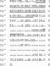
Zebrafish C1qR-like protein is distantly related to the human complement receptor C1qR precursor protein (hC1qR1). Lectin homology domain is underlined; a line above a section indicates EGF-Ca domain; identical and similar amino acids are shaded. GeneWorks 2.5 software (Oxford Molecular Group, Campbell, CA) was used for the alignment.
Figure 7.
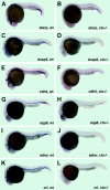
Expression of vascular-specific genes in cloche mutants at 22 to 24 hpf. (A-B) etsrp expression in the vasculature of wild-type siblings (A) and clo_–/_– embryos (B). Note that only a few _etsrp_-expressing cells in the cardinal vein plexus region are remaining in clo mutant embryos. (C-D) dusp5 expression in wild-type siblings (C) and clo_–/_– embryos (D). Note that only a few cells with faint dusp5 expression in the cardinal vein plexus region are remaining in clo mutant embryos. (E-F) cdh5 mRNA expression in wild-type siblings (E) and clo_–/_– embryos (F). Note that only a few _cdh5_-expressing cells in the cardinal vein plexus region are remaining in clo mutant embryos. (G-H) aqp8 expression in wild-type siblings (G) and clo_–/_– embryos (H). Note that only a few _aqp8_-expressing cells in the cardinal vein plexus region are remaining in clo mutant embryos. (I-J) admr expression in wild-type siblings (I) and clo_–/_– embryos (J). Note that only a few cells with a very faint admr expression in the cardinal vein plexus region are remaining in clo mutant embryos. (K-L) crl expression in wild-type siblings (K) and clo_–/_– embryos (L). Note that no _crl_-expressing cells are remaining in clo mutant embryos.
Similar articles
- Microarray analysis of zebrafish cloche mutant using amplified cDNA and identification of potential downstream target genes.
Qian F, Zhen F, Ong C, Jin SW, Meng Soo H, Stainier DY, Lin S, Peng J, Wen Z. Qian F, et al. Dev Dyn. 2005 Jul;233(3):1163-72. doi: 10.1002/dvdy.20444. Dev Dyn. 2005. PMID: 15937927 - SCL/Tal-1 transcription factor acts downstream of cloche to specify hematopoietic and vascular progenitors in zebrafish.
Liao EC, Paw BH, Oates AC, Pratt SJ, Postlethwait JH, Zon LI. Liao EC, et al. Genes Dev. 1998 Mar 1;12(5):621-6. doi: 10.1101/gad.12.5.621. Genes Dev. 1998. PMID: 9499398 Free PMC article. - The zebrafish gene cloche acts upstream of a flk-1 homologue to regulate endothelial cell differentiation.
Liao W, Bisgrove BW, Sawyer H, Hug B, Bell B, Peters K, Grunwald DJ, Stainier DY. Liao W, et al. Development. 1997 Jan;124(2):381-9. doi: 10.1242/dev.124.2.381. Development. 1997. PMID: 9053314 - The cloche and spadetail genes differentially affect hematopoiesis and vasculogenesis.
Thompson MA, Ransom DG, Pratt SJ, MacLennan H, Kieran MW, Detrich HW 3rd, Vail B, Huber TL, Paw B, Brownlie AJ, Oates AC, Fritz A, Gates MA, Amores A, Bahary N, Talbot WS, Her H, Beier DR, Postlethwait JH, Zon LI. Thompson MA, et al. Dev Biol. 1998 May 15;197(2):248-69. doi: 10.1006/dbio.1998.8887. Dev Biol. 1998. PMID: 9630750 - Characterization of a weak allele of zebrafish cloche mutant.
Ma N, Huang Z, Chen X, He F, Wang K, Liu W, Zhao L, Xu X, Liao W, Ruan H, Luo S, Zhang W. Ma N, et al. PLoS One. 2011;6(11):e27540. doi: 10.1371/journal.pone.0027540. Epub 2011 Nov 23. PLoS One. 2011. PMID: 22132109 Free PMC article.
Cited by
- Etv2 and fli1b function together as key regulators of vasculogenesis and angiogenesis.
Craig MP, Grajevskaja V, Liao HK, Balciuniene J, Ekker SC, Park JS, Essner JJ, Balciunas D, Sumanas S. Craig MP, et al. Arterioscler Thromb Vasc Biol. 2015 Apr;35(4):865-76. doi: 10.1161/ATVBAHA.114.304768. Epub 2015 Feb 26. Arterioscler Thromb Vasc Biol. 2015. PMID: 25722433 Free PMC article. - SH2 domain protein E and ABL signaling regulate blood vessel size.
Schumacher JA, Wright ZA, Rufin Florat D, Anand SK, Dasyani M, Batta SPR, Laverde V, Ferrari K, Klimkaite L, Bredemeier NO, Gurung S, Koller GM, Aguera KN, Chadwick GP, Johnson RD, Davis GE, Sumanas S. Schumacher JA, et al. PLoS Genet. 2024 Jan 8;20(1):e1010851. doi: 10.1371/journal.pgen.1010851. eCollection 2024 Jan. PLoS Genet. 2024. PMID: 38190417 Free PMC article. - Global analysis of hematopoietic and vascular endothelial gene expression by tissue specific microarray profiling in zebrafish.
Covassin L, Amigo JD, Suzuki K, Teplyuk V, Straubhaar J, Lawson ND. Covassin L, et al. Dev Biol. 2006 Nov 15;299(2):551-62. doi: 10.1016/j.ydbio.2006.08.020. Epub 2006 Aug 10. Dev Biol. 2006. PMID: 16999953 Free PMC article. - The "Ets" factor: vessel formation in zebrafish--the missing link?
Patterson LJ, Patient R. Patterson LJ, et al. PLoS Biol. 2006 Jan;4(1):e24. doi: 10.1371/journal.pbio.0040024. PLoS Biol. 2006. PMID: 16535776 Free PMC article. - A web based resource characterizing the zebrafish developmental profile of over 16,000 transcripts.
Ouyang M, Garnett AT, Han TM, Hama K, Lee A, Deng Y, Lee N, Liu HY, Amacher SL, Farber SA, Ho SY. Ouyang M, et al. Gene Expr Patterns. 2008 Feb;8(3):171-80. doi: 10.1016/j.gep.2007.10.011. Epub 2007 Nov 7. Gene Expr Patterns. 2008. PMID: 18068546 Free PMC article.
References
- Ema M, Rossant J. Cell fate decisions in early blood vessel formation. Trends Cardiovasc Med. 2003;13: 254-259. - PubMed
- Choi K, Kennedy M, Kazarov A, Papadimitriou JC, Keller G. A common precursor for hematopoietic and endothelial cells. Development. 1998; 125: 725-732. - PubMed
- Drake CJ. Embryonic and adult vasculogenesis. Birth Defects Res Part C Embryo Today. 2003;69: 73-82. - PubMed
- Isogai S, Horiguchi M, Weinstein BM. The vascular anatomy of the developing zebrafish: an atlas of embryonic and early larval development. Dev Biol. 2001;230: 278-301. - PubMed
Publication types
MeSH terms
Substances
LinkOut - more resources
Full Text Sources
Other Literature Sources
Molecular Biology Databases
Miscellaneous


