Functional analysis of human microtubule-based motor proteins, the kinesins and dyneins, in mitosis/cytokinesis using RNA interference - PubMed (original) (raw)
Functional analysis of human microtubule-based motor proteins, the kinesins and dyneins, in mitosis/cytokinesis using RNA interference
Changjun Zhu et al. Mol Biol Cell. 2005 Jul.
Abstract
Microtubule (MT)-based motor proteins, kinesins and dyneins, play important roles in multiple cellular processes including cell division. In this study, we describe the generation and use of an Escherichia coli RNase III-prepared human kinesin/dynein esiRNA library to systematically analyze the functions of all human kinesin/dynein MT motor proteins. Our results indicate that at least 12 kinesins are involved in mitosis and cytokinesis. Eg5 (a member of the kinesin-5 family), Kif2A (a member of the kinesin-13 family), and KifC1 (a member of the kinesin-14 family) are crucial for spindle formation; KifC1, MCAK (a member of the kinesin-13 family), CENP-E (a member of the kinesin-7 family), Kif14 (a member of the kinesin-3 family), Kif18 (a member of the kinesin-8 family), and Kid (a member of the kinesin-10 family) are required for chromosome congression and alignment; Kif4A and Kif4B (members of the kinesin-4 family) have roles in anaphase spindle dynamics; and Kif4A, Kif4B, MKLP1, and MKLP2 (members of the kinesin-6 family) are essential for cytokinesis. Using immunofluorescence analysis, time-lapse microscopy, and rescue experiments, we investigate the roles of these 12 kinesins in detail.
Figures
Figure 1.
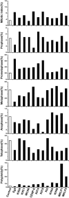
Quantitation of mitotic and cytokinetic defects in HeLa cells treated with kinesin esiRNAs. HeLa cells were transfected with 100 nM of control (luciferase) esiRNA or kinesin esiRNA. Two days posttransfection, cells were fixed and stained with anti-α-tubulin antibody and DAPI. Mitotic index and the percentage of cells at different stages of mitosis and cytokinesis were scored using a fluorescence microscope. For mitotic index and polyploidy analyses, more than 500 mitotic and interphase cells were counted. For mitosis analyses, more than 150 mitotic cells were counted.
Figure 2.
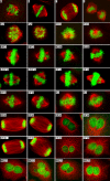
Typical abnormal mitotic and cytokinetic phenotype(s) observed in HeLa cells treated with kinesin esiRNAs. HeLa cells were transfected with kinesin esiRNAs as in Figure 1. Two days posttransfection, cells were fixed, stained with anti-α-tubulin antibody (red) and DAPI (green) and photographed using a fluorescence microscope. Cells transfected with control (luciferase) esiRNA undergo normal mitosis and cytokinesis (A, prometaphase; B, metaphase; C, anaphase; and D, telophase). Cells transfected with Eg5 or Kif2A esiRNA exhibit monopolar spindles. Cells transfected with KifC1 show multipolar spindles and misaligned chromosomes. Cells transfected with MCAK, CENP-E, Kif14, Kif18, or Kid show misaligned chromosomes. Cells transfected with Kif4A or Kif4B show abnormal anaphase spindle morphology and binucleated cells. Cell transfected with MKLP1 or MKLP2 show binucleated/multinucleated cells.
Figure 3.
Eg5, Kif2A, and KifC1 are essential for bipolar spindle formation. HeLa cells were transfected with 100 nM of control, Eg5, Kif2A, or KifC1 esiRNA. Two days posttransfection, cells were fixed and stained with anti-pericentrin antibodies (green), ADA (red), anti-α-tubulin antibody (blue), and DAPI (DNA, white). Two representative cells in each experiment are shown. Bar, 5 μm.
Figure 4.
CENP-E, Kif14, Kif18, and Kid are involved in regulating chromosome congression and alignment. (A) HeLa cells were transfected with 100 nM of control, CENP-E, Kif14, Kif18, or Kid esiRNA. Two days posttransfection, cells were fixed and stained with anti-INCENP antibodies (green), ADA (red), anti-α-tubulin antibody (blue), and DAPI (white). A representative metaphase cell from each experiment is shown. Bar, 5 μm. (B) HeLa cells were transfected with esiRNAs as in A. Two days posttransfection, cells were permeabilized, fixed, and then stained with anti-INCENP antibodies (green), ADA (red), and anti-α-tubulin antibody (blue). Top: a representative cell from each experiment. Bottom (a-e): higher magnification views of the images in the top panel. White bar, 5 μm and yellow bar, 1 μm. (C) HeLa cells were transfected with esiRNAs as in A. Thirty-six hours posttransfection, cells were incubated with 100 μM monastrol for an additional 12 h. Then cells were fixed and stained with ADA (green), anti-α-tubulin antibody (red), and DAPI (blue). A representative cell with a monopolar spindle is shown for each experiment. Bar, 5 μm.
Figure 5.
Analysis of interkinetochore distance on sister chromatids. HeLa cells were transfected with 100 nM of control, CENP-E, Kif14, Kif18, or Kid esiRNA as in Figure 4 or treated with 100 ng/ml nocadazole or 33 nM taxol. Two days posttransfection or 12 h after drug treatment, cells were fixed and immunostained with anti-INCENP antibody (green), ADA (red), and DAPI (blue). Left panel shows a representative immunostained cell from each experiment. Kinetochore pairs on sister chromatids were observed by central INCENP staining and CREST signal at either side in esiRNA- or drug-treated cells (top right corner images; scale bar, 1.25 μm). Interkinetochore distances were measured using Image J software (
). More than 100 kinetochore pairs were measured in each experiment, and the average interkinetochore distance was calculated using the SPSS 11.5 program (histograms, right panel). White bar, 5 μm and yellow bar, 1.25 μm.
Figure 6.
Effects of depleting MKLP1 or MKLP2 by esiRNA in HeLa cells. (A) HeLa cells were mock-transfected (H2O) or transfected with 100 nM of control (luciferase), MKLP1 or MKLP2 esiRNA. Two days posttransfection, cells lysates were subjected to SDS-PAGE, transferred to PVDF membrane and then immunoblotted with anti-MKLP1, anti-MKLP2, or anti-α-tubulin antibodies. (B) HeLa cells were transfected with esiRNA as in A. Two days posttransfection, cells were fixed and stained with propidium iodide (PI). DNA content was measured by flow cytometry. (C) HeLa cells were transfected with esiRNA as in A. Two days posttransfection, cells were fixed and stained with anti-MKLP1 antibodies (red), anti-MKLP2 antibodies (green), anti-α-tubulin antibody (blue), and DAPI (white).
Figure 7.
Rescue of cytokinetic defects in MKLP1 esiRNA-treated cells by ectopic expression of GFP-MKLP1. (A) HeLa cells were transfected with pEGFP or pEGFP-MKLP1 plasmid. Twelve hours later, cells were transfected with 100 nM MKLP1 esiRNA. Two days after esiRNA transfection, cells were fixed and stained with DAPI. The percentage of polyploid cells was scored using a fluorescence microscope. Histograms show the percentages of polyploidy in pEGFP- or pEGFP-MKLP1–untransfected cells (GFP negative) and pEGFP- or pEGFP-MKLP1–transfected cells (GFP positive). (B) HeLa cells were transfected with plasmids and esiRNA as in A. Two days posttransfection, cell lysates were subjected to SDS-PAGE, transferred to PVDF membrane, and immunoblotted with anti-MKLP1 antibodies or anti-α-tubulin.
Similar articles
- The roles of microtubule-based motor proteins in mitosis: comprehensive RNAi analysis in the Drosophila S2 cell line.
Goshima G, Vale RD. Goshima G, et al. J Cell Biol. 2003 Sep 15;162(6):1003-16. doi: 10.1083/jcb.200303022. J Cell Biol. 2003. PMID: 12975346 Free PMC article. - The KinI kinesin Kif2a is required for bipolar spindle assembly through a functional relationship with MCAK.
Ganem NJ, Compton DA. Ganem NJ, et al. J Cell Biol. 2004 Aug 16;166(4):473-8. doi: 10.1083/jcb.200404012. Epub 2004 Aug 9. J Cell Biol. 2004. PMID: 15302853 Free PMC article. - The marine natural product adociasulfate-2 as a tool to identify the MT-binding region of kinesins.
Brier S, Carletti E, DeBonis S, Hewat E, Lemaire D, Kozielski F. Brier S, et al. Biochemistry. 2006 Dec 26;45(51):15644-53. doi: 10.1021/bi061395n. Epub 2006 Dec 1. Biochemistry. 2006. PMID: 17176086 - Chromokinesin: Kinesin superfamily regulating cell division through chromosome and spindle.
Zhong A, Tan FQ, Yang WX. Zhong A, et al. Gene. 2016 Sep 1;589(1):43-48. doi: 10.1016/j.gene.2016.05.026. Epub 2016 May 16. Gene. 2016. PMID: 27196062 Review. - Chromokinesins.
Almeida AC, Maiato H. Almeida AC, et al. Curr Biol. 2018 Oct 8;28(19):R1131-R1135. doi: 10.1016/j.cub.2018.07.017. Curr Biol. 2018. PMID: 30300593 Free PMC article. Review.
Cited by
- Expression of the cytokinesis regulator PRC1 results in p53-pathway activation in A549 cells but does not directly regulate gene expression in the nucleus.
Hanselmann S, Gertzmann D, Shin WJ, Ade CP, Gaubatz S. Hanselmann S, et al. Cell Cycle. 2023 Feb;22(4):419-432. doi: 10.1080/15384101.2022.2122258. Epub 2022 Sep 22. Cell Cycle. 2023. PMID: 36135961 Free PMC article. - Kinesin-5 motors are required for organization of spindle microtubules in Silvetia compressa zygotes.
Peters NT, Kropf DL. Peters NT, et al. BMC Plant Biol. 2006 Aug 31;6:19. doi: 10.1186/1471-2229-6-19. BMC Plant Biol. 2006. PMID: 16945151 Free PMC article. - Environmentally-relevant exposure to diethylhexyl phthalate (DEHP) alters regulation of double-strand break formation and crossover designation leading to germline dysfunction in Caenorhabditis elegans.
Cuenca L, Shin N, Lascarez-Lagunas LI, Martinez-Garcia M, Nadarajan S, Karthikraj R, Kannan K, Colaiácovo MP. Cuenca L, et al. PLoS Genet. 2020 Jan 9;16(1):e1008529. doi: 10.1371/journal.pgen.1008529. eCollection 2020 Jan. PLoS Genet. 2020. PMID: 31917788 Free PMC article. - Kinesin-14 motor protein KIFC1 participates in DNA synthesis and chromatin maintenance.
Wei YL, Yang WX. Wei YL, et al. Cell Death Dis. 2019 May 24;10(6):402. doi: 10.1038/s41419-019-1619-9. Cell Death Dis. 2019. PMID: 31127080 Free PMC article. - Targeting mitotic pathways for endocrine-related cancer therapeutics.
Agarwal S, Varma D. Agarwal S, et al. Endocr Relat Cancer. 2017 Sep;24(9):T65-T82. doi: 10.1530/ERC-17-0080. Epub 2017 Jun 14. Endocr Relat Cancer. 2017. PMID: 28615236 Free PMC article. Review.
References
- Andrews, P. D., Ovechkina, Y., Morrice, N., Wagenbach, M., Duncan, K., Wordeman, L., and Swedlow, J. R. (2004). Aurora B regulates MCAK at the mitotic centromere. Dev. Cell 6, 253–268. - PubMed
- Antonio, C., Ferby, I., Wilhelm, H., Jones, M., Karsenti, E., Nebreda, A. R., and Vernos, I. (2000). Xkid, a chromokinesin required for chromosome alignment on the metaphase plate. Cell 102, 425–435. - PubMed
- Blangy, A., Lane, H. A., d'Herin, P., Harper, M., Kress, M., and Nigg, E. A. (1995). Phosphorylation by p34cdc2 regulates spindle association of human Eg5, a kinesin-related motor essential for bipolar spindle formation in vivo. Cell 83, 1159–1169. - PubMed
- Carvalho, A., Carmena, M., Sambade, C., Earnshaw, W. C., and Wheatley, S. P. (2003). Survivin is required for stable checkpoint activation in taxol-treated HeLa cells. J. Cell Sci. 116, 2987–2998. - PubMed
Publication types
MeSH terms
Substances
LinkOut - more resources
Full Text Sources
Other Literature Sources
Molecular Biology Databases
Research Materials
Miscellaneous


