Meta-analysis of neuroimaging studies of the Wisconsin card-sorting task and component processes - PubMed (original) (raw)
Meta-analysis of neuroimaging studies of the Wisconsin card-sorting task and component processes
Bradley R Buchsbaum et al. Hum Brain Mapp. 2005 May.
Abstract
A quantitative meta-analysis using the activation likelihood estimation (ALE) method was used to investigate the brain basis of the Wisconsin Card-Sorting Task (WCST) and two hypothesized component processes, task switching and response suppression. All three meta-analyses revealed distributed frontoparietal activation patterns consistent with the status of the WCST as an attention-demanding executive task. The WCST was associated with extensive bilateral clusters of reliable cross-study activity in the lateral prefrontal cortex, anterior cingulate cortex, and inferior parietal lobule. Task switching revealed a similar, although less robust, frontoparietal pattern with additional clusters of activity in the opercular region of the ventral prefrontal cortex, bilaterally. Response-suppression tasks, represented by studies of the go/no-go paradigm, showed a large and highly right-lateralized region of activity in the right prefrontal cortex. The activation patterns are interpreted as reflecting a neural fractionation of the cognitive components that must be integrated during the performance of the WCST.
Figures
Figure 1
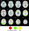
Axial slices showing significant ALE activation in WCST (green), task switching (red), and the conjunction of WCST and task switching (yellow) overlaid on the International Consortium for Brain Mapping (ICBM) single subject template.
Figure 2
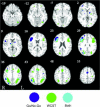
Axial slices showing significant ALE activation in WCST (green), go/no‐go (blue), and the conjunction of WCST and go/no‐go (blue) overlaid on the ICBM single subject template.
Figure 3
Three‐dimensional surface rendered views of all three task meta‐analyses including all possible conjunctions. 1, WCST (green); 2, task switching (red); 3, go/no‐go (blue); 4, WCST and task switching (yellow); 5, WCST and go/no‐go (cyan); 6, task switching and go/no‐go (violet); 7, WCST and task switching and go/no‐go (white). Image rendering was created with SUMA (and associated AFNI program 3dVol2Surf; [Saad et al.,2004]) using a surface representation of the ICBM single‐subject brain that was created with the FreeSurfer software package [Dale et al.,1999; Fischl et al.,1999].
Similar articles
- Using fMRI to decompose the neural processes underlying the Wisconsin Card Sorting Test.
Lie CH, Specht K, Marshall JC, Fink GR. Lie CH, et al. Neuroimage. 2006 Apr 15;30(3):1038-49. doi: 10.1016/j.neuroimage.2005.10.031. Epub 2006 Jan 18. Neuroimage. 2006. PMID: 16414280 - N-back working memory paradigm: a meta-analysis of normative functional neuroimaging studies.
Owen AM, McMillan KM, Laird AR, Bullmore E. Owen AM, et al. Hum Brain Mapp. 2005 May;25(1):46-59. doi: 10.1002/hbm.20131. Hum Brain Mapp. 2005. PMID: 15846822 Free PMC article. - Are the neural correlates of stopping and not going identical? Quantitative meta-analysis of two response inhibition tasks.
Swick D, Ashley V, Turken U. Swick D, et al. Neuroimage. 2011 Jun 1;56(3):1655-65. doi: 10.1016/j.neuroimage.2011.02.070. Epub 2011 Mar 3. Neuroimage. 2011. PMID: 21376819 - Executive functions and neurocognitive aging: dissociable patterns of brain activity.
Turner GR, Spreng RN. Turner GR, et al. Neurobiol Aging. 2012 Apr;33(4):826.e1-13. doi: 10.1016/j.neurobiolaging.2011.06.005. Epub 2011 Jul 24. Neurobiol Aging. 2012. PMID: 21791362 Review. - Reliable differences in brain activity between young and old adults: a quantitative meta-analysis across multiple cognitive domains.
Spreng RN, Wojtowicz M, Grady CL. Spreng RN, et al. Neurosci Biobehav Rev. 2010 Jul;34(8):1178-94. doi: 10.1016/j.neubiorev.2010.01.009. Epub 2010 Jan 28. Neurosci Biobehav Rev. 2010. PMID: 20109489 Review.
Cited by
- Decreased neural activity and neural connectivity while performing a set-shifting task after inhibiting repetitive transcranial magnetic stimulation on the left dorsal prefrontal cortex.
Gerrits NJHM, van den Heuvel OA, van der Werf YD. Gerrits NJHM, et al. BMC Neurosci. 2015 Jul 22;16:45. doi: 10.1186/s12868-015-0181-3. BMC Neurosci. 2015. PMID: 26199083 Free PMC article. Clinical Trial. - Visual-motor association learning in undergraduate students as a function of the autism-spectrum quotient.
Parkington KB, Clements RJ, Landry O, Chouinard PA. Parkington KB, et al. Exp Brain Res. 2015 Oct;233(10):2883-95. doi: 10.1007/s00221-015-4358-x. Epub 2015 Jun 24. Exp Brain Res. 2015. PMID: 26105755 - Microstructural abnormalities of the brain white matter in attention-deficit/hyperactivity disorder.
Chen L, Huang X, Lei D, He N, Hu X, Chen Y, Li Y, Zhou J, Guo L, Kemp GJ, Gong QY. Chen L, et al. J Psychiatry Neurosci. 2015 Jul;40(4):280-7. doi: 10.1503/jpn.140199. J Psychiatry Neurosci. 2015. PMID: 25853285 Free PMC article. - Influence of Exposure at Different Altitudes on the Executive Function of Plateau Soldiers-Evidence From ERPs and Neural Oscillations.
Wei X, Ni X, Zhao S, Chi A. Wei X, et al. Front Physiol. 2021 Apr 16;12:632058. doi: 10.3389/fphys.2021.632058. eCollection 2021. Front Physiol. 2021. PMID: 33935798 Free PMC article. - Lesion mapping of cognitive control and value-based decision making in the prefrontal cortex.
Gläscher J, Adolphs R, Damasio H, Bechara A, Rudrauf D, Calamia M, Paul LK, Tranel D. Gläscher J, et al. Proc Natl Acad Sci U S A. 2012 Sep 4;109(36):14681-6. doi: 10.1073/pnas.1206608109. Epub 2012 Aug 20. Proc Natl Acad Sci U S A. 2012. PMID: 22908286 Free PMC article.
References
- Andres P (2003): Frontal cortex as the central executive of working memory: time to revise our view. Cortex 39: 871–895. - PubMed
- Aron AR, Monsell S, Sahakian BJ, Robbins TW (2004a): A componential analysis of task‐switching deficits associated with lesions of left and right frontal cortex. Brain 127: 1561–1573. - PubMed
- Aron AR, Robbins TW, Poldrack RA (2004b): Inhibition and the right inferior frontal cortex. Trends Cogn Sci 8: 170–177. - PubMed
- Asahi S, Okamoto Y, Okada G, Yamawaki S, Yokota N (2004): Negative correlation between right prefrontal activity during response inhibition and impulsiveness: a fMRI study. Eur Arch Psychiatry Clin Neurosci 254: 245–251. - PubMed
- Baddeley A (1992): Working memory. Science 255: 556–559. - PubMed
MeSH terms
LinkOut - more resources
Full Text Sources
Medical


