Impairment of starvation-induced and constitutive autophagy in Atg7-deficient mice - PubMed (original) (raw)
. 2005 May 9;169(3):425-34.
doi: 10.1083/jcb.200412022. Epub 2005 May 2.
Satoshi Waguri, Takashi Ueno, Junichi Iwata, Shigeo Murata, Isei Tanida, Junji Ezaki, Noboru Mizushima, Yoshinori Ohsumi, Yasuo Uchiyama, Eiki Kominami, Keiji Tanaka, Tomoki Chiba
Affiliations
- PMID: 15866887
- PMCID: PMC2171928
- DOI: 10.1083/jcb.200412022
Impairment of starvation-induced and constitutive autophagy in Atg7-deficient mice
Masaaki Komatsu et al. J Cell Biol. 2005.
Abstract
Autophagy is a membrane-trafficking mechanism that delivers cytoplasmic constituents into the lysosome/vacuole for bulk protein degradation. This mechanism is involved in the preservation of nutrients under starvation condition as well as the normal turnover of cytoplasmic component. Aberrant autophagy has been reported in several neurodegenerative disorders, hepatitis, and myopathies. Here, we generated conditional knockout mice of Atg7, an essential gene for autophagy in yeast. Atg7 was essential for ATG conjugation systems and autophagosome formation, amino acid supply in neonates, and starvation-induced bulk degradation of proteins and organelles in mice. Furthermore, Atg7 deficiency led to multiple cellular abnormalities, such as appearance of concentric membranous structure and deformed mitochondria, and accumulation of ubiquitin-positive aggregates. Our results indicate the important role of autophagy in starvation response and the quality control of proteins and organelles in quiescent cells.
Figures
Figure 1.
Generation of _Atg7_F/F mice. (A) Schematic representation of the targeting vector and the targeted allele of Atg7 gene. The coding exons numbered in accordance with the initiation site as exon 1 are depicted by black boxes. Green and red boxes indicate Atg7 cDNA fragment (aa 1786–2097) and Atg7 cDNA fragment (aa 1669–1698) with stop codon, respectively. The open triangles denote loxP sequence. A probe for Southern blot analysis is shown as a gray ellipse. Arrows indicate the positions of PCR primers. The asterisk denotes the essential cysteine residue on exon 14. EcoRV, EcoRV sites; neo, neomycin-resistant gene cassette; DT-A, diphtheria toxin gene. (B) Southern blot analysis of genomic DNAs extracted from mice tails. Wild-type and Flox alleles are detected as 17.5- and 7.5-kb bands, respectively. (C) Immunoblot of Atg7 in MEFs. The lysates of MEFs of indicated genotypes were immunoblotted with Atg7 and actin.
Figure 2.
The phenotypes of _Atg7_-deficient mice. (A) PCR analysis of genomic DNA extracted from wild-type, Atg7 +/−, and Atg7 −/− mice tail. The amplified fragments derived from wild and mutant alleles are indicated. (B) Expression of Atg7 transcript. Atg7 transcript was detected by RT-PCR analysis. The region amplified was between exons 12 and 13. G3PDH cDNA was amplified as an internal control. (C) ATG conjugation systems in Atg7 −/− mice liver. The liver homogenate was centrifuged at 800 g for 10 min and the postnuclear supernatant (PNS) was immunoblotted with antibodies against Atg7, Atg5, LC3, and actin as a loading control. The bottom panel of Atg5 blotting is the long exposure of the top panel to detect free Atg5. Data shown are representative of three separate experiments. (D and E) Deficiency of autophagosome formation in Atg7 −/− heart. Atg7 +/− (D) and Atg7 −/− (E) mice expressing GFP-LC3 were obtained by caesarean delivery and analyzed by fluorescence microscopy. Representative results obtained from each neonatal heart at 3 h after caesarean delivery. (F) Morphology of Atg7 +/− and Atg7 −/− mice. (G) Kaplan-Meier curves of survival of newborn mice. Control and Atg7 −/− mice were delivered by caesarean section, and their survival was followed up to 26 h.
Figure 3.
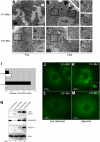
Impaired autophagosome formation in _Atg7_-deficient liver. (A–H) Electron micrographs of liver from Atg7 F/+:Mx1 (A–D) and Atg7 F/F:Mx1 (E–H) mice fed ad libitum (A and E) or fasted for 1 d (B and F). (C and D) Early stages of autophagic vacuoles observed in B are highlighted. (E and F) Autophagosome was not induced in mutant hepatocytes upon fasting. Insets show higher magnification views of glycogen granules. (G and H) Occasionally observed autophagosome-like structures in mutant hepatocytes. Bars: (A, B, E, and F) 5 μm; (C, D, G, and H) 0.5 μm. (I) Number of autophagosomes per hepatocyte (n = 20) in each genotype was counted and their averages are shown. (J–M) Immunofluorescent analysis of LC3 in primary cultured hepatocytes. Hepatocytes isolated from Atg7 F/+:Mx1 (J and K) and Atg7 F/F:Mx1 mice (L and M) were cultured in Williams' E (J and L) or Hanks' solution (K and M). Inset highlights the cup-like structure of LC3 observed in K. (N) Immunoblot analysis of Atg8 homologues in the liver. Atg7 F/+:Mx1 (lanes 1 and 2) and Atg7 F/F:Mx1 mice (lanes 3 and 4) were fed ad libitum (lanes 1 and 3) or fasted for 1 d (lanes 2 and 4), and then PNS fractions of liver were analyzed by immunoblotting with anti-LC3, GABARAP, GATE-16, and actin antibodies. Asterisk denotes a nonspecific band. Data shown are representative of three separate experiments.
Figure 4.
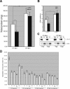
Fasting response of _Atg7_-deficient liver. (A and B) Livers from Atg7 F/+:Mx1 and Atg7 F/F:Mx1 mice fed ad libitum (Fed) or fasted for 1 d (Fast) at 20 d after pIpC injection were dissected, and the amount of total protein (A) and SDH activity (B) per liver were measured. Data are mean ± SD values of five mice in each group; *, P < 0.01. (C) Cytochrome c levels in the cytosolic and mitochondria/lysosomal fractions of the liver at 20 d after injection. Equal amount of PNS fractions were centrifuged at 8,000 g for 10 min and the pellets were used as the mitochondrial/lysosomal fraction (ML). The supernatants were further centrifuged at 100,000 g for 1 h and the supernatant was used as the cytosolic fraction (C). Actin was blotted as control. Data shown are representative of two separate experiments. (D) Turnover of long-lived protein. Hepatocytes from Atg7 F/+:Mx1 and Atg7 F/F:Mx1 mice were isolated and labeled with [14C]leucine for 24 h, and degradation of long-lived protein in deprived or nondeprived condition was measured. Monomethylamine (MA) and/or E64d and pepstatin (E/P) or epoxomicin (epoxo) was added as indicated. Data are the mean ± SD of triplicate experiments.
Figure 5.
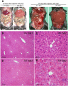
Atg7 deficiency in the liver causes hepatomegaly and hepatic cell swelling. (A) The gross anatomical views of representative mice at the indicated day after pIpC injection. (B–E) Histology of representative livers with Atg7 deficiency. Hematoxylin and eosin staining of Atg7 F/+:Mx1 (B and C) and Atg7 F/F:Mx1 (D and E) liver at 90 d after pIpC injection.
Figure 6.
Electron micrographs of hepatic cells with _A_tg7 deficiency. (A and B) Note the presence of concentric membranous structures in the mutant cells (arrowheads). Higher magnification view (B) shows the membranous elements are continuous with the rough ER (arrowheads). (C and D) The mutant cells contained a high number of peroxisomes (arrows) and deformed mitochondria (asterisks). Insets show higher magnification views. Nuc, nucleus. Bars: (A) 5 μm; (B) 0.5 μm; (C and D) 1 μm.
Figure 7.
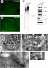
Accumulation of ubiquitin-positive aggregates in _Atg7_-deficient liver. (A and B) Immunofluorescent detection of ubiquitin in the Atg7 F/+:Mx1 (A) and Atg7 F/F:Mx1 (B) liver. (C–F) Immunoelectron micrographs of ubiquitin in a representative mutant liver. The high-magnification view shows ubiquitin particles near the lipid dropletlike structure (D–F). Bars, 0.5 μm. (G) Immunoblot analysis of the liver. PNS fractions of the liver at 90 d after injection were immunoblotted with the indicated antibodies. Data shown are representative of three separate experiments.
Similar articles
- Autophagy-physiology and pathophysiology.
Uchiyama Y, Shibata M, Koike M, Yoshimura K, Sasaki M. Uchiyama Y, et al. Histochem Cell Biol. 2008 Apr;129(4):407-20. doi: 10.1007/s00418-008-0406-y. Epub 2008 Mar 5. Histochem Cell Biol. 2008. PMID: 18320203 Free PMC article. Review. - [Autophagic-lysosomal system: physiology and pathology].
Komatsu M, Kominami E. Komatsu M, et al. Nihon Shinkei Seishin Yakurigaku Zasshi. 2006 Apr;26(2):75-81. Nihon Shinkei Seishin Yakurigaku Zasshi. 2006. PMID: 16722464 Review. Japanese. - Loss of autophagy in the central nervous system causes neurodegeneration in mice.
Komatsu M, Waguri S, Chiba T, Murata S, Iwata J, Tanida I, Ueno T, Koike M, Uchiyama Y, Kominami E, Tanaka K. Komatsu M, et al. Nature. 2006 Jun 15;441(7095):880-4. doi: 10.1038/nature04723. Epub 2006 Apr 19. Nature. 2006. PMID: 16625205 - β-cell autophagy: A novel mechanism regulating β-cell function and mass: Lessons from β-cell-specific Atg7-deficient mice.
Fujitani Y, Ebato C, Uchida T, Kawamori R, Watada H. Fujitani Y, et al. Islets. 2009 Sep-Oct;1(2):151-3. doi: 10.4161/isl.1.2.9057. Islets. 2009. PMID: 21099263 - The role of autophagy during the early neonatal starvation period.
Kuma A, Hatano M, Matsui M, Yamamoto A, Nakaya H, Yoshimori T, Ohsumi Y, Tokuhisa T, Mizushima N. Kuma A, et al. Nature. 2004 Dec 23;432(7020):1032-6. doi: 10.1038/nature03029. Epub 2004 Nov 3. Nature. 2004. PMID: 15525940
Cited by
- The Role of Autophagy in Skeletal Muscle Diseases.
Xia Q, Huang X, Huang J, Zheng Y, March ME, Li J, Wei Y. Xia Q, et al. Front Physiol. 2021 Mar 25;12:638983. doi: 10.3389/fphys.2021.638983. eCollection 2021. Front Physiol. 2021. PMID: 33841177 Free PMC article. Review. - Sarcopenia and cachexia: the adaptations of negative regulators of skeletal muscle mass.
Sakuma K, Yamaguchi A. Sakuma K, et al. J Cachexia Sarcopenia Muscle. 2012 Jun;3(2):77-94. doi: 10.1007/s13539-011-0052-4. Epub 2012 Jan 12. J Cachexia Sarcopenia Muscle. 2012. PMID: 22476916 Free PMC article. - Inhibition of autophagic turnover in β-cells by fatty acids and glucose leads to apoptotic cell death.
Mir SU, George NM, Zahoor L, Harms R, Guinn Z, Sarvetnick NE. Mir SU, et al. J Biol Chem. 2015 Mar 6;290(10):6071-85. doi: 10.1074/jbc.M114.605345. Epub 2014 Dec 29. J Biol Chem. 2015. PMID: 25548282 Free PMC article. - Autophagy modulates dynamics of connexins at the plasma membrane in a ubiquitin-dependent manner.
Bejarano E, Girao H, Yuste A, Patel B, Marques C, Spray DC, Pereira P, Cuervo AM. Bejarano E, et al. Mol Biol Cell. 2012 Jun;23(11):2156-69. doi: 10.1091/mbc.E11-10-0844. Epub 2012 Apr 11. Mol Biol Cell. 2012. PMID: 22496425 Free PMC article. - Differential use of autophagy by primary dendritic cells specialized in cross-presentation.
Mintern JD, Macri C, Chin WJ, Panozza SE, Segura E, Patterson NL, Zeller P, Bourges D, Bedoui S, McMillan PJ, Idris A, Nowell CJ, Brown A, Radford KJ, Johnston AP, Villadangos JA. Mintern JD, et al. Autophagy. 2015;11(6):906-17. doi: 10.1080/15548627.2015.1045178. Autophagy. 2015. PMID: 25950899 Free PMC article.
References
- Baumeister, W., J. Walz, F. Zuhl, and E. Seemuller. 1998. The proteasome: paradigm of a self-compartmentalizing protease. Cell. 92:367–380. - PubMed
- Bence, N.F., R.M. Sampat, and R.R. Kopito. 2001. Impairment of the ubiquitin-proteasome system by protein aggregation. Science. 292:1552–1555. - PubMed
- Bursch, W. 2001. The autophagosomal-lysosomal compartment in programmed cell death. Cell Death Differ. 8:569–581. - PubMed
- Ciechanover, A., and P. Brundin. 2003. The ubiquitin proteasome system in neurodegenerative diseases: sometimes the chicken, sometimes the egg. Neuron. 40:427–446. - PubMed
- Dunn, W.A., Jr. 1994. Autophagy and related mechanisms of lysosome-mediated protein degradation. Trends Cell Biol. 4:139–143. - PubMed
Publication types
MeSH terms
Substances
LinkOut - more resources
Full Text Sources
Other Literature Sources
Molecular Biology Databases


