Clinical evaluation of autoantibodies to a novel PM/Scl peptide antigen - PubMed (original) (raw)
Comparative Study
doi: 10.1186/ar1729. Epub 2005 Apr 1.
Affiliations
- PMID: 15899056
- PMCID: PMC1174964
- DOI: 10.1186/ar1729
Comparative Study
Clinical evaluation of autoantibodies to a novel PM/Scl peptide antigen
Michael Mahler et al. Arthritis Res Ther. 2005.
Abstract
Anti-PM/Scl antibodies represent a specific serological marker for a subset of patients with scleroderma (Scl) and polymyositis (PM), and especially with the PM/Scl overlap syndrome (PM/Scl). Anti-PM/Scl reactivity is found in 24% of PM/Scl patients and is found in 3-10% of Scl and PM patients. The PM/Scl autoantigen complex comprises 11-16 different polypeptides. Many of those proteins can serve as targets of the anti-PM/Scl B-cell response, but most frequently the PM/Scl-100 and PM/Scl-75 polypeptides are targeted. In the present study we investigated the clinical relevance of a major alpha helical PM/Scl-100 epitope (PM1-alpha) using a newly developed peptide-based immunoassay and compared the immunological properties of this peptide with native and recombinant PM/Scl antigens. In a technical comparison, we showed that an ELISA based on the PM1-alpha peptide is more sensitive than common techniques to detect anti-PM/Scl antibodies such as immunoblot, indirect immunofluorescence on HEp-2 cells and ELISA with recombinant PM/Scl polypeptides. We found no statistical evidence of a positive association between anti-PM1-alpha and other antibodies, with the exception of known PM/Scl components. In our cohort a negative correlation could be found with anti-Scl-70 (topoisomerase I), anti-Jo-1 (histidyl tRNA synthetase) and anti-centromere proteins. In a multicenter evaluation we demonstrated that the PM1-alpha peptide represents a sensitive and reliable substrate for the detection of a subclass of anti-PM/Scl antibodies. In total, 22/40 (55%) PM/Scl patients, 27/205 (13.2%) Scl patients and 3/40 (7.5%) PM patients, but only 5/288 (1.7%) unrelated controls, tested positive for the anti-PM1-alpha peptide antibodies. These data indicate that anti-PM1-alpha antibodies appear to be exclusively present in sera from PM/Scl patients, from Scl patients and, to a lesser extent, from PM patients. The anti-PM1-alpha ELISA thus offers a new serological marker to diagnose and discriminate different systemic autoimmune disorders.
Figures
Figure 1
Correlation diagrams of PM1-α, PM/Scl-75a, PM/Scl-75c and PM/Scl-100. A panel of sera tested previously for reactivity to recombinant polymyositis/scleroderma (PM/Scl) components (PM/Scl-75a, PM/Scl-75c and PM/Scl-100) was assayed for anti-PM1-α peptide reactivity in an ELISA [18]. Correlation diagrams are shown comparing the peptide ELISA with the recombinant proteins (a)–(c) for all sera (n = 81) and (b)–(f) for only the sera of PM/Scl patients (n = 36).
Figure 2
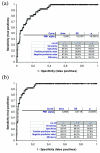
Receiver operating characteristic analysis of the PM1-α ELISA. Results obtained from three centers and based on 567 patients including polymyositis/scleroderma (PM/Scl) patients (n = 40), Scl patients (n = 205) and PM patients (n = 40) as well as other controls were used to calculate a receiver operating characteristic analysis (a) for all control samples and (b) for unrelated controls (without Scl and PM). The curve shows a clear discrimination between PM/Scl patient samples and various controls as emphasized by an area under the curve value of 0.901 (all controls) and 0.958 (unrelated controls). The differentiation between PM/Scl patients and controls was significantly improved when Scl patients and PM patients were excluded from the control group (b). SE, standard error.
Figure 3
Reactivity of polymyositis/scleroderma (PM/Scl) patients and controls in the PM1-α ELISA. Results obtained from three centers and based on 567 patients including PM/Scl patients (n = 40), Scl patients (n = 205) and PM patients (n = 40) as well as other controls were used to calculate comparative descriptive analysis. The diagram shows a significantly increased reactivity of the PM/Scl sera compared with the control groups. Comparative descriptives show vertical box-plots for each sample, side by side for comparison. The blue line series shows parametric statistics: diamond, mean and the requested confidence interval around the mean; notched line, requested parametric percentile range. The notched box and whiskers show non-parametric statistics: notched box, median, lower and upper quartiles, and confidence interval around the median; dotted line, connects the nearest observations within 1.5 interquartile ranges (IQR) of the lower and upper quartiles. + and ○, possible outliers – observations more than 1.5 IQR (near outliers) and more than 3.0 IQR (far outliers) from the quartiles. Vertical lines, requested nonparametric percentile range. SLE, systemic lupus erythematosus; HCV, hepatitis C virus; RA, rheumatoid arthritis.
Similar articles
- Anti-PM/Scl antibodies are found in Japanese patients with various systemic autoimmune conditions besides myositis and scleroderma.
Muro Y, Hosono Y, Sugiura K, Ogawa Y, Mimori T, Akiyama M. Muro Y, et al. Arthritis Res Ther. 2015 Mar 11;17(1):57. doi: 10.1186/s13075-015-0573-x. Arthritis Res Ther. 2015. PMID: 25885224 Free PMC article. - PM1-Alpha ELISA: the assay of choice for the detection of anti-PM/Scl autoantibodies?
Mahler M, Fritzler MJ. Mahler M, et al. Autoimmun Rev. 2009 Mar;8(5):373-8. doi: 10.1016/j.autrev.2008.12.001. Epub 2008 Dec 25. Autoimmun Rev. 2009. PMID: 19103309 Review. - Serological and clinical characterization of anti-dsDNA and anti-PM/Scl double-positive patients.
Mahler M, Greidinger EL, Szmyrka M, Kromminga A, Fritzler MJ. Mahler M, et al. Ann N Y Acad Sci. 2007 Aug;1109:311-21. doi: 10.1196/annals.1398.037. Ann N Y Acad Sci. 2007. PMID: 17785320 - Novel aspects of autoantibodies to the PM/Scl complex: clinical, genetic and diagnostic insights.
Mahler M, Raijmakers R. Mahler M, et al. Autoimmun Rev. 2007 Aug;6(7):432-7. doi: 10.1016/j.autrev.2007.01.013. Epub 2007 Feb 20. Autoimmun Rev. 2007. PMID: 17643929 Review. - C1D is a major autoantibody target in patients with the polymyositis-scleroderma overlap syndrome.
Schilders G, Egberts WV, Raijmakers R, Pruijn GJ. Schilders G, et al. Arthritis Rheum. 2007 Jul;56(7):2449-54. doi: 10.1002/art.22710. Arthritis Rheum. 2007. PMID: 17599775
Cited by
- The changing landscape of the clinical value of the PM/Scl autoantibody system.
Mahler M, Fritzler MJ. Mahler M, et al. Arthritis Res Ther. 2009;11(2):106. doi: 10.1186/ar2646. Epub 2009 Mar 26. Arthritis Res Ther. 2009. PMID: 19351430 Free PMC article. - Anti-PM/Scl antibodies are found in Japanese patients with various systemic autoimmune conditions besides myositis and scleroderma.
Muro Y, Hosono Y, Sugiura K, Ogawa Y, Mimori T, Akiyama M. Muro Y, et al. Arthritis Res Ther. 2015 Mar 11;17(1):57. doi: 10.1186/s13075-015-0573-x. Arthritis Res Ther. 2015. PMID: 25885224 Free PMC article. - Myositis-related interstitial lung disease and antisynthetase syndrome.
Solomon J, Swigris JJ, Brown KK. Solomon J, et al. J Bras Pneumol. 2011 Jan-Feb;37(1):100-9. doi: 10.1590/s1806-37132011000100015. J Bras Pneumol. 2011. PMID: 21390438 Free PMC article. Review. - Case report: Clinical, genetic and immunological characterization of a novel XK variant in a patient with McLeod syndrome.
Dambietz CA, Doescher A, Heming M, Schirmacher A, Schlüter B, Schulte-Mecklenbeck A, Thomas C, Wiendl H, Meyer Zu Hörste G, Wiethoff S. Dambietz CA, et al. Front Genet. 2024 Aug 21;15:1421952. doi: 10.3389/fgene.2024.1421952. eCollection 2024. Front Genet. 2024. PMID: 39233738 Free PMC article. - Scleromyositis: A distinct novel entity within the systemic sclerosis and autoimmune myositis spectrum. Implications for care and pathogenesis.
Giannini M, Ellezam B, Leclair V, Lefebvre F, Troyanov Y, Hudson M, Senécal JL, Geny B, Landon-Cardinal O, Meyer A. Giannini M, et al. Front Immunol. 2023 Jan 26;13:974078. doi: 10.3389/fimmu.2022.974078. eCollection 2022. Front Immunol. 2023. PMID: 36776390 Free PMC article. Review.
References
- Tan EM. Antinuclear antibodies: diagnostic markers for autoimmune diseases and probes for cell biology. Adv Immunol. 1989;44:93–151. - PubMed
- Reimer G, Steen VD, Penning CA, Medsger TA, Jr, Tan EM. Correlates between autoantibodies to nucleolar antigens and clinical features in patients with systemic sclerosis (scleroderma) Arthritis Rheum. 1988;31:525–532. - PubMed
Publication types
MeSH terms
Substances
LinkOut - more resources
Full Text Sources
Medical
Molecular Biology Databases
Research Materials
Miscellaneous


