Lateral prefrontal cortex: architectonic and functional organization - PubMed (original) (raw)
Review
Lateral prefrontal cortex: architectonic and functional organization
Michael Petrides. Philos Trans R Soc Lond B Biol Sci. 2005.
Abstract
A comparison of the architecture of the human prefrontal cortex with that of the macaque monkey showed a very similar architectonic organization in these two primate species. There is no doubt that the prefrontal cortical areas of the human brain have undergone considerable development, but it is equally clear that the basic architectonic organization is the same in the two species. Thus, a comparative approach to the study of the functional organization of the primate prefrontal cortex is more likely to reveal the essential aspects of the various complex control processes that are the domain of frontal function. The lateral frontal cortex appears to be functionally organized along both a rostral-caudal axis and a dorsal-ventral axis. The most caudal frontal region, the motor region on the precentral gyrus, is involved in fine motor control and direct sensorimotor mappings, whereas the caudal lateral prefrontal region is involved in higher order control processes that regulate the selection among multiple competing responses and stimuli based on conditional operations. Further rostrally, the mid-lateral prefrontal region plays an even more abstract role in cognitive control. The mid-lateral prefrontal region is itself organized along a dorsal-ventral axis of organization, with the mid-dorsolateral prefrontal cortex being involved in the monitoring of information in working memory and the mid-ventrolateral prefrontal region being involved in active judgments on information held in posterior cortical association regions that are necessary for active retrieval and encoding of information.
Figures
Figure 1
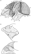
(a) Cytoarchitectonic map of the lateral surface of the cerebral cortex of the monkey by Brodmann (1905). Note that the orbital frontal cortex is also partially shown as an extension of the ventral part of the lateral surface. (b) Cytoarchitectonic map of the lateral and orbital prefrontal cortex of the macaque monkey by Walker (1940).
Figure 2
(a) Cytoarchitectonic map of the lateral surface of the human cerebral cortex by Brodmann (1909). Note that the orbital frontal cortex is also shown as an extension of the ventral part of the lateral surface. (b) Cytoarchitectonic map of the lateral surface of the human cerebral cortex by Sarkissov et al. (1955).
Figure 3
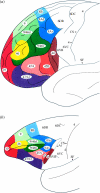
Cytoarchitectonic map of the lateral surface of the prefrontal cortex of (a) the human brain and (b) the macaque monkey brain by Petrides & Pandya (1994). Abbreviations: Ai, the inferior arcuate sulcus; CS, central sulcus; SF, Sylvian fissure.
Figure 4
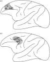
Schematic diagram of the monkey brain illustrating the rostral–caudal axis of lateral frontal cortex organization. Some of the interactions of the caudal lateral frontal region with post-rolandic cortical regions are shown by the thick dashed lines and interactions within the lateral frontal cortex are shown by the thin dashed lines. Abbreviations: A, auditory processing in the superior temporal gyrus; CC, corpus callosum; CDL, caudolateral frontal region; CG, cingulate gyrus; CVL, caudal ventrolateral frontal region; K, kinaesthetic processing in the superior parietal lobule; MDL, mid-dorsolateral prefrontal region; MR, motor region; MTL, medial temporal lobe region; MVL, mid-ventrolateral prefrontal region; S, body-centred (i.e. somato-centric) amodal processing in rostral inferior parietal lobule; SMA, supplementary motor area; SP, spatial processing in lateral and medial posterior parietal cortex; V, visual processing in prestriate cortex and the occipito-temporal cortical region. The numbers refer to architectonic areas in the lateral frontal cortex. Note that the posterior bank of the inferior branch of the arcuate sulcus is displayed opened in order to illustrate area 44 that lies within the inferior bank of the arcuate sulcus.
Figure 5
Schematic illustration of (a) the mid-dorsolateral (MDL) prefrontal lesion and (b) the caudal dorsolateral prefrontal lesion, which involved the cortex within and around the dorsal arcuate sulcus, i.e. the periarcuate (PA) region. These lesions in the monkey were used to study fundamental differences in function along the rostral–caudal axis of lateral frontal cortex. The numbers refer to the architectonic areas involved in these lesions.
Figure 6
(Left): schematic diagram of the experimental arrangement in the visual–visual object conditional task administered to monkeys. On each trial, one of the two white perspex boxes is lit and the other remains unlit by remotely turning on a light bulb that is inside them. One of two conditional stimuli is then presented in front of the opaque screen hiding the experimenter and the animal responds by pushing back one of the two boxes. The reward is delivered via the tubes that are attached to the boxes. (Right): performance of animals with mid-dorsolateral prefrontal lesions (MDL), animals with periarcuate lesions (PA) and normal control animals (NC). Solid circles indicate the scores of individual animals in each group. The animals with MDL and NC lesions reached criterion (90% correct performance across three consecutive days of testing, i.e. 90 trials) within a mean of 300 and 330 trials, respectively. None of the animals with PA lesions was able to reach criterion within the limits of testing (i.e. 1020 trials) and the mean level of correct performance achieved during the last 3 days of testing (i.e. last 90 trials) was 58% correct. Data from Petrides (1985).
Figure 7
(Upper panel): schematic diagram of the experimental arrangement in the self-ordered monitoring working memory condition and the recognition memory condition administered to the monkeys. The upper displays illustrate the presentation trials and the lower displays the test trials in both the monitoring and recognition conditions. (Lower panel): postoperative performance of animals with mid-dorsolateral frontal lesions (MDL), animals with periarcuate lesions (PA) and normal control animals (NC). The mean per cent correct performance over the four postoperative testing blocks (20 days of testing per block) is shown. Solid circles indicate the scores of individual animals in each group. In the monitoring condition, the animals with MDL lesions were severely impaired, whereas the animals with PA lesions performed as well as the NC animals. Both groups with lesions performed as well as the normal control animals in the recognition memory condition. Data from Petrides (1991).
Figure 8
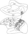
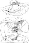
Schematic diagram of the monkey brain illustrating the dorsal–ventral axis of lateral frontal cortex organization. Some of the interactions postulated to underlie the mid-dorsolateral (MDL) and the mid-ventrolateral (MVL) prefrontal region functional organization. Abbreviations: A, auditory processing in superior temporal gyrus; CC, corpus callosum; CG, cingulate gyrus; ec, entorhinal cortex; M, multimodal processing in superior temporal sulcus; MTL, medial temporal lobe region; S, body-centred (i.e. somato-centric) amodal processing in rostral inferior parietal lobule; SP, spatial processing in posterior parietal cortex; V, visual object processing in rostral inferotemporal cortex.
Figure 9
(a) An example of a pair of abstract designs used in the functional neuroimaging experiment requiring different types of mnemonic decisions on such visual stimuli. (b) Increased activity in the right mid-ventrolateral prefrontal cortex (area 47/12) during the making of active judgments on the familiarity of stimuli (comparison: familiarity/novelty decision condition minus control condition). (c) Note the additional increase of activity in the right mid-dorsolateral prefrontal cortex (areas 46 and 9/46) in the monitoring condition minus control condition comparison. The observed increase of activity in the mid-ventrolateral prefrontal cortex (area 47/12) was expected because an active decision was included in the monitoring condition. (d) Note that only the mid-dorsolateral prefrontal cortex (areas 46 and 9/46) showed increased activity when the comparison was the monitoring condition minus the familiarity/novelty decision condition. This comparison isolated the mid-dorsolateral prefrontal involvement in monitoring because the active memory decision, which leads to increased activity in the mid-ventrolateral prefrontal cortex, was common to both the familiarity/novelty decision condition and the monitoring condition, while the monitoring requirement was present only in the monitoring condition. Abbreviations: CS, central sulcus; HS, horizontal sulcus; IFS, inferior frontal sulcus; INFS, intermediate frontal sulcus; LF, lateral fissure. Data from Petrides et al. (2002).
Similar articles
- Multimodal architectonic subdivision of the caudal ventrolateral prefrontal cortex of the macaque monkey.
Gerbella M, Belmalih A, Borra E, Rozzi S, Luppino G. Gerbella M, et al. Brain Struct Funct. 2007 Dec;212(3-4):269-301. doi: 10.1007/s00429-007-0158-9. Epub 2007 Sep 25. Brain Struct Funct. 2007. PMID: 17899184 - Functional specialization of areas along the anterior-posterior axis of the primate prefrontal cortex.
Riley MR, Qi XL, Constantinidis C. Riley MR, et al. Cereb Cortex. 2017 Jul 1;27(7):3683-3697. doi: 10.1093/cercor/bhw190. Cereb Cortex. 2017. PMID: 27371761 Free PMC article. - Specialized systems for the processing of mnemonic information within the primate frontal cortex.
Petrides M. Petrides M. Philos Trans R Soc Lond B Biol Sci. 1996 Oct 29;351(1346):1455-61; discussion 1461-2. doi: 10.1098/rstb.1996.0130. Philos Trans R Soc Lond B Biol Sci. 1996. PMID: 8941957 Review. - Lateral prefrontal cortex is organized into parallel dorsal and ventral streams along the rostro-caudal axis.
Blumenfeld RS, Nomura EM, Gratton C, D'Esposito M. Blumenfeld RS, et al. Cereb Cortex. 2013 Oct;23(10):2457-66. doi: 10.1093/cercor/bhs223. Epub 2012 Aug 9. Cereb Cortex. 2013. PMID: 22879354 Free PMC article. - The role of the inferior frontal junction area in cognitive control.
Brass M, Derrfuss J, Forstmann B, von Cramon DY. Brass M, et al. Trends Cogn Sci. 2005 Jul;9(7):314-6. doi: 10.1016/j.tics.2005.05.001. Trends Cogn Sci. 2005. PMID: 15927520 Review.
Cited by
- Restoring large-scale brain networks in PTSD and related disorders: a proposal for neuroscientifically-informed treatment interventions.
Lanius RA, Frewen PA, Tursich M, Jetly R, McKinnon MC. Lanius RA, et al. Eur J Psychotraumatol. 2015 Mar 31;6:27313. doi: 10.3402/ejpt.v6.27313. eCollection 2015. Eur J Psychotraumatol. 2015. PMID: 25854674 Free PMC article. - Neural basis of recollection in first-episode major depression.
van Eijndhoven P, van Wingen G, Fernández G, Rijpkema M, Pop-Purceleanu M, Verkes RJ, Buitelaar J, Tendolkar I. van Eijndhoven P, et al. Hum Brain Mapp. 2013 Feb;34(2):283-94. doi: 10.1002/hbm.21439. Epub 2012 Jan 3. Hum Brain Mapp. 2013. PMID: 22753179 Free PMC article. - Linking Resting-State Networks in the Prefrontal Cortex to Executive Function: A Functional Near Infrared Spectroscopy Study.
Zhao J, Liu J, Jiang X, Zhou G, Chen G, Ding XP, Fu G, Lee K. Zhao J, et al. Front Neurosci. 2016 Oct 7;10:452. doi: 10.3389/fnins.2016.00452. eCollection 2016. Front Neurosci. 2016. PMID: 27774047 Free PMC article. - Functional Connectome Controllability in Patients with Mild Cognitive Impairment after Repetitive Transcranial Magnetic Stimulation of the Dorsolateral Prefrontal Cortex.
Papallo S, Di Nardo F, Siciliano M, Esposito S, Canale F, Cirillo G, Cirillo M, Trojsi F, Esposito F. Papallo S, et al. J Clin Med. 2024 Sep 10;13(18):5367. doi: 10.3390/jcm13185367. J Clin Med. 2024. PMID: 39336854 Free PMC article. - Unravelling the intrinsic functional organization of the human lateral frontal cortex: a parcellation scheme based on resting state fMRI.
Goulas A, Uylings HB, Stiers P. Goulas A, et al. J Neurosci. 2012 Jul 25;32(30):10238-52. doi: 10.1523/JNEUROSCI.5852-11.2012. J Neurosci. 2012. PMID: 22836258 Free PMC article.
References
- Amunts K, Schleicher A, Burgel U, Mohlberg H, Uylings H.B.M, Zilles K. Broca's region revisited: cytoarchitecture and intersubject variability. J. Comp. Neurol. 1999;412:319–341. - PubMed
- Andersen R, Gnadt J.W. Role of posterior parietal cortex in saccadic eye movements. In: Wurtz R, Goldberg M, editors. The neurobiology of saccadic eye movements. Elsevier; Amsterdam: 1989. pp. 315–335.
- Andersen R.A, Asanuma C, Essick G, Siegel R.M. Corticocortical connections of anatomically and physiologically defined subdivisions within the inferior parietal lobule. J. Comp. Neurol. 1990;296:65–113. - PubMed
- Bachevalier J, Mishkin M. Visual recognition impairment follows ventromedial but not dorsolateral prefrontal lesions in monkeys. Behav. Brain Res. 1986;20:249–261. - PubMed
- Bailey P, Bonin G. University of Illinois Press; Urbana: 1951. The isocortex of man.
Publication types
MeSH terms
LinkOut - more resources
Full Text Sources
