Hyaluronidase induces a transcapillary pressure gradient and improves the distribution and uptake of liposomal doxorubicin (Caelyx) in human osteosarcoma xenografts - PubMed (original) (raw)
Hyaluronidase induces a transcapillary pressure gradient and improves the distribution and uptake of liposomal doxorubicin (Caelyx) in human osteosarcoma xenografts
L Eikenes et al. Br J Cancer. 2005.
Abstract
Liposomal drug delivery enhances the tumour selective localisation and may improve the uptake compared to free drug. However, the drug distribution within the tumour tissue may still be heterogeneous. Degradation of the extracellular matrix is assumed to improve the uptake and penetration of drugs. The effect of the ECM-degrading enzyme hyaluronidase on interstitial fluid pressure and microvascular pressure were measured in human osteosarcoma xenografts by the wick-in-needle and micropipette technique, respectively. The tumour uptake and distribution of liposomal doxorubicin were studied on tumour sections by confocal laser scanning microscopy. The drugs were injected i.v. 1 h after the hyaluronidase pretreatment. Intratumoral injection of hyaluronidase reduced interstitial fluid pressure in a nonlinear dose-dependent manner. Maximum interstitial fluid pressure reduction of approximately 50% was found after injection of 1500 U hyaluronidase. Neither intratumoral nor i.v. injection of hyaluronidase induced any changes in the microvascular pressure. Thus, hyaluronidase induced a transcapillary pressure gradient, resulting in a four-fold increase in the tumour uptake and improving the distribution of the liposomal doxorubicin. Hyaluronidase reduces a major barrier for drug delivery by inducing a transcapillary pressure gradient, and administration of hyaluronidase adjuvant with liposomal doxorubicin may thus improve the therapeutic outcome.
Figures
Figure 1
Maximum reduction (%) in IFP 1 h after intratumoral injection of PBS (_n_=5), 150 U (_n_=6), 500 U (_n_=5), 1500 U (_n_=6) and 3000 U (_n_=6) hyaluronidase in o.t. tumours. Each value is the mean of n tumours, the bars represent s.e. *Data significantly different from untreated controls.
Figure 2
Interstitial fluid pressure response as a function of time after intratumoral injection of 150 U () (_n_=4), 500 U (Δ) (_n_=5), 1500 U (∇)(_n_=4) and 3000 U (□) (_n_=6) hyaluronidase in o.t. tumours. Interstitial fluid pressure is normalised to IFP before the injection. Each curve is the mean of n measurements.
Figure 3
Maximum reduction in IFP after intratumoral injection of hyaluronidase as a function of tumour volume. Each symbol represents one measurement, the solid line represents linear regression analysis and was found to be significant for all doses except 150 U.
Figure 4
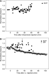
Normalised pressure after (A) intratumoral and (B) i.v. injection of 1500 U hyaluronidase in s.c. tumours. Microvascular pressure (•) was not measured continuously, and the data are based on different time intervals for _n_=5 (intratumoral injection) and _n_=7 (i.v. injection) mice. The bars represent s.e. at some time points. The IFP (○) graph is the mean of _n_=2 measurements.
Figure 5
Tumour tissue hyaluronan (blue) in o.t. osteosarcoma xenografts relative to capillaries (red) and tumour cells (green) treated with PBS (A) and hyaluronidase (1500 U) (B, C).
Figure 6
Distribution of liposomal doxorubicin in osteosarcoma xenografts treated with liposomal doxorubicin alone (16 mg kg− 1) (A) or liposomal doxorubicin combined with hyaluronidase (1500 U) (B). Representative images of doxorubicin (green) relative to capillaries (red) are presented from the rim to the centre of the tumour sections.
Figure 7
Doxorubicin fluorescence intensity profile in osteosarcoma xenografts treated with liposomal doxorubicin alone (16 mg kg− 1) (○) or combined with hyaluronidase (1500 U) (•). Each symbol is the mean of 5–20 images per section and 10 sections per tumour in three tumours. Bars represent s.e. The increase in the doxorubicin uptake in the hyaluronidase-treated tumours is significant throughout the whole tumour (P<0.05).
Figure 8
Number of images as a function of fluorescence intensity for osteosarcoma xenografts given liposomal doxorubicin alone (16 mg kg− 1) (□) or combined with hyaluronidase (1500 U) (▪). Each value is the mean of 5–20 images per section and 10 sections per tumour in three tumours. Bars represent s.e. *Data significantly different from Caelyx™ alone.
Figure 9
Intracellular distribution of doxorubicin (green) (A), DNA (red) (B) and colocalisation of doxorubicin and DNA (C) in osteosarcoma xenografts given liposomal doxorubicin alone (16 mg kg− 1). Bar=20 _μ_m.
Similar articles
- Radiation improves the distribution and uptake of liposomal doxorubicin (caelyx) in human osteosarcoma xenografts.
Davies Cde L, Lundstrøm LM, Frengen J, Eikenes L, Bruland S ØS, Kaalhus O, Hjelstuen MH, Brekken C. Davies Cde L, et al. Cancer Res. 2004 Jan 15;64(2):547-53. doi: 10.1158/0008-5472.can-03-0576. Cancer Res. 2004. PMID: 14744768 - Collagenase increases the transcapillary pressure gradient and improves the uptake and distribution of monoclonal antibodies in human osteosarcoma xenografts.
Eikenes L, Bruland ØS, Brekken C, Davies Cde L. Eikenes L, et al. Cancer Res. 2004 Jul 15;64(14):4768-73. doi: 10.1158/0008-5472.CAN-03-1472. Cancer Res. 2004. PMID: 15256445 - Pharmacokinetics of pegylated liposomal Doxorubicin: review of animal and human studies.
Gabizon A, Shmeeda H, Barenholz Y. Gabizon A, et al. Clin Pharmacokinet. 2003;42(5):419-36. doi: 10.2165/00003088-200342050-00002. Clin Pharmacokinet. 2003. PMID: 12739982 Review. - Polyethylene glycol-coated (pegylated) liposomal doxorubicin. Rationale for use in solid tumours.
Gabizon A, Martin F. Gabizon A, et al. Drugs. 1997;54 Suppl 4:15-21. doi: 10.2165/00003495-199700544-00005. Drugs. 1997. PMID: 9361957 Review.
Cited by
- Real-time visualization and quantitation of vascular permeability in vivo: implications for drug delivery.
Pink DB, Schulte W, Parseghian MH, Zijlstra A, Lewis JD. Pink DB, et al. PLoS One. 2012;7(3):e33760. doi: 10.1371/journal.pone.0033760. Epub 2012 Mar 29. PLoS One. 2012. PMID: 22479438 Free PMC article. - Improvement of intratumor microdistribution of PEGylated liposome via tumor priming by metronomic S-1 dosing.
Doi Y, Abu Lila AS, Matsumoto H, Okada T, Shimizu T, Ishida T. Doi Y, et al. Int J Nanomedicine. 2016 Oct 25;11:5573-5582. doi: 10.2147/IJN.S119069. eCollection 2016. Int J Nanomedicine. 2016. PMID: 27822036 Free PMC article. - Extracellular matrix in cancer progression and therapy.
He X, Lee B, Jiang Y. He X, et al. Med Rev (2021). 2022 Apr 26;2(2):125-139. doi: 10.1515/mr-2021-0028. eCollection 2022 Apr. Med Rev (2021). 2022. PMID: 37724245 Free PMC article. Review. - Overcoming the challenges of cancer drug resistance through bacterial-mediated therapy.
Zargar A, Chang S, Kothari A, Snijders AM, Mao JH, Wang J, Hernández AC, Keasling JD, Bivona TG. Zargar A, et al. Chronic Dis Transl Med. 2020 Jan 8;5(4):258-266. doi: 10.1016/j.cdtm.2019.11.001. eCollection 2019 Dec. Chronic Dis Transl Med. 2020. PMID: 32055785 Free PMC article. - Improving the distribution of Doxil® in the tumor matrix by depletion of tumor hyaluronan.
Kohli AG, Kivimäe S, Tiffany MR, Szoka FC. Kohli AG, et al. J Control Release. 2014 Oct 10;191:105-14. doi: 10.1016/j.jconrel.2014.05.019. Epub 2014 May 20. J Control Release. 2014. PMID: 24852095 Free PMC article.
References
- Albanell J, Baselga J (2000) Systemic therapy emergencies. Semin Oncol 27: 347–361 - PubMed
- Alexandrakis G, Brown EB, Tong RT, McKee TD, Campbell RB, Boucher Y, Jain RK (2004) Two-photon fluorescence correlation microscopy reveals the two-phase nature of transport in tumors. Nat Med 10: 203–207 - PubMed
- Baumgartner G (1987) Hyaluronidase in the therapy of malignant diseases. Wien Klin Wochenschr Suppl 174: 1–22 - PubMed
- Beckenlehner K, Bannke S, Spruss T, Bernhardt G, Schönenberger H, Schiess W (1992) Hyaluronidase enhances the activity of adriamycin in breast cancer models in vitro and in vivo. J Cancer Res Clin Oncol 118: 591–596 - PubMed
Publication types
MeSH terms
Substances
LinkOut - more resources
Full Text Sources
Other Literature Sources


