Plasmacytoid predendritic cells initiate psoriasis through interferon-alpha production - PubMed (original) (raw)
Plasmacytoid predendritic cells initiate psoriasis through interferon-alpha production
Frank O Nestle et al. J Exp Med. 2005.
Abstract
Psoriasis is one of the most common T cell-mediated autoimmune diseases in humans. Although a role for the innate immune system in driving the autoimmune T cell cascade has been proposed, its nature remains elusive. We show that plasmacytoid predendritic cells (PDCs), the natural interferon (IFN)-alpha-producing cells, infiltrate the skin of psoriatic patients and become activated to produce IFN-alpha early during disease formation. In a xenograft model of human psoriasis, we demonstrate that blocking IFN-alpha signaling or inhibiting the ability of PDCs to produce IFN-alpha prevented the T cell-dependent development of psoriasis. Furthermore, IFN-alpha reconstitution experiments demonstrated that PDC-derived IFN-alpha is essential to drive the development of psoriasis in vivo. These findings uncover a novel innate immune pathway for triggering a common human autoimmune disease and suggest that PDCs and PDC-derived IFN-alpha represent potential early targets for the treatment of psoriasis.
Figures
Figure 1.
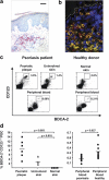
PDC infiltration in the skin of psoriasis patients. (a) Immunohistochemical analysis of BDCA-2 expression in psoriatic plaque lesions. A representative staining (n = 8) at 400× is shown. Bar, 40 μm. (b) CLSM of psoriatic plaque lesions stained for BDCA-2 (green), CD123 (red), and CD3 (blue). A representative analysis shows PDCs costaining for BDCA-2 and CD123 (yellow), T cells staining for CD3 (blue), and CD123 single-positive endothelial cells (*, red). Dashed lines indicate the border between the epidermis (top) and dermis (bottom). (c and d) Flow cytometric quantification of PDCs in dermal single cell suspensions isolated from psoriatic plaque lesions, uninvolved skin, normal skin, and peripheral blood of psoriatic patients and healthy individuals according to their BDCA-2+ CD123high phenotype. Representative flow cytometry analyses are shown in panel c and the percentages of BDCA-2+ CD123high PDCs are indicated in the plots. The results of multiple donors are plotted in panel d. Horizontal bars represent the mean.
Figure 2.
Local activation of PDCs in psoriatic plaque lesions. Flow cytometry analysis of CD86, CD80, and CD83 expression by BDCA-2+ CD123+ PDCs in dermal single cell suspensions of plaque lesions, uninvolved skin, and peripheral blood derived from the same psoriasis patient. Open histograms represent staining of DC activation markers; closed histograms represent isotype control. Data are representative of three independent experiments. Differences in the percentages of CD86high, CD80+, and CD83+ expression on PDCs between plaque lesions and uninvolved skin (P = 0.007, P = 0.02, and P = 0.005, respectively) and between plaque lesions and peripheral blood (P = 0.006, P = 0.02, and P = 0.005, respectively) were significant.
Figure 3.
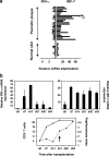
Quantification of IFN-α and IRF-7 mRNA levels in primary plaque lesions and developing psoriasis. (a) Normalized IFN-α and IRF-7 mRNA expression (fg/25 ng of total mRNA) in primary psoriatic plaque lesions compared with normal skin and measured by quantitative real-time RT-PCR. The increased IRF-7 mRNA expression levels in psoriatic skin lesions (n = 24, mean = 32.09) compared with normal skin (n = 11, mean = 5.026) were significant (P < 0.0001, unpaired Student's t test). (b, top) Kinetics of normalized human IFN-α (gene copies per picogram of human mRNA) and IRF-7 (fg/10 pg of human mRNA) expression during psoriasis development in the AGR−/− xenograft model. (b, bottom) Human CD3+ T cells (open circles) and papillomatosis index (closed circles) during the development of psoriasis in the AGR−/− model. The arrow indicates the onset of a psoriatic phenotype defined by the typical epidermal hyperplasia involving both an increase in papillomatosis and acanthosis (not depicted) indices. Data represent the mean of three independent experiments.
Figure 4.
IFN-α production in developing psoriatic lesions is mediated by PDCs. Flow cytometric analysis of surface BDCA-2 and intracellular IFN-α2 expression in dermal single cell suspensions derived from developing psoriatic lesions (across the margin zone of advancing psoriatic plaques) and uninvolved skin. In developing psoriatic lesions, intracellular IFN-α expression was detected among BDCA-2+ PDCs but completely absent in dermal cells lacking BDCA-2, a cell fraction which mainly consists of T cells and myeloid APCs. Data are representative of three independent experiments. Percentages of positive cells are indicated in each quadrant. Differences in the percentages of PDCs positively stained for IFN-α in comparison with matched isotype control, as well as differences in the percentages of IFN-α–expressing BDCA-2+ PDCs compared with non-PDCs, were significant (P < 0.003, unpaired Student's t test).
Figure 5.
IFN-α/β signaling is required for the development of psoriatic skin lesions. Human CD3+ T cell count (a) and papillomatosis index in skin grafts before transplantation (uninvolved prepsoriatic skin at day 0) and 35 d after transplantation subsequent to the administration of isotype-matched control Ab or neutralizing anti–IFN-α/β receptor mAb (b). Values of primary psoriatic plaques derived from the graft donors are provided in comparison. Error bars in panel a represent one SD. Dots in panel b represent independently grafted mice or patient samples. p-values calculated by the unpaired Student's t test are indicated.
Figure 6.
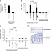
PDC-derived IFN-α is necessary and sufficient to drive the development of psoriatic skin lesions. (a) Normalized human IFN-α mRNA expression in skin grafts harvested 13 d after transplantation subsequent to the administration of isotype-control Ab or anti–BDCA-2 mAb. The mean plus one SD of two independent experiments is given. Human CD3+ T cell count (b) and papillomatosis index (c) in skin grafts before transplantation (uninvolved skin at day 0) and 35 d after transplantation subsequent to the administration of isotype-matched control Ab, anti–BDCA-2 mAb, or anti–BDCA-2 plus recombinant human IFN-α. Dots represent independently grafted mice. In panel b, the mean of these experiments plus one SD is given. p-values were calculated using the unpaired Student's t test. As a control, normal skin (from healthy donors) was transplanted with or without systemic injections of recombinant human IFN-α. (d) Histologic analysis of the skin grafts harvested at day 35 after transplantation. The development of a psoriatic phenotype is inhibited by the administration of anti–BDCA-2 and fully restored by the addition of recombinant human IFN-α. The dashed line indicates the border between the epidermis and the dermis.
Similar articles
- CpG-DNA-induced IFN-alpha production involves p38 MAPK-dependent STAT1 phosphorylation in human plasmacytoid dendritic cell precursors.
Takauji R, Iho S, Takatsuka H, Yamamoto S, Takahashi T, Kitagawa H, Iwasaki H, Iida R, Yokochi T, Matsuki T. Takauji R, et al. J Leukoc Biol. 2002 Nov;72(5):1011-9. J Leukoc Biol. 2002. PMID: 12429724 - Monocyte-mediated inhibition of TLR9-dependent IFN-α induction in plasmacytoid dendritic cells questions bacterial DNA as the active ingredient of bacterial lysates.
Poth JM, Coch C, Busch N, Boehm O, Schlee M, Janke M, Zillinger T, Schildgen O, Barchet W, Hartmann G. Poth JM, et al. J Immunol. 2010 Dec 15;185(12):7367-73. doi: 10.4049/jimmunol.1001798. Epub 2010 Nov 5. J Immunol. 2010. PMID: 21057083 - Psoriasis triggered by toll-like receptor 7 agonist imiquimod in the presence of dermal plasmacytoid dendritic cell precursors.
Gilliet M, Conrad C, Geiges M, Cozzio A, Thürlimann W, Burg G, Nestle FO, Dummer R. Gilliet M, et al. Arch Dermatol. 2004 Dec;140(12):1490-5. doi: 10.1001/archderm.140.12.1490. Arch Dermatol. 2004. PMID: 15611427 - Immune functions and recruitment of plasmacytoid dendritic cells in psoriasis.
Albanesi C, Scarponi C, Bosisio D, Sozzani S, Girolomoni G. Albanesi C, et al. Autoimmunity. 2010 Apr;43(3):215-9. doi: 10.3109/08916930903510906. Autoimmunity. 2010. PMID: 20166874 Review. - Monocyte-derived interferon-alpha primed dendritic cells in the pathogenesis of psoriasis: new pieces in the puzzle.
Farkas A, Kemény L. Farkas A, et al. Int Immunopharmacol. 2012 Jun;13(2):215-8. doi: 10.1016/j.intimp.2012.04.003. Epub 2012 Apr 19. Int Immunopharmacol. 2012. PMID: 22522054 Review.
Cited by
- Selective Blockade of Interferon-α and -β Reveals Their Non-Redundant Functions in a Mouse Model of West Nile Virus Infection.
Sheehan KC, Lazear HM, Diamond MS, Schreiber RD. Sheehan KC, et al. PLoS One. 2015 May 26;10(5):e0128636. doi: 10.1371/journal.pone.0128636. eCollection 2015. PLoS One. 2015. PMID: 26010249 Free PMC article. - The significance of macrophage polarization subtypes for animal models of tissue fibrosis and human fibrotic diseases.
Wermuth PJ, Jimenez SA. Wermuth PJ, et al. Clin Transl Med. 2015 Feb 7;4:2. doi: 10.1186/s40169-015-0047-4. eCollection 2015. Clin Transl Med. 2015. PMID: 25852818 Free PMC article. - Topical nanoparticles interfering with the DNA-LL37 complex to alleviate psoriatic inflammation in mice and monkeys.
Liang H, Yan Y, Wu J, Ge X, Wei L, Liu L, Chen Y. Liang H, et al. Sci Adv. 2020 Jul 31;6(31):eabb5274. doi: 10.1126/sciadv.abb5274. eCollection 2020 Jul. Sci Adv. 2020. PMID: 32923608 Free PMC article. - Cross Talk between Proliferative, Angiogenic, and Cellular Mechanisms Orchestred by HIF-1α in Psoriasis.
Torales-Cardeña A, Martínez-Torres I, Rodríguez-Martínez S, Gómez-Chávez F, Cancino-Díaz JC, Vázquez-Sánchez EA, Cancino-Díaz ME. Torales-Cardeña A, et al. Mediators Inflamm. 2015;2015:607363. doi: 10.1155/2015/607363. Epub 2015 Jun 7. Mediators Inflamm. 2015. PMID: 26136626 Free PMC article. Review. - Recent Progress in Dendritic Cell-Based Cancer Immunotherapy.
Matsuo K, Yoshie O, Kitahata K, Kamei M, Hara Y, Nakayama T. Matsuo K, et al. Cancers (Basel). 2021 May 20;13(10):2495. doi: 10.3390/cancers13102495. Cancers (Basel). 2021. PMID: 34065346 Free PMC article. Review.
References
- Lebwohl, M. 2003. Psoriasis. Lancet. 361:1197–1204. - PubMed
- Lew, W., A.M. Bowcock, and J.G. Krueger. 2004. Psoriasis vulgaris: cutaneous lymphoid tissue supports T-cell activation and ‘Type 1’ inflammatory gene expression. Trends Immunol. 25:295–305. - PubMed
- Adorini, L., and F. Sinigaglia. 1997. Pathogenesis and immunotherapy of autoimmune diseases. Immunol. Today. 18:209–211. - PubMed
- Davidson, A., and B. Diamond. 2001. Autoimmune diseases. N. Engl. J. Med. 345:340–350. - PubMed
Publication types
MeSH terms
Substances
LinkOut - more resources
Full Text Sources
Other Literature Sources
Medical


