The functional organization of the intraparietal sulcus in humans and monkeys - PubMed (original) (raw)
Review
The functional organization of the intraparietal sulcus in humans and monkeys
Christian Grefkes et al. J Anat. 2005 Jul.
Abstract
In macaque monkeys, the posterior parietal cortex (PPC) is concerned with the integration of multimodal information for constructing a spatial representation of the external world (in relation to the macaque's body or parts thereof), and planning and executing object-centred movements. The areas within the intraparietal sulcus (IPS), in particular, serve as interfaces between the perceptive and motor systems for controlling arm and eye movements in space. We review here the latest evidence for the existence of the IPS areas AIP (anterior intraparietal area), VIP (ventral intraparietal area), MIP (medial intraparietal area), LIP (lateral intraparietal area) and CIP (caudal intraparietal area) in macaques, and discuss putative human equivalents as assessed with functional magnetic resonance imaging. The data suggest that anterior parts of the IPS comprising areas AIP and VIP are relatively well preserved across species. By contrast, posterior areas such as area LIP and CIP have been found more medially in humans, possibly reflecting differences in the evolution of the dorsal visual stream and the inferior parietal lobule. Despite interspecies differences in the precise functional anatomy of the IPS areas, the functional relevance of this sulcus for visuomotor tasks comprising target selections for arm and eye movements, object manipulation and visuospatial attention is similar in humans and macaques, as is also suggested by studies of neurological deficits (apraxia, neglect, Bálint's syndrome) resulting from lesions to this region.
Figures
Fig. 1
Lateral view of a macaque brain with the intraparietal sulcus (IPS) opened up. The arrangement of the IPS areas is schematically depicted as defined in various electrophysiological experiments (see text for references). The bottom of the IPS is indicated by a dashed line. cs, central sulcus; pos, parieto-occipital sulcus; ls, lunate sulcus; sf, Sylvian fissure (lateral sulcus); sts, superior temporal sulcus.
Fig. 2
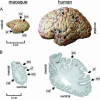
Post-mortem anatomy of a macaque and a human brain (specimens taken from the Zilles collection, C. & O. Vogt Institute of Brain Research, Düsseldorf University, Germany). (A) The sulcal anatomy of a macaque brain is much less complicated than the highly gyrified cortex of a human brain. The IPS in humans often has various side branches (indicated here by asterisks in A), and shows a complex folding pattern as demonstrated on the cell-stained coronal sections through posterior portions of the IPS (B). Note the highly gyrified medial aspect of the human IPS (right section) with a large unlabelled intraparietal gyrus. cal, calcarine sulcus; spl; superior parietal lobule; ipl; inferior parietal lobule; other abbreviations as defined in Fig. 1.
Fig. 3
Putative human AIP equivalent as assessed with fMRI (Grefkes et al. 2002). (A) Subjects had to encode and subsequently to recognize the shape of 3D objects either by visual inspection (as presented on a video screen) or by tactile manipulation (using their right hand).(B) When 3D information had to be transferred between the visual and the sensorimotor systems for successful object recognition (cross-modal transfer), increased neural activity was measured in the lateral anterior aspect of left intraparietal sulcus (P < 0.05, small volume corrected for multiple comparisons, n = 12). That region was significantly less active when objects were encoded and recognized within the same modality (visual, tactile). L, left; R, right; A, anterior; P, posterior; pocs, postcentral sulcus; other abbreviations as in Fig. 1. (Adapted from Grefkes et al. 2002, with permission.)
Fig. 4
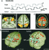
Putative human VIP equivalent as assessed with fMRI (Bremmer et al. 2001). (A) Subjects experienced either a visual (large random dot pattern), a tactile (air flow across forehead) or an auditory (binaural beats) motion stimulus, or a stationary modality-specific control. (B) A conjunction analysis (P < 0.05, corrected for multiple comparisons, n = 8) testing for increased neural activity common to all three conditions conveying motion information revealed significant activations in three circumscribed regions, one of which was located in parietal cortex (labelled ‘1’). The superimposition of the functional images with the mean anatomical volume showed that the area activated lay in the depth of either intraparietal sulcus (IPS). (Adapted from Bremmer et al. 2001, with kind permission.)
Fig. 5
Putative human MIP equivalent as assessed with fMRI (Grefkes et al. 2004). (A) Electrophysiological MIP experiments in the macaque brain (Eskandar & Assad, 2002). MIP neurons were recorded while the monkey performed a visuomotor joystick task. MIP neurons responded irrespective of whether a visual movement feedback was present on the video screen but much less when joystick movements were absent (playback trials). (From Eskandar & Assad, 2002, with permission.) (B) Adopted protocol used for the fMRI study with human subjects (Grefkes et al. 2004). Fixating volunteers were asked to guide the black square from the white to the black circle using an MRI-compatible joystick. (C) Visually directed joystick movements (with or without movement feedback) elicited significantly higher activity in medial IPS than the control conditions consisting of stereotype-intransitive joystick movements. (Adapted from Grefkes et al. 2004, with permission.)
Fig. 6
Putative human LIP equivalent as assessed with fMRI (Koyama et al. 2004). (A) The oculomotor task consisted of horizontal saccades in response to the shift of the fixation target. Upper row: horizontal target displacement. Lower rows: eye movement recordings in horizontal and vertical direction. (B) BOLD signal associated with saccades in monkeys. Increased neural activity is found in the lateral wall of IPS (left image: coronal section; right image: horizontal section). (C) BOLD signal associated with saccades in humans. Increased neural activity is found in the medial wall of IPS (horizontal section). (D) Saccade-related activity superimposed on 3D renderings of a macaque brain (left) and a human brain (right). Monkey area LIP is situated in lateral IPS, the putative human LIP equivalent is located in medial aspects of posterior IPS. (Adapted from Koyama et al. 2004, with kind permission.)
Fig. 7
Putative human CIP equivalent as assessed with fMRI (Shikata et al. 2001). (A) Electrophysiological recordings from a texture gradient-sensitive neuron in macaque area CIP (adapted from Tsutsui et al. 2002, with permission). Responses to three orientations [180° tilt, frontoparallel (FP), 0° tilt) are shown for dot texture patterns (dot-TP) and line texture patterns (line-TP) aligned at the onset of sample stimulus presentation. Insets above rasters indicate stimuli presented. The neuronal responses are selective to texture gradients of patterns defining 0° tilt (or right-side-nearer orientation). (B) Protocol of the fMRI study with human subjects (Shikata et al. 2001). After a cue stimulus (‘O’ for orientation discrimination, ‘C’ for colour discrimination), sample and test stimuli composed of dot-TPs and different colours were sequentially presented. Subjects were asked to press a button as soon as the matching stimulus appeared. (C) Surface orientation discrimination vs. colour discrimination significantly activated two regions in human IPS bilaterally (single subject brain, P < 0.05, corrected; detailed images thresholded at P < 0.001, uncorrected). The posterior activation (putative human CIP, hCIP) is stronger positively modulated by the orientation discrimination performance of the subjects than the anterior region (putative human AIP, hAIP). (Adapted from Shikata et al. 2001, with kind permission.)
Similar articles
- Human medial intraparietal cortex subserves visuomotor coordinate transformation.
Grefkes C, Ritzl A, Zilles K, Fink GR. Grefkes C, et al. Neuroimage. 2004 Dec;23(4):1494-506. doi: 10.1016/j.neuroimage.2004.08.031. Neuroimage. 2004. PMID: 15589113 - Localization of human intraparietal areas AIP, CIP, and LIP using surface orientation and saccadic eye movement tasks.
Shikata E, McNamara A, Sprenger A, Hamzei F, Glauche V, Büchel C, Binkofski F. Shikata E, et al. Hum Brain Mapp. 2008 Apr;29(4):411-21. doi: 10.1002/hbm.20396. Hum Brain Mapp. 2008. PMID: 17497631 Free PMC article. - Parietal inputs to dorsal versus ventral premotor areas in the macaque monkey: evidence for largely segregated visuomotor pathways.
Tanné-Gariépy J, Rouiller EM, Boussaoud D. Tanné-Gariépy J, et al. Exp Brain Res. 2002 Jul;145(1):91-103. doi: 10.1007/s00221-002-1078-9. Epub 2002 Apr 30. Exp Brain Res. 2002. PMID: 12070749 - The role of parietal cortex in visuomotor control: what have we learned from neuroimaging?
Culham JC, Cavina-Pratesi C, Singhal A. Culham JC, et al. Neuropsychologia. 2006;44(13):2668-84. doi: 10.1016/j.neuropsychologia.2005.11.003. Epub 2005 Dec 9. Neuropsychologia. 2006. PMID: 16337974 Review. - From thought to action: the parietal cortex as a bridge between perception, action, and cognition.
Gottlieb J. Gottlieb J. Neuron. 2007 Jan 4;53(1):9-16. doi: 10.1016/j.neuron.2006.12.009. Neuron. 2007. PMID: 17196526 Review.
Cited by
- Reorganization of integration and segregation networks in brain-based visual impairment.
Diez I, Troyas C, Bauer CM, Sepulcre J, Merabet LB. Diez I, et al. Neuroimage Clin. 2024 Oct 15;44:103688. doi: 10.1016/j.nicl.2024.103688. Online ahead of print. Neuroimage Clin. 2024. PMID: 39432973 Free PMC article. - Individualized functional magnetic resonance imaging neuromodulation enhances visuospatial perception: a proof-of-concept study.
Allam A, Allam V, Reddy S, Rohren EM, Sheth SA, Froudarakis E, Papageorgiou TD. Allam A, et al. Philos Trans R Soc Lond B Biol Sci. 2024 Dec 2;379(1915):20230083. doi: 10.1098/rstb.2023.0083. Epub 2024 Oct 21. Philos Trans R Soc Lond B Biol Sci. 2024. PMID: 39428879 Free PMC article. - Functional Connectivity Profiles of Ten Sub-Regions within the Premotor and Supplementary Motor Areas: Insights into Neurophysiological Integration.
Alahmadi A. Alahmadi A. Diagnostics (Basel). 2024 Sep 9;14(17):1990. doi: 10.3390/diagnostics14171990. Diagnostics (Basel). 2024. PMID: 39272774 Free PMC article. - Human intraparietal sulcal morphology relates to individual differences in language and memory performance.
Santacroce F, Cachia A, Fragueiro A, Grande E, Roell M, Baldassarre A, Sestieri C, Committeri G. Santacroce F, et al. Commun Biol. 2024 May 2;7(1):520. doi: 10.1038/s42003-024-06175-9. Commun Biol. 2024. PMID: 38698168 Free PMC article. - Information-based rhythmic transcranial magnetic stimulation to accelerate learning during auditory working memory training: a proof-of-concept study.
Whittaker HT, Khayyat L, Fortier-Lavallée J, Laverdière M, Bélanger C, Zatorre RJ, Albouy P. Whittaker HT, et al. Front Neurosci. 2024 Apr 4;18:1355565. doi: 10.3389/fnins.2024.1355565. eCollection 2024. Front Neurosci. 2024. PMID: 38638697 Free PMC article.
References
- Binkofski F, Dohle C, Posse S, Stephan KM, Hefter H, Seitz RJ, Freund HJ. Human anterior intraparietal area subserves prehension: a combined lesion and functional MRI activation study. Neurology. 1998;50:1253–1259. - PubMed
- Bisley JW, Goldberg ME. The role of the parietal cortex in the neural processing of saccadic eye movements. Adv Neurol. 2003a;93:141–157. - PubMed
- Bisley JW, Goldberg ME. Neuronal activity in the lateral intraparietal area and spatial attention. Science. 2003b;299:81–86. - PubMed
- Blatt GJ, Andersen RA, Stoner GR. Visual receptive field organization and cortico-cortical connections of the lateral intraparietal area (area LIP) in the macaque. J Comp Neurol. 1990;299:421–445. - PubMed
Publication types
MeSH terms
LinkOut - more resources
Full Text Sources
Medical
Miscellaneous


