Spontaneous atopic dermatitis in mice expressing an inducible thymic stromal lymphopoietin transgene specifically in the skin - PubMed (original) (raw)
Spontaneous atopic dermatitis in mice expressing an inducible thymic stromal lymphopoietin transgene specifically in the skin
Jane Yoo et al. J Exp Med. 2005.
Abstract
The cytokine thymic stromal lymphopoietin (TSLP) has recently been implicated in the pathogenesis of atopic dermatitis (AD) and other allergic diseases in humans. To further characterize its role in this disease process, transgenic mice were generated that express a keratinocyte-specific, tetracycline-inducible TSLP transgene. Skin-specific overexpression of TSLP resulted in an AD-like phenotype, with the development of eczematous lesions containing inflammatory dermal cellular infiltrates, a dramatic increase in Th2 CD4+ T cells expressing cutaneous homing receptors, and elevated serum levels of IgE. These transgenic mice demonstrate that TSLP can initiate a cascade of allergic inflammation in the skin and provide a valuable animal model for future study of this common disease.
Figures
Figure 1.
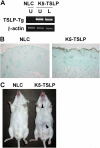
Skin-targeted transgenic expression of TSLP causes eczematous-like skin lesions accompanied by pronounced lymphadenopathy and splenomegaly. (A) RT-PCR analysis of TSLP transgene expression in the skin of a K5-TSLP mouse and a normal littermate control (NLC) after 3 wk of dox treatment. From the K5-TSLP mouse, total RNA isolated from affected lesional skin (L) and unaffected skin (U) were analyzed separately. (B) An immunohistochemical localization of TSLP in the skin of a K5-TSLP mouse as compared with NLC. TSLP in a K5-TSLP mouse was expressed in the epidermis and its basement membrane (brown). (C) Images showing a K5-TSLP mouse and NLC after 3 wk of dox treatment. Arrowheads mark eczematous lesions on the snout and trunk. Images are representative of >10 K5-TSLP mice analyzed.
Figure 2.
Histological observations of the skin in K5-TSLP mice. Paraffin-embedded sections of lesional skin from a K5-TSLP mouse and an NLC were stained with H&E. (A) Skin sections showing pronounced acanthosis (arrow) and dermal infiltration (arrowhead) at 10×. (B) Images of lesional skin from a K5-TSLP mouse showing epidermal spongiosis (asterisk) and dermal infiltration by mast cells (arrows) and eosinophils (arrowheads) at 100×.
Figure 3.
Immunohistochemical localizations of infiltrated macrophages and Langerhans cells in the skin of a K5-TSLP mouse. As compared with the NLC, a larger number of macrophages and Langerhans cells (BM8+) are infiltrated into affected skin areas of a K5-TSLP mouse.
Figure 4.
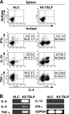
The CD4**+** T cell response in K5-TSLP mice is Th2 cell polarized. (A) Intracellular proallergic cytokine staining of splenic CD4+ T cells isolated from a K5-TSLP mouse and an NLC after 5 wk of dox treatment. The percentages of cells in each quadrant are shown. Data are representative of more than five animals analyzed in this manner. (B) RT-PCR analysis of cytokine gene expression in the lesional skin of a K5-TSLP mouse and skin from an NLC.
Figure 5.
CD4**+** T cells in the K5-TSLP mice express cutaneous homing receptors. Flow cytometry analysis of homing receptor expression by CD4+ T cells isolated from a K5-TSLP mouse and an NLC after 5 wk of dox treatment. Data are representative of more than five animals analyzed in this manner. (A) P-ligand (P-lig) and E-ligand (E-lig) expression was assessed by binding to P- and E-selectin–IgM fusion proteins in conjunction with intracellular cytokine staining to detect expression of IL-4. The percentages of cells in each quadrant are shown. Per., peripheral. (B) RT-PCR analysis of chemokine expression in the skin of a K5-TSLP mouse and NLC after 3 wk of dox treatment. From the K5-TSLP mouse, total RNA isolated from affected lesional skin (L) and unaffected skin (U) were analyzed separately. The levels of mRNA of CCL17, CCL22, and β-actin were determined by semiquantitative RT-PCR analysis with threefold serial dilution of the template cDNA. (C) CCR4 and CCR7 expression assessed by binding to CCL22- and CCL19-IgG3 fusion proteins in conjunction with staining for L-selectin (L-sel). The percentages of cells in each quadrant are shown.
Figure 6.
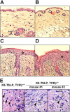
Histological observations of the skin in different genotypes of mice that were crossed between K5-TSLP and TCRβ KO mice. (A) A wild-type mouse without the transgene for K5-TSLP and KO alleles for TCRβ. (B) A mouse without the transgene for K5-TSLP but with KO alleles for TCRβ. (C) A mouse with the transgene for K5-TSLP but without KO alleles for TCRβ. (D) A mouse with the transgene for K5-TSLP and KO alleles for TCRβ. Skin sections show pronounced acanthosis (asterisks) and infiltration in K5-TSLP mice with and without KO alleles for TCRβ at 10× (C and D), suggesting that skin symptoms of AD occur in K5-TSLP mice, regardless of whether αβ T cells are present. All paraffin-embedded sections were stained with H&E. (E) H&E-stained skin sections from K5-TSLP, TCRβ+/− and K5-TSLP, TCRβ−/− mice at 100×, showing the presence of eosinophils (closed arrowheads) and mast cells (open arrowheads) in skin from both sets of mice.
Similar articles
- Ginsenoside Rh2 Ameliorates Atopic Dermatitis in NC/Nga Mice by Suppressing NF-kappaB-Mediated Thymic Stromal Lymphopoietin Expression and T Helper Type 2 Differentiation.
Ko E, Park S, Lee JH, Cui CH, Hou J, Kim MH, Kim SC. Ko E, et al. Int J Mol Sci. 2019 Dec 4;20(24):6111. doi: 10.3390/ijms20246111. Int J Mol Sci. 2019. PMID: 31817146 Free PMC article. - Thymic stromal lymphopoietin-induced interleukin-17A is involved in the development of IgE-mediated atopic dermatitis-like skin lesions in mice.
Mizutani N, Sae-Wong C, Kangsanant S, Nabe T, Yoshino S. Mizutani N, et al. Immunology. 2015 Dec;146(4):568-81. doi: 10.1111/imm.12528. Epub 2015 Sep 24. Immunology. 2015. PMID: 26310839 Free PMC article. - Prostaglandin E2 (PGE2)-EP2 signaling negatively regulates murine atopic dermatitis-like skin inflammation by suppressing thymic stromal lymphopoietin expression.
Sawada Y, Honda T, Nakamizo S, Nakajima S, Nonomura Y, Otsuka A, Egawa G, Yoshimoto T, Nakamura M, Narumiya S, Kabashima K. Sawada Y, et al. J Allergy Clin Immunol. 2019 Nov;144(5):1265-1273.e9. doi: 10.1016/j.jaci.2019.06.036. Epub 2019 Jul 11. J Allergy Clin Immunol. 2019. PMID: 31301371 - Thymic stromal lymphopoietin and OX40 ligand pathway in the initiation of dendritic cell-mediated allergic inflammation.
Liu YJ. Liu YJ. J Allergy Clin Immunol. 2007 Aug;120(2):238-44; quiz 245-6. doi: 10.1016/j.jaci.2007.06.004. J Allergy Clin Immunol. 2007. PMID: 17666213 Review. - The Role of TSLP in Atopic Dermatitis: From Pathogenetic Molecule to Therapeutical Target.
Luo J, Zhu Z, Zhai Y, Zeng J, Li L, Wang D, Deng F, Chang B, Zhou J, Sun L. Luo J, et al. Mediators Inflamm. 2023 Apr 15;2023:7697699. doi: 10.1155/2023/7697699. eCollection 2023. Mediators Inflamm. 2023. PMID: 37096155 Free PMC article. Review.
Cited by
- Keratinocyte-specific deletion of SHARPIN induces atopic dermatitis-like inflammation in mice.
Sundberg JP, Pratt CH, Goodwin LP, Silva KA, Kennedy VE, Potter CS, Dunham A, Sundberg BA, HogenEsch H. Sundberg JP, et al. PLoS One. 2020 Jul 20;15(7):e0235295. doi: 10.1371/journal.pone.0235295. eCollection 2020. PLoS One. 2020. PMID: 32687504 Free PMC article. - MyD88 is critically involved in immune tolerance breakdown at environmental interfaces of Foxp3-deficient mice.
Rivas MN, Koh YT, Chen A, Nguyen A, Lee YH, Lawson G, Chatila TA. Rivas MN, et al. J Clin Invest. 2012 May;122(5):1933-47. doi: 10.1172/JCI40591. Epub 2012 Apr 2. J Clin Invest. 2012. PMID: 22466646 Free PMC article. - From gene identifications to therapeutic targets for asthma: Focus on great potentials of TSLP, ORMDL3, and GSDMB.
Zhang Y. Zhang Y. Chin Med J Pulm Crit Care Med. 2023 Sep 14;1(3):139-147. doi: 10.1016/j.pccm.2023.08.001. eCollection 2023 Sep. Chin Med J Pulm Crit Care Med. 2023. PMID: 39171126 Free PMC article. Review. - Mediators of Chronic Pruritus in Atopic Dermatitis: Getting the Itch Out?
Mollanazar NK, Smith PK, Yosipovitch G. Mollanazar NK, et al. Clin Rev Allergy Immunol. 2016 Dec;51(3):263-292. doi: 10.1007/s12016-015-8488-5. Clin Rev Allergy Immunol. 2016. PMID: 25931325 Review. - Keratinocyte-derived cytokine TSLP promotes growth and metastasis of melanoma by regulating the tumor-associated immune microenvironment.
Yao W, German B, Chraa D, Braud A, Hugel C, Meyer P, Davidson G, Laurette P, Mengus G, Flatter E, Marschall P, Segaud J, Guivarch M, Hener P, Birling MC, Lipsker D, Davidson I, Li M. Yao W, et al. JCI Insight. 2022 Nov 8;7(21):e161438. doi: 10.1172/jci.insight.161438. JCI Insight. 2022. PMID: 36107619 Free PMC article.
References
- Oranje, A.P., and F.B. de Waard-van der Spek. 2002. Atopic dermatitis: review 2000 to January 2001. Curr. Opin. Pediatr. 14:410–413. - PubMed
- Kang, K., and S.R. Stevens. 2003. Pathophysiology of atopic dermatitis. Clin. Dermatol. 21:116–121. - PubMed
- Eichenfield, L.F., J.M. Hanifin, T.A. Luger, S.R. Stevens, and H.B. Pride. 2003. Consensus conference of pediatric atopic dermatitis. J. Am. Acad. Dermatol. 49:1088–1095. - PubMed
- Gupta, M.A., and A.K. Gupta. 2003. Psychiatric and psychological co-morbidity in patients with dermatologic disorders: epidemiology and management. Am. J. Clin. Dermatol. 4:833–842. - PubMed
Publication types
MeSH terms
Substances
LinkOut - more resources
Full Text Sources
Other Literature Sources
Molecular Biology Databases
Research Materials


