RhoC is dispensable for embryogenesis and tumor initiation but essential for metastasis - PubMed (original) (raw)
RhoC is dispensable for embryogenesis and tumor initiation but essential for metastasis
Anne Hakem et al. Genes Dev. 2005.
Abstract
The Rho proteins are Ras-related guanosine triphosphatases (GTPases) that function in cytoskeletal reorganization, cell migration, and stress fiber and focal adhesion formation. Overexpression of RhoC enhances the ability of melanoma cells to exit the blood and colonize the lungs. However, in vivo confirmation of RhoC's role in metastasis has awaited a RhoC-deficient mouse model. Here we report the generation of RhoC-deficient mice and show that RhoC is dispensable for embryonic and post-natal development. We demonstrate that loss of RhoC does not affect tumor development but decreases tumor cell motility and metastatic cell survival leading to a drastic inhibition of metastasis.
Figures
Figure 1.
Generation of _RhoC_-deficient mice. (A) Schematic representations of the wild-type RhoC locus, the targeting construct (floxed 2-3 Neo), and the mutant RhoC_Δ_2-3 allele. Exons are indicated by filled boxes and Loxp sites are indicated by triangles. The 5′-flanking probe used for Southern hybridizations is indicated. (K) Kpn 1 site. (B) Identification of ES cell clones bearing the floxed RhoC allele (RhoCfl2-3). Clones that had undergone homologous recombination at the RhoC locus were first identified by PCR (not shown) and then confirmed by Southern blotting using the 5′ probe indicated in A. Four independent ES clones were subsequently transfected with Cre recombinase to generate ES cell clones lacking RhoC exon 2, exon 3, and the neomycin gene. (C) Southern blot analysis of tail DNA from RhoC+/+, RhoC+/- and _RhoC_-/- littermate mice hybridized to the 5′ probe in A.(D) Northern blot of total RNA from RhoC+/- or _RhoC_-/- MEFs. The wild-type (Wt) RhoC transcript (1.4 kb) appears in RNA derived from RhoC+/- MEFs but not _RhoC_-/- MEFs.
Figure 2.
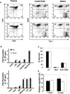
RhoC is not essential in T- or B-cell development, activation, apoptosis, or migration. (A) Flow cytometric analyses of thymic, lymph node, and splenic total T- and B-lymphocyte populations in wild-type (upper panels) or RhoC-/- (lower panels) mice. Results representative of seven independent experiments are shown. (B) Proliferative responses of T and B lymphocytes to stimuli. Purified T and B cells from wild-type (Wt; black bars) and RhoC-/- (white bars) mice were stimulated for 48 h with anti-CD3 with or without costimulation by anti-CD28 and/or IL-2, or PMA plus ionomycine (Iono) for T cells; or anti-IgM, anti-CD40, anti-IgM plus anti-CD40, LPS, or CpG for B cells. [3H]-thymidine incorporation was then assessed. (C) Thymocyte sensitivity to apoptosis. Triplicate cultures of thymocytes from wild-type (black bars) or RhoC-/- (white bars) mice were treated with dexamethasone (DEX) to induce mitochondria-mediated apoptosis, or with anti-CD95 to induce death-receptor-mediated apoptosis. Cell viability was determined 24 h later by Annexin V FITC/PI staining. (UT) Untreated control. (D) Cell migration. T and B cells and thymocytes (∼106 cells) from wild-type (black bars) or RhoC-/- (white bars) mice were placed in the upper chamber of Transwell motility plates. Data shown are the mean percentages of duplicate sample of cells that migrated to the lower chamber after 24 h. For B, C and D, data are presented as the mean ± SD and are representative of three independent experiments.
Figure 3.
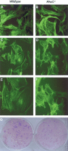
RhoC is required for stress fiber formation but not transformation. (A,B) Normal culture conditions. Wild-type and RhoC-/- MEFs were cultured in DMEM containing 10% FCS for 48 h, fixed, and stained with FITC-phalloidin. Normal cytoskeletal structures can be seen even in the absence of RhoC. (C,D) Stress conditions. Wild-type and RhoC-/- MEFs were cultured in medium lacking FCS for 48 h, fixed, and stained as in A and B. Abnormal cytoskeletal structures are present in stressed RhoC-/- MEFs. Magnification: A-D,20×.(E,F) Higher-magnification (40×) view of the cells in C and D. RhoC-/- MEFs clearly show a defect in stress fiber formation. (G) E1A/Ras transformation. Wild-type and RhoC-/- MEFs were transformed with Ras/E1A retrovirus as described in Materials and Methods. No differences in the number of colonies and time of appearance of colonies were observed in the absence of RhoC. Data shown are representative of two independent experiments each.
Figure 4.
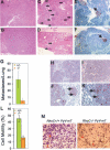
RhoC is essential for metastasis but not for primary tumor formation. (A-F) Histological examination of H&E-stained RhoC+/+ PyV-mT (A,C) and RhoC-/- PyV-mT (B,D) tissue samples. (A,B) Mammary adenocarcinoma. (C,D) Lung metastases (arrows). (E,F) TUNEL assays of sections of primary tumors from RhoC+/+ PyV-mT (E) and RhoC-/- PyV-mT (F) mice. Red arrows indicate TUNEL-positive cells. (G) Metastases in H&E-stained lung sections from RhoC+/+ PyV-mT (n = 12) and RhoC-/- PyV-mT (n = 16) mice. Metastases were counted on five lobes of the lung. Results shown are the mean number of metastases/lung ± SD. (H-K) Lung sections from RhoC+/+ PyV-mT (H,I) and RhoC-/- PyV-mT (J,K) mice were immunostained to detect cleaved caspase3. Arrows indicate cells positive for cleaved caspase3. (L) In vitro migration efficiency. Cells from RhoC+/+ PyV-mT (green) and RhoC-/- PyV-mT (orange) tumors were assayed for motility in Transwell assays as described in Materials and Methods. Data are expressed as the mean percentage (duplicate samples) of the initial seeded population that was able to migrate into the lower chamber. Five different tumors for each genotype were analyzed. (M) In vitro invasiveness. Tumor cells from RhoC+/+ PyV-mT and RhoC-/- PyV-mT mice were assessed for invasiveness using Transwell invasion chambers as described in Materials and Methods. Cells present in the polycarbonate membranes at 20 h post-seeding were stained with crystal violet. Representative results for RhoC+/+ PyV-mT (left panel) and RhoC-/- PyV-mT (right panel) tumors are shown.
Similar articles
- Inhibition of invasion and metastasis of hepatocellular carcinoma cells via targeting RhoC in vitro and in vivo.
Wang W, Wu F, Fang F, Tao Y, Yang L. Wang W, et al. Clin Cancer Res. 2008 Nov 1;14(21):6804-12. doi: 10.1158/1078-0432.CCR-07-4820. Clin Cancer Res. 2008. PMID: 18980974 - RhoA and RhoC proteins promote both cell proliferation and cell invasion of human oesophageal squamous cell carcinoma cell lines in vitro and in vivo.
Faried A, Faried LS, Kimura H, Nakajima M, Sohda M, Miyazaki T, Kato H, Usman N, Kuwano H. Faried A, et al. Eur J Cancer. 2006 Jul;42(10):1455-65. doi: 10.1016/j.ejca.2006.02.012. Epub 2006 Jun 5. Eur J Cancer. 2006. PMID: 16750623 - Rho GTPases: promising cellular targets for novel anticancer drugs.
Fritz G, Kaina B. Fritz G, et al. Curr Cancer Drug Targets. 2006 Feb;6(1):1-14. Curr Cancer Drug Targets. 2006. PMID: 16475973 Review. - [Transgenic mice for analysis of neoplasm transformation processes of T and B lymphocytes].
Kobzdej M, Strzadała L. Kobzdej M, et al. Postepy Hig Med Dosw. 1996;50(2):113-29. Postepy Hig Med Dosw. 1996. PMID: 8848421 Review. Polish.
Cited by
- Identification of a novel actin-binding domain within the Rho guanine nucleotide exchange factor TEM4.
Mitin N, Rossman KL, Der CJ. Mitin N, et al. PLoS One. 2012;7(7):e41876. doi: 10.1371/journal.pone.0041876. Epub 2012 Jul 24. PLoS One. 2012. PMID: 22911862 Free PMC article. - RhoC regulates radioresistance via crosstalk of ROCK2 with the DNA repair machinery in cervical cancer.
Pranatharthi A, Thomas P, Udayashankar AH, Bhavani C, Suresh SB, Krishna S, Thatte J, Srikantia N, Ross CR, Srivastava S. Pranatharthi A, et al. J Exp Clin Cancer Res. 2019 Sep 5;38(1):392. doi: 10.1186/s13046-019-1385-7. J Exp Clin Cancer Res. 2019. PMID: 31488179 Free PMC article. - Neuronal polarity.
Tahirovic S, Bradke F. Tahirovic S, et al. Cold Spring Harb Perspect Biol. 2009 Sep;1(3):a001644. doi: 10.1101/cshperspect.a001644. Cold Spring Harb Perspect Biol. 2009. PMID: 20066106 Free PMC article. Review. - Therapeutic intervention based on protein prenylation and associated modifications.
Gelb MH, Brunsveld L, Hrycyna CA, Michaelis S, Tamanoi F, Van Voorhis WC, Waldmann H. Gelb MH, et al. Nat Chem Biol. 2006 Oct;2(10):518-28. doi: 10.1038/nchembio818. Nat Chem Biol. 2006. PMID: 16983387 Free PMC article. Review. - Formin-like 3 regulates RhoC/FAK pathway and actin assembly to promote cell invasion in colorectal carcinoma.
Zeng YF, Xiao YS, Liu Y, Luo XJ, Wen LD, Liu Q, Chen M. Zeng YF, et al. World J Gastroenterol. 2018 Sep 14;24(34):3884-3897. doi: 10.3748/wjg.v24.i34.3884. World J Gastroenterol. 2018. PMID: 30228782 Free PMC article.
References
- Clark E.A., Golub, T.R., Lander, E.S., and Hynes, R.O. 2000. Genomic analysis of metastasis reveals an essential role for RhoC. Nature 406: 532-535. - PubMed
- Etienne-Manneville S. and Hall, A. 2002. Rho GTPases in cell biology. Nature 420: 629-635. - PubMed
Publication types
MeSH terms
Substances
LinkOut - more resources
Full Text Sources
Other Literature Sources
Molecular Biology Databases


