Overexpression of insulin receptor substrate-2 in human and murine hepatocellular carcinoma - PubMed (original) (raw)
Overexpression of insulin receptor substrate-2 in human and murine hepatocellular carcinoma
Mathieu Boissan et al. Am J Pathol. 2005 Sep.
Abstract
De-regulations in insulin and insulin-like growth factor (IGF) pathways may contribute to hepatocellular carcinoma. Although intracellular insulin receptor substrate-2 (IRS-2) is the main effector of insulin signaling in the liver, its role in hepatocarcinogenesis is unknown. Here, we show that IRS-2 was overexpressed in two murine models of hepatocarcinogenesis: administration of diethylnitrosamine and hepatic overexpression of SV40 large T antigen. In both models, IRS-2 overexpression was detected in preneoplastic lesions and at higher levels in tumoral nodules. IRS-2 overexpression associated with IGF-2 and IRS-1 overexpression and with GSK-3beta inhibition. Increased expression of IRS-2 was also detected in human hepatocellular carcinoma specimens and hepatoma cell lines. In murine and human hepatoma cells, IRS-2 protein induction associated with increased IRS-2 mRNA levels. The functionality of IRS-2 was demonstrated in Hep 3 B cells, in which IRS-2 tyrosine phosphorylation and its association with phosphatidylinositol-3 kinase were induced by IGF-2. Moreover, down-regulation of IRS-2 expression increased apoptosis in these cells. In conclusion, we demonstrate that IRS-2 is overexpressed in human and murine hepatocellular carcinoma. The emergence of IRS-2 overexpression at preneoplastic stages during experimental hepatocarcinogenesis and its protective effect against apoptosis suggest that IRS-2 contributes to liver tumor progression.
Figures
Figure 1
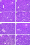
Histology of the liver from DEN-injected and ASV mice. Histological findings in liver sections stained with hematoxylin and phloxin: A, normal liver in a 3-month-old noninjected male; B, liver in a 3-month-old DEN-injected male with no evidence of either architectural or cytological modifications; C, liver in a 6-month-old DEN-injected male showing a basophilic foci of altered hepatocytes (arrows); D, HCC in a 12-month-old DEN-injected male; E, normal liver in a 3-month-old control female; F, liver in a 2-month-old ASV male with hepatocellular atypias and mitosis; G, liver in a 3-month-old ASV male with a clear foci of altered hepatocytes (arrows) and atypias around the foci; H, HCC in a 6-month-old ASV male. Original magnifications, ×200.
Figure 2
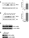
IRS-2 overexpression in HCC from DEN-injected and ASV mice. Protein extracts (40 μg) prepared from HCC (T) and nontumoral (NT) tissues from 12- and 13-month-old DEN-injected mice (A) and from 6-month-old ASV males (ASV 1 to 6) (B) were analyzed by Western blotting for IRS-2 protein expression. The liver was entirely tumoral in 6-month-old ASV males and the normal liver from an age-matched female was used as a control. Blots were reprobed with an anti-Akt antibody to ensure equivalent protein loading. Quantitative values are means ±SEM; *P < 0.05, compared with nontumoral tissue. Total RNA (2 μg) extracted from HCC (T) and nontumoral (NT) tissues from 12- and 13-month-old DEN-injected mice (C) and from 6-month-old ASV males (ASV 1 to 3) (D) and a control female were analyzed by semiquantitative RT-PCR using murine IRS-2 primers. β-Actin PCR products were run in parallel to ensure that equivalent amounts of cDNA were amplified.
Figure 3
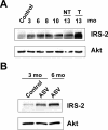
Time course of IRS-2 expression in murine hepatocarcinogenesis. Protein extracts were prepared at different stages of DEN and ASV hepatocarcinogenesis and analyzed for IRS-2 protein expression by Western blotting. Blots were reprobed with an anti-Akt antibody to ensure equivalent protein loading. A: IRS-2 expression in the liver from 3- to 13-month-old males injected with DEN in comparison with normal liver from a 6-month-old noninjected male. NT, nontumoral tissue; T, tumoral. B: IRS-2 expression in the liver from 3- and 6-month-old ASV mice in comparison with normal liver from a 3-month-old control female. At 6 months of age, the liver sample was a HCC.
Figure 4
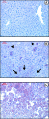
Immunohistochemical detection of IRS-2 in the liver from control and DEN-injected mice. Liver tissue sections from control and DEN-injected mice were immunolabeled with an anti-IRS-2 antibody (reddish-brown labeling) and counterstained with hematoxylin. Histologically normal liver from a 3-month-old noninjected male (A) and from a 3-month-old DEN-injected male (B). IRS-2 immunoreactivity is detected in isolated hepatocytes (arrowheads) and in a small cluster (arrows). C: HCC nodules from a 12-month-old DEN-injected male. Original magnifications, ×200.
Figure 5
Charaterization of the insulin/IGF signaling pathway in IRS-2-overexpressing HCC from DEN-injected mice. Total RNA and protein extracts were prepared from HCC (T) and nontumoral (NT) liver tissues obtained from 12- and 13-month-old mice injected with DEN. A: RNAs (2 μg) were analyzed by semiquantitative RT-PCR using IGF-2 primers. B: Protein extracts (40 μg) of six HCC-overexpressing IRS-2 were analyzed by Western blotting for IRS-1, phospho-Ser9, and total GSK-3β, phospho-Ser473 and total Akt, and phospho-Thr410 PKCζ protein expressions.
Figure 6
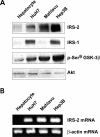
IRS-2 overexpression in human hepatoma cell lines. Protein extracts and total RNA were prepared from human hepatocytes maintained in primary culture and HCC cell lines (HuH7, Mahlavu, Hep3B) cultured for 17 hours in serum-free medium. A: Protein extracts (40 μg) were analyzed for IRS-2, IRS-1, and phospho-Ser9 GSK-3β protein expressions by Western blotting. Blots were reprobed with an anti-Akt antibody to ensure equivalent protein loading. B: RNA samples (2 μg) were analyzed by semiquantitative RT-PCR using human IRS-2 primers. β-Actin PCR products were run in parallel to ensure that equivalent amounts of cDNA were amplified.
Figure 7
Functional analyses of IRS-2 in Hep3B cells. A: Hep3B cells were stimulated for 5 minutes with IGF-2 (10−8 mol/L) and lysed. Protein extracts (500 μg) were immunoprecipitated with an anti-IRS-2 antibody and analyzed by Western blotting with anti-phosphotyrosine (top) and anti-p85 (middle) antibodies. Blots were reprobed with an anti-IRS-2 antibody (bottom) to ensure that equivalent protein amounts were immunoprecipitated. B: Hep3B cells were transiently transfected with an IRS-2 or an unrelated control siRNA duplex. Twenty-four hours after transfection, cells were examined for IRS-2 protein expression by Western blotting (top) and for apoptosis by flow cytometry after staining with annexin V-FITC conjugate and PI (bottom). Data are representative of two independent experiments. Blots were reprobed with an anti-β-actin antibody to ensure equivalent protein loading.
Figure 8
IRS-2 overexpression in human HCC specimens. Protein extracts and total RNA were prepared from human HCC (T) and nontumoral (NT) liver tissues from seven patients. A: IRS-2 and IRS-1 protein expressions were evaluated by Western blotting analysis. Blots were reprobed with an anti-Akt antibody to ensure equivalent protein loading. B: IRS-2 mRNA expression was evaluated by semiquantitative RT-PCR. β-Actin PCR products were run in parallel to ensure that equivalent amounts of cDNA were amplified.
Similar articles
- Depressing time: Waiting, melancholia, and the psychoanalytic practice of care.
Salisbury L, Baraitser L. Salisbury L, et al. In: Kirtsoglou E, Simpson B, editors. The Time of Anthropology: Studies of Contemporary Chronopolitics. Abingdon: Routledge; 2020. Chapter 5. In: Kirtsoglou E, Simpson B, editors. The Time of Anthropology: Studies of Contemporary Chronopolitics. Abingdon: Routledge; 2020. Chapter 5. PMID: 36137063 Free Books & Documents. Review. - Comparison of Two Modern Survival Prediction Tools, SORG-MLA and METSSS, in Patients With Symptomatic Long-bone Metastases Who Underwent Local Treatment With Surgery Followed by Radiotherapy and With Radiotherapy Alone.
Lee CC, Chen CW, Yen HK, Lin YP, Lai CY, Wang JL, Groot OQ, Janssen SJ, Schwab JH, Hsu FM, Lin WH. Lee CC, et al. Clin Orthop Relat Res. 2024 Dec 1;482(12):2193-2208. doi: 10.1097/CORR.0000000000003185. Epub 2024 Jul 23. Clin Orthop Relat Res. 2024. PMID: 39051924 - Blocking interleukin-1 receptor type 1 (IL-1R1) signaling in hepatocytes slows down diethylnitrosamine-induced liver tumor growth in obese mice.
Gehrke N, Hofmann LJ, Straub BK, Ridder DA, Waisman A, Kaps L, Galle PR, Schattenberg JM. Gehrke N, et al. Hepatol Commun. 2024 Nov 29;8(12):e0568. doi: 10.1097/HC9.0000000000000568. eCollection 2024 Dec 1. Hepatol Commun. 2024. PMID: 39621069 Free PMC article. - Ephrin-A1 expression contributes to the malignant characteristics of {alpha}-fetoprotein producing hepatocellular carcinoma.
Iida H, Honda M, Kawai HF, Yamashita T, Shirota Y, Wang BC, Miao H, Kaneko S. Iida H, et al. Gut. 2005 Jun;54(6):843-51. doi: 10.1136/gut.2004.049486. Gut. 2005. PMID: 15888795 Free PMC article. - Tamoxifen for adults with hepatocellular carcinoma.
Naing C, Ni H, Aung HH. Naing C, et al. Cochrane Database Syst Rev. 2024 Aug 12;8(8):CD014869. doi: 10.1002/14651858.CD014869.pub2. Cochrane Database Syst Rev. 2024. PMID: 39132750 Review.
Cited by
- Role of insulin receptor substrates in the progression of hepatocellular carcinoma.
Sakurai Y, Kubota N, Takamoto I, Obata A, Iwamoto M, Hayashi T, Aihara M, Kubota T, Nishihara H, Kadowaki T. Sakurai Y, et al. Sci Rep. 2017 Jul 14;7(1):5387. doi: 10.1038/s41598-017-03299-3. Sci Rep. 2017. PMID: 28710407 Free PMC article. - Insulin resistance disrupts epithelial repair and niche-progenitor Fgf signaling during chronic liver injury.
Manzano-Núñez F, Arámbul-Anthony MJ, Galán Albiñana A, Leal Tassias A, Acosta Umanzor C, Borreda Gascó I, Herrera A, Forteza Vila J, Burks DJ, Noon LA. Manzano-Núñez F, et al. PLoS Biol. 2019 Jan 29;17(1):e2006972. doi: 10.1371/journal.pbio.2006972. eCollection 2019 Jan. PLoS Biol. 2019. PMID: 30695023 Free PMC article. - Insulin Receptor Substrate 1 Is Involved in the Phycocyanin-Mediated Antineoplastic Function of Non-Small Cell Lung Cancer Cells.
Hao S, Li Q, Liu Y, Li F, Yang Q, Wang J, Wang C. Hao S, et al. Molecules. 2021 Aug 4;26(16):4711. doi: 10.3390/molecules26164711. Molecules. 2021. PMID: 34443299 Free PMC article. - Expression and function of the insulin receptor substrate proteins in cancer.
Mardilovich K, Pankratz SL, Shaw LM. Mardilovich K, et al. Cell Commun Signal. 2009 Jun 17;7:14. doi: 10.1186/1478-811X-7-14. Cell Commun Signal. 2009. PMID: 19534786 Free PMC article. - IGF-1R contributes to stress-induced hepatocellular damage in experimental cholestasis.
Cadoret A, Rey C, Wendum D, Elriz K, Tronche F, Holzenberger M, Housset C. Cadoret A, et al. Am J Pathol. 2009 Aug;175(2):627-35. doi: 10.2353/ajpath.2009.081081. Epub 2009 Jul 23. Am J Pathol. 2009. PMID: 19628767 Free PMC article.
References
- Tanaka S, Sugimachi K, Maehara S, Harimoto N, Shirabe K, Wands JR. Oncogenic signal transduction and therapeutic strategy for hepatocellular carcinoma. Surgery. 2002;131:S142–S147. - PubMed
- Scharf JG, Braulke T. The role of the IGF axis in hepatocarcinogenesis. Horm Metab Res. 2003;35:685–693. - PubMed
- Bannasch P, Klimek F, Mayer D. Early bioenergetic changes in hepatocarcinogenesis: preneoplastic phenotypes mimic responses to insulin and thyroid hormone. J Bioenerg Biomembr. 1997;29:303–313. - PubMed
- Kaburagi Y, Yamauchi T, Yamamoto-Honda R, Ueki K, Tobe K, Akanuma Y, Yazaki Y, Kadowaki T. The mechanism of insulin-induced signal transduction mediated by the insulin receptor substrate family. Endocr J. 1999;46:S25–S34. - PubMed
- White MF. IRS proteins and the common path to diabetes. Am J Physiol. 2002;283:E413–E422. - PubMed
Publication types
MeSH terms
Substances
LinkOut - more resources
Full Text Sources
Other Literature Sources
Medical
Molecular Biology Databases
Miscellaneous
