The let-7 MicroRNA family members mir-48, mir-84, and mir-241 function together to regulate developmental timing in Caenorhabditis elegans - PubMed (original) (raw)
The let-7 MicroRNA family members mir-48, mir-84, and mir-241 function together to regulate developmental timing in Caenorhabditis elegans
Allison L Abbott et al. Dev Cell. 2005 Sep.
Abstract
The microRNA let-7 is a critical regulator of developmental timing events at the larval-to-adult transition in C. elegans. Recently, microRNAs with sequence similarity to let-7 have been identified. We find that doubly mutant animals lacking the let-7 family microRNA genes mir-48 and mir-84 exhibit retarded molting behavior and retarded adult gene expression in the hypodermis. Triply mutant animals lacking mir-48, mir-84, and mir-241 exhibit repetition of L2-stage events in addition to retarded adult-stage events. mir-48, mir-84, and mir-241 function together to control the L2-to-L3 transition, likely by base pairing to complementary sites in the hbl-1 3' UTR and downregulating hbl-1 activity. Genetic analysis indicates that mir-48, mir-84, and mir-241 specify the timing of the L2-to-L3 transition in parallel to the heterochronic genes lin-28 and lin-46. These results indicate that let-7 family microRNAs function in combination to affect both early and late developmental timing decisions.
Figures
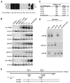
Figure 1. Isolation of Deletion Alleles of the let-7 Family MicroRNA Genes mir-48, mir-84, and mir-241
(A) Alignment of mature ~22 nt microRNA sequences of the four C. elegans let-7 family members and the human let-7 RNA (hs let-7). Shaded boxes and asterisks indicate bases conserved in two or three worm family members, respectively. (B) Temporal expression patterns of let-7 family microRNA genes, mir-48, mir-84, mir-241, and let-7. Northern blot analysis of RNA isolated from populations of staged worms. As a control for staging of the worms during early larval development, lin-4 expression is observed during the late L1 stage. Northern blots indicate that miR-241, miR-48, and miR-84 reached half-maximal expression at about the L3 stage, while let-7 RNA reached half-maximal expression at about the L4 stage. Each blot was stripped and probed for U6 snRNA expression to standardize loading of RNA samples. The numbers underneath each band indicate the signal of the corresponding microRNA normalized to that of U6 snRNA presented relative to maximal expression for each microRNA. (C) List of deletion mutations isolated in the mir-48, mir-84, and mir-241 genes. (D) Northern blot analysis of RNA isolated from wild-type or mutant animals and hybridized with probes to the mature ~22 nt microRNA sequences for miR-48, miR-241, and miR-84. The deletion mutations result in the loss of the corresponding ~22 nt mature microRNA for miR-48, miR-241, and miR-84. (E) The mir-48 and mir-241 locus on chromosome V. The mature microRNA sequences are located within a 1.8 kb genomic region on cosmid F56A12. Deletions at this locus are shown below. The deficiency nDf51 removes both the mir-48 and mir-241 mature microRNA sequences. For rescue experiments, we injected the plasmid pAAS50, which contains a 5073 bp fragment of genomic DNA encompassing both the mir-48 and mir-241 mature microRNA sequences. For control experiments, we injected the plasmid pAAS60, which was generated from pAAS50 but lacked the sequences corresponding to the mir-48 and mir-241 mature microRNA sequences.
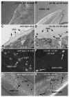
Figure 2. mir-48; mir-84 Double Mutants Display Supernumerary Molting Behavior in Adult Worms
(A and B) Nomarski DIC images of a mir-48; mir-84 animal with a (A) fully formed vulva and with embryos visible inside of the worm to show that the worm is in the adult stage and (B) unshed cuticle surrounding the anterior region of the worm. (C and D) Electron micrographs of adult-stage cuticle in a (C) wild-type animal and a (D) mir-48; mir-84 adult. Asterisks indicate cuticles. The scale bar is 300 nm. (C) Normal single cuticle with alae in a wild-type adult. (D) Two cuticles, both with alae structures (arrows), are visible in a mir-48; mir-84 adult. (E and F) Fluorescence micrographs of a (E) wild-type and a (F) mir-48; mir-84 worm at the L4 molt stage that carry the col-19::gfp transgene maIs105. (E) Fluorescence micrograph of a wild-type L4 molt-stage worm. Expression of col-19::gfp is observed in nuclei of the hyp7 syncytium (arrowheads) and hypodermal seam cells (arrows) in a wild-type L4 molt-stage-worm. (F) Expression of col-19::gfp is observed in hypodermal seam cells (arrows) in a mir-48; mir-84 L4 molt-stage worm. col-19::gfp expression is reduced or absent in hyp7. (G and H) Nomarski DIC images of the hypodermis of the animals in (E) and (F), respectively, showing seam cell (arrows) and hyp7 (arrowheads) nuclei.
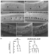
Figure 3. Extra Seam Cells Generated as a Consequence of Reiteration of the L2-Stage Developmental Program in Animals Lacking mir-48, mir-84, and mir-241 MicroRNA Activity
Nomarski DIC images of the hypodermis. (A and B) Arrows indicate seam cells. Normal seam cell number is observed in a (A) wild-type and (B) lin-46 mutant animal at the L4 stage. (C) Extra seam cells in a mir-48 mir-241; mir-84 triple mutant at the L4 stage. (D) Enhanced extra-seam-cell phenotype of a lin-46 mir-48 mir-241; mir-84 mutant animal at the L4 stage. (E–G) Sequential Nomarski DIC images of a mir-48 mir-241; mir-84 mutant animal (E) during the late L2 stage, (F) during the L2 molt, and (G) during the early L3 stage. The same V seam cell lineage in a single animal is shown in all three images. (E) A single seam cell prior to cell division (arrow). (F) The seam cell in (E) divided during the L2 molt, resulting in two daughters. (G) Each of the daughters from (F) executed another round of cell divisions, resulting in four daughter nuclei in the early-L3 stage. The posterior daughter of each pair of daughter nuclei retains the seam cell identity, and the anterior daughter fuses with the hyp7 syncytium (Sulston and Horvitz, 1977). (H) Diagram of Vn (V1–V4, V6) lineage of a normal N2 wild-type animal (adapted from Sulston and Horvitz, 1977) and a mir-48 mir-241; mir-84 triple mutant animal. Reiteration of the L2-stage proliferative program is observed in V lineage seam cells of mir-48 mir-241; mir-84 triple mutants.
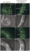
Figure 4. mir-48, mir-84, and mir-241 Are Necessary for the Downregulation of hbl-1::gfp::hbl-1 in the hyp7 Syncytium
(A–D) Larval stage and genotype are indicated for each panel. All fluorescent images were taken with identical exposure times. Fluorescent micrographs of a (A and C) wild-type and a (B and D) mir-48 mir-241; mir-84 mutant animal carrying the hbl-1::gfp::hbl-1 containing transgene ctIs39 (Fay et al., 1999). (A and B) hbl-1::gfp::hbl-1 is expressed at the L1 stage in hyp7 in a (A) wild-type and (B) mir-48 mir-241; mir-84 mutant animal. Little or no expression is visible in the seam cells. Arrows indicate examples of seam cells, and arrowheads indicate examples of hyp7 nuclei. (C) hbl-1::gfp::hbl-1 expression is undetectable in hyp7 in a L3-stage wild-type animal. (D) hbl-1::gfp::hbl-1 expression is elevated in hyp7 in a L3-stage mir-48 mir-241; mir-84 animal. (A′–D′) Corresponding Nomarski DIC images for images shown in (A)–(D). Arrows indicate examples of seam cells, and arrowheads indicate examples of hyp7 nuclei.
Figure 5. Model for the Early Role of mir-48, mir-84, and mir-241 in the Heterochronic Gene Pathway at the L2-to-L3 Transition and the Late Role for let-7 in the L4-to-Adult Transition
See text for details. Our data indicate that mir-48, mir-84, and mir-241 act in parallel with lin-28 and lin-46 and upstream of hbl-1 to control specification of L3-stage events. Both the lin-28 and lin-46 pathway and the mir-48, mir-84, mir-241 pathway converge on hbl-1 to specify the L2-to-L3 transition. let-7 functions later to control specification of adult-stage events through its downstream effectors, hbl-1 and lin-41. mir-48, mir-84, and mir-241 control the exit from the molting cycle and may contribute to the repression of hbl-1 and lin-41 at the L4-to-adult transition.
Similar articles
- Regulatory mutations of mir-48, a C. elegans let-7 family MicroRNA, cause developmental timing defects.
Li M, Jones-Rhoades MW, Lau NC, Bartel DP, Rougvie AE. Li M, et al. Dev Cell. 2005 Sep;9(3):415-22. doi: 10.1016/j.devcel.2005.08.002. Dev Cell. 2005. PMID: 16139229 - acn-1, a C. elegans homologue of ACE, genetically interacts with the let-7 microRNA and other heterochronic genes.
Metheetrairut C, Ahuja Y, Slack FJ. Metheetrairut C, et al. Cell Cycle. 2017 Oct 2;16(19):1800-1809. doi: 10.1080/15384101.2017.1344798. Epub 2017 Sep 21. Cell Cycle. 2017. PMID: 28933985 Free PMC article. - An elegant miRror: microRNAs in stem cells, developmental timing and cancer.
Nimmo RA, Slack FJ. Nimmo RA, et al. Chromosoma. 2009 Aug;118(4):405-18. doi: 10.1007/s00412-009-0210-z. Epub 2009 Apr 3. Chromosoma. 2009. PMID: 19340450 Free PMC article. Review. - Roles of microRNAs in the Caenorhabditis elegans nervous system.
Meng L, Chen L, Li Z, Wu ZX, Shan G. Meng L, et al. J Genet Genomics. 2013 Sep 20;40(9):445-52. doi: 10.1016/j.jgg.2013.07.002. Epub 2013 Aug 7. J Genet Genomics. 2013. PMID: 24053946 Review.
Cited by
- Exploiting Drosophila genetics to understand microRNA function and regulation.
Dai Q, Smibert P, Lai EC. Dai Q, et al. Curr Top Dev Biol. 2012;99:201-235. doi: 10.1016/B978-0-12-387038-4.00008-2. Curr Top Dev Biol. 2012. PMID: 22365740 Free PMC article. Review. - Downregulation of miR-27a* and miR-532-5p and upregulation of miR-146a and miR-155 in LPS-induced RAW264.7 macrophage cells.
Cheng Y, Kuang W, Hao Y, Zhang D, Lei M, Du L, Jiao H, Zhang X, Wang F. Cheng Y, et al. Inflammation. 2012 Aug;35(4):1308-13. doi: 10.1007/s10753-012-9443-8. Inflammation. 2012. PMID: 22415194 - Gene Regulation and Cellular Metabolism: An Essential Partnership.
Carthew RW. Carthew RW. Trends Genet. 2021 Apr;37(4):389-400. doi: 10.1016/j.tig.2020.09.018. Epub 2020 Oct 19. Trends Genet. 2021. PMID: 33092903 Free PMC article. Review. - MicroRNA in carcinogenesis & cancer diagnostics: a new paradigm.
Ahmad J, Hasnain SE, Siddiqui MA, Ahamed M, Musarrat J, Al-Khedhairy AA. Ahmad J, et al. Indian J Med Res. 2013 Apr;137(4):680-94. Indian J Med Res. 2013. PMID: 23703335 Free PMC article. Review. - C. elegans Runx/CBFβ suppresses POP-1 TCF to convert asymmetric to proliferative division of stem cell-like seam cells.
van der Horst SEM, Cravo J, Woollard A, Teapal J, van den Heuvel S. van der Horst SEM, et al. Development. 2019 Nov 18;146(22):dev180034. doi: 10.1242/dev.180034. Development. 2019. PMID: 31740621 Free PMC article.
References
- Abrahante JE, Daul AL, Li M, Volk ML, Tennessen JM, Miller EA, Rougvie AE. The Caenorhabditis elegans hunchback-like gene lin-57/hbl-1 controls developmental time and is regulated by microRNAs. Dev. Cell. 2003;4:625–637. - PubMed
- Ambros V. A hierarchy of regulatory genes controls a larva-to-adult developmental switch in C. elegans. Cell. 1989;57:49–57. - PubMed
- Ambros V, Horvitz HR. Heterochronic mutants of the nematode Caenorhabditis elegans. Science. 1984;226:409–416. - PubMed
- Ambros V, Lee RC, Lavanway A, Williams PT, Jewell D. MicroRNAs and other tiny endogenous RNAs in C. elegans. Curr Biol. 2003;13:807–818. - PubMed
Publication types
MeSH terms
Substances
Grants and funding
- GM34028/GM/NIGMS NIH HHS/United States
- 5F32GM065721-02/GM/NIGMS NIH HHS/United States
- R01 GM034028/GM/NIGMS NIH HHS/United States
- GM067031/GM/NIGMS NIH HHS/United States
- R01 GM067031/GM/NIGMS NIH HHS/United States
- WT_/Wellcome Trust/United Kingdom
- F32 GM065721/GM/NIGMS NIH HHS/United States
LinkOut - more resources
Full Text Sources
Other Literature Sources
Molecular Biology Databases
Research Materials


