Simulating movement of tRNA into the ribosome during decoding - PubMed (original) (raw)
Simulating movement of tRNA into the ribosome during decoding
Kevin Y Sanbonmatsu et al. Proc Natl Acad Sci U S A. 2005.
Abstract
Decoding is the key step during protein synthesis that enables information transfer from RNA to protein, a process critical for the survival of all organisms. We have used large-scale (2.64 x 10(6) atoms) all-atom simulations of the entire ribosome to understand a critical step of decoding. Although the decoding problem has been studied for more than four decades, the rate-limiting step of cognate tRNA selection has only recently been identified. This step, known as accommodation, involves the movement inside the ribosome of the aminoacyl-tRNA from the partially bound "A/T" state to the fully bound "A/A" state. Here, we show that a corridor of 20 universally conserved ribosomal RNA bases interacts with the tRNA during the accommodation movement. Surprisingly, the tRNA is impeded by the A-loop (23S helix 92), instead of enjoying a smooth transition to the A/A state. In particular, universally conserved 23S ribosomal RNA bases U2492, C2556, and C2573 act as a 3D gate, causing the acceptor stem to pause before allowing entrance into the peptidyl transferase center. Our simulations demonstrate that the flexibility of the acceptor stem of the tRNA, in addition to flexibility of the anticodon arm, is essential for tRNA selection. This study serves as a template for simulating conformational changes in large (>10(6) atoms) biological and artificial molecular machines.
Figures
Fig. 1.
Accommodation simulation overview and aminoacyl-tRNA-50S interactions. (a) Time evolution of cognate tRNA accommodation into the ribosome for one of seven 2-ns simulations. Explicit waters, ions, and the top portion of the 70S ribosome are not shown so that the tRNAs are visible. The 23S rRNA (white), 50S proteins (light green), 16S rRNA (purple), 30S proteins (pink), mRNA (dark green), aminoacyl-tRNA (yellow) with Phe amino acid (dark green), peptidyl-tRNA (cyan), and 23S rRNA 2553 (red) are shown. The schematics are similar to that of Moazed and Noller (5) and depict the process of accommodation. A, aminoacyl site; P, peptidyl site. Stage 1 is represented by the initial production structure, after 1.6 ns of equilibration (t = 0 ns). Stage 2 shows the relaxation of the aminoacyl-tRNA body (i.e., all but the 3′-CCA portion) (t = 0.292 ns). In stage 3, the aminoacyl-tRNA body is accommodated (t = 0.65 ns). During stage 4, the aminoacyl-tRNA 3′-CCA end is accommodated into the peptidyl transferase center (2.2 ns). (b) Secondary structure diagrams color-coded by the region of tRNA that participates in the aminoacyl-tRNA-50S interaction. Green, interactions with TΨC loop; blue, interactions with D loop; magenta, interactions with 3′-CCA end. ASL, anticodon stem loop.
Fig. 2.
Stages of accommodation. (a) Aminoacyl-tRNA interaction regions (within 3.5 Å) colored according to the accommodation stage. (Inset) The context of the accommodation wall on the 50S ribosomal subunit. (b) Time evolution of parameters that describe the deformation of aminoacyl-tRNA. Average values (black) of θ1, φ1, and φ2 (averaged over seven trajectories) and corresponding variances (yellow) are shown. θ1 is the angle between O3′ atoms at positions 33, 40, and 55 of the tRNA. φ1 is the dihedral angle between O3′ atoms at positions 33, 40, 55 and 1. φ2 is the dihedral angle between O3′ atoms at positions 69, 71, and 73 and the O of the Phe. (c) Snapshots from stage 1 (t = 0 ns), stage 2 (t = 0.488 ns), stage 3 (t = 0.650 ns), and stage 4 (t = 2.2 ns).
Fig. 3.
Time evolution of probability densities, p, of interactions (blue, no interaction; red, strong interaction). p is the number of interactions normalized by the number of simulations (defined in Fig. 7). Nucleotide/residue positions for the aminoacyl-tRNA, 23S rRNA, and 50S proteins participating in aminoacyl-tRNA interactions are shown along the x axis. Diagonal contours for the A-loop correspond to the aminoacyl-tRNA 3′-CCA end interacting with consecutive A-loop nucleotides as a function of time. Distinct regions are separated by vertical black bars.
Fig. 4.
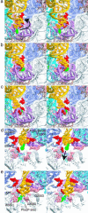
Close-up stereoview of the accommodation of the aminoacyl-tRNA 3′-CCA end by the peptidyl transferase center of the large ribosomal subunit. The aminoacyl-tRNA (yellow) and Phe amino acid (green), peptidyl-tRNA (cyan), 50S proteins (pink), 23S H69 and H71 (light blue), H92 A-loop (purple), H90 (light purple), H89 (dark blue), gate nucleotides (U2492, C2556, and C2573, red), and peptidyl transferase center nucleotides (A2451, C2452, G2553, and U2585, pink/peach) are shown. (a) The 3′-CCA end begins its interaction with the A-loop (t = 0.345 ns). (b) 3′-CCA end continues to move through the A-loop (t = 0.592 ns). (c) The 3′-CCA end enters the peptidyl transferase center (t = 0.670 ns). (d) View from the peptidyl transferase center (t = 0.676 ns). Strong interactions between the 3′-CCA end and universally conserved 23S rRNA U2492, C2556, and C2573 occur. (e) The 3′-CCA end reaches the target A/A state configuration where the CCA end forms the C75:G2553 base pair and close interaction with A2451 and P-tRNA Phe (t = 2.2 ns).
Similar articles
- Alignment/misalignment hypothesis for tRNA selection by the ribosome.
Sanbonmatsu KY. Sanbonmatsu KY. Biochimie. 2006 Aug;88(8):1075-89. doi: 10.1016/j.biochi.2006.07.002. Epub 2006 Jul 26. Biochimie. 2006. PMID: 16890341 Review. - Accommodation of aminoacyl-tRNA into the ribosome involves reversible excursions along multiple pathways.
Whitford PC, Geggier P, Altman RB, Blanchard SC, Onuchic JN, Sanbonmatsu KY. Whitford PC, et al. RNA. 2010 Jun;16(6):1196-204. doi: 10.1261/rna.2035410. Epub 2010 Apr 28. RNA. 2010. PMID: 20427512 Free PMC article. - Mutational analysis reveals two independent molecular requirements during transfer RNA selection on the ribosome.
Cochella L, Brunelle JL, Green R. Cochella L, et al. Nat Struct Mol Biol. 2007 Jan;14(1):30-6. doi: 10.1038/nsmb1183. Epub 2006 Dec 10. Nat Struct Mol Biol. 2007. PMID: 17159993 - Mutations at the accommodation gate of the ribosome impair RF2-dependent translation termination.
Burakovsky DE, Sergiev PV, Steblyanko MA, Kubarenko AV, Konevega AL, Bogdanov AA, Rodnina MV, Dontsova OA. Burakovsky DE, et al. RNA. 2010 Sep;16(9):1848-53. doi: 10.1261/rna.2185710. Epub 2010 Jul 28. RNA. 2010. PMID: 20668033 Free PMC article. - Functions and interplay of the tRNA-binding sites of the ribosome.
Márquez V, Wilson DN, Nierhaus KH. Márquez V, et al. Biochem Soc Trans. 2002 Apr;30(2):133-40. Biochem Soc Trans. 2002. PMID: 12023840 Review.
Cited by
- Molecular dynamics simulations of sarcin-ricin rRNA motif.
Spacková N, Sponer J. Spacková N, et al. Nucleic Acids Res. 2006 Feb 2;34(2):697-708. doi: 10.1093/nar/gkj470. Print 2006. Nucleic Acids Res. 2006. PMID: 16456030 Free PMC article. - Yeast ribosomal protein L10 helps coordinate tRNA movement through the large subunit.
Petrov AN, Meskauskas A, Roshwalb SC, Dinman JD. Petrov AN, et al. Nucleic Acids Res. 2008 Nov;36(19):6187-98. doi: 10.1093/nar/gkn643. Epub 2008 Sep 29. Nucleic Acids Res. 2008. PMID: 18824477 Free PMC article. - Ribosomal protein L3 functions as a 'rocker switch' to aid in coordinating of large subunit-associated functions in eukaryotes and Archaea.
Meskauskas A, Dinman JD. Meskauskas A, et al. Nucleic Acids Res. 2008 Nov;36(19):6175-86. doi: 10.1093/nar/gkn642. Epub 2008 Oct 2. Nucleic Acids Res. 2008. PMID: 18832371 Free PMC article. - The multiscale coarse-graining method. IV. Transferring coarse-grained potentials between temperatures.
Krishna V, Noid WG, Voth GA. Krishna V, et al. J Chem Phys. 2009 Jul 14;131(2):024103. doi: 10.1063/1.3167797. J Chem Phys. 2009. PMID: 19603966 Free PMC article. - Structure of a mitochondrial ribosome with minimal RNA.
Sharma MR, Booth TM, Simpson L, Maslov DA, Agrawal RK. Sharma MR, et al. Proc Natl Acad Sci U S A. 2009 Jun 16;106(24):9637-42. doi: 10.1073/pnas.0901631106. Epub 2009 Jun 3. Proc Natl Acad Sci U S A. 2009. PMID: 19497863 Free PMC article.
References
- Rodnina, M. V. & Wintermeyer, W. (2001) Annu. Rev. Biochem. 70, 415-435. - PubMed
- Ninio, J. (1974) J. Mol. Biol. 84, 297-313. - PubMed
- Moazed, D. & Noller, H. F. (1989) Nature 342, 142-148. - PubMed
Publication types
MeSH terms
Substances
LinkOut - more resources
Full Text Sources
Other Literature Sources


