MicroRNAs 221 and 222 inhibit normal erythropoiesis and erythroleukemic cell growth via kit receptor down-modulation - PubMed (original) (raw)
Comparative Study
. 2005 Dec 13;102(50):18081-6.
doi: 10.1073/pnas.0506216102. Epub 2005 Dec 5.
Laura Fontana, Elvira Pelosi, Rosanna Botta, Desirée Bonci, Francesco Facchiano, Francesca Liuzzi, Valentina Lulli, Ornella Morsilli, Simona Santoro, Mauro Valtieri, George Adrian Calin, Chang-Gong Liu, Antonio Sorrentino, Carlo M Croce, Cesare Peschle
Affiliations
- PMID: 16330772
- PMCID: PMC1312381
- DOI: 10.1073/pnas.0506216102
Comparative Study
MicroRNAs 221 and 222 inhibit normal erythropoiesis and erythroleukemic cell growth via kit receptor down-modulation
Nadia Felli et al. Proc Natl Acad Sci U S A. 2005.
Abstract
MicroRNAs (miRs) are small noncoding RNAs that regulate gene expression primarily through translational repression. In erythropoietic (E) culture of cord blood CD34+ progenitor cells, the level of miR 221 and 222 is gradually and sharply down-modulated. Hypothetically, this decline could promote erythropoiesis by unblocking expression of key functional proteins. Indeed, (i) bioinformatic analysis suggested that miR 221 and 222 target the 3' UTR of kit mRNA; (ii) the luciferase assay confirmed that both miRs directly interact with the kit mRNA target site; and (iii) in E culture undergoing exponential cell growth, miR down-modulation is inversely related to increasing kit protein expression, whereas the kit mRNA level is relatively stable. Functional studies show that treatment of CD34+ progenitors with miR 221 and 222, via oligonucleotide transfection or lentiviral vector infection, causes impaired proliferation and accelerated differentiation of E cells, coupled with down-modulation of kit protein: this phenomenon, observed in E culture releasing endogenous kit ligand, is magnified in E culture supplemented with kit ligand. Furthermore, transplantation experiments in NOD-SCID mice reveal that miR 221 and 222 treatment of CD34+ cells impairs their engraftment capacity and stem cell activity. Finally, miR 221 and 222 gene transfer impairs proliferation of the kit+ TF-1 erythroleukemic cell line. Altogether, our studies indicate that the decline of miR 221 and 222 during exponential E growth unblocks kit protein production at mRNA level, thus leading to expansion of early erythroblasts. Furthermore, the results on kit+ erythroleukemic cells suggest a potential role of these miRs in cancer therapy.
Figures
Fig. 1.
Expression of miR 221 and 222 and kit in unilineage E±KL culture. (A Top) Growth curve and KL release in HPC E culture. Shown are mean values from seven independent experiments. (A Middle) Growth curve of HPC E culture supplemented with KL. Shown are mean values from three separate experiments. (A Bottom) E maturation in E and E+KL culture: percentage of late (polychromatophilic + orthochromatic) erythroblasts is presented. (B) miR 221 and 222 expression in HPC E culture. (Upper) Microarray results, as compared with normalized day 0 level. (Lower) Northern blot results. Representative experiments are presented. (C) kit expression in HPC E culture. (Upper) Representative immunoblotting of kit protein. (Lower) Real-time PCR of kit mRNA level (mean ± SEM values from four separate experiments). (D) miR 221 and 222 expression versus kit protein level in E culture (Upper). (Lower) Inverse correlation of miR 221 and 222 vs. kit (_r_2 = 0.96, P < 0.01 in both cases).
Fig. 2.
kit mRNA 3′ UTR site targeted by miR 221 and 222
Fig. 3.
miR 221 and 222 directly interact with kit 3′ UTR, as evaluated by luciferase targeting assay. Shown are mean ± SEM values from four separate experiments. **, P < 0.01 when compared with control.
Fig. 4.
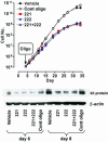
miR 221 and 222 overexpression impairs cell growth in HPC E+KL culture. Growth curve (Upper) and kit protein expression (Lower) in E+KL culture transfected on day 4 with miR 221 and/or 222 oligonucleotide, as compared with vehicle and control oligonucleotide. A representative experiment of four independent experiments is presented.
Fig. 5.
Inhibition of cell growth in TF-1 cell line and unilineage E+KL culture upon infection with Tween-221 and Tween-222 vectors. (A Top) Growth curve of kit+ TF-1 cells infected with Tween-221 or Tween-222 vectors, as compared with empty Tween control vector. (A Middle) Western blot of kit protein and Northern blot of miR 222 in Tween-222-infected TF-1 cells. Similar results were obtained in Tween-221-infected cells (data not shown). (A Bottom) Growth curve of kit-HL-60 cells, infected with Tween-221 or Tween-222 vectors, as compared with empty Tween control vector. A representative experiment of six independent experiments is presented. (B) Growth curve (Top) and maturation of late erythroblasts (Middle) in E+KL culture of HPCs transduced with Tween-221 or -222 vectors, as compared with Tween control vector. (B Bottom) Western blot histogram of kit protein and Northern blot of miR 222 in Tween-222-infected cells at day 10 of culture, as compared with control value; similar results were obtained for Tween-221-infected cells (data not shown). A representative experiment of four independent experiments is presented.
Similar articles
- PLZF-mediated control on c-kit expression in CD34(+) cells and early erythropoiesis.
Spinello I, Quaranta MT, Pasquini L, Pelosi E, Petrucci E, Pagliuca A, Castelli G, Mariani G, Diverio D, Foà R, Testa U, Labbaye C. Spinello I, et al. Oncogene. 2009 Jun 11;28(23):2276-88. doi: 10.1038/onc.2009.87. Epub 2009 May 4. Oncogene. 2009. PMID: 19421145 - miR-22 Inhibits CD34+ Cell Expansion Through Decreasing β-Catenin in Osteoblasts.
Yang Y, Zhang Y, Miao Z, Zou J, Luo J. Yang Y, et al. Hum Gene Ther. 2017 Jan;28(1):135-145. doi: 10.1089/hum.2016.104. Epub 2016 Sep 19. Hum Gene Ther. 2017. PMID: 27762627 - Mechanism of human Hb switching: a possible role of the kit receptor/miR 221-222 complex.
Gabbianelli M, Testa U, Morsilli O, Pelosi E, Saulle E, Petrucci E, Castelli G, Giovinazzi S, Mariani G, Fiori ME, Bonanno G, Massa A, Croce CM, Fontana L, Peschle C. Gabbianelli M, et al. Haematologica. 2010 Aug;95(8):1253-60. doi: 10.3324/haematol.2009.018259. Epub 2010 Mar 19. Haematologica. 2010. PMID: 20305142 Free PMC article. - Regulation of the MIR155 host gene in physiological and pathological processes.
Elton TS, Selemon H, Elton SM, Parinandi NL. Elton TS, et al. Gene. 2013 Dec 10;532(1):1-12. doi: 10.1016/j.gene.2012.12.009. Epub 2012 Dec 14. Gene. 2013. PMID: 23246696 Review. - Role of Tat-interacting protein of 110 kDa and microRNAs in the regulation of hematopoiesis.
Liu Y, He JJ. Liu Y, et al. Curr Opin Hematol. 2016 Jul;23(4):325-30. doi: 10.1097/MOH.0000000000000246. Curr Opin Hematol. 2016. PMID: 27071021 Review.
Cited by
- FOSL1 is a key regulator of a super-enhancer driving TCOF1 expression in triple-negative breast cancer.
He Q, Hu J, Huang H, Wu T, Li W, Ramakrishnan S, Pan Y, Chan KM, Zhang L, Yang M, Wang X, Chin YR. He Q, et al. Epigenetics Chromatin. 2024 Nov 10;17(1):34. doi: 10.1186/s13072-024-00559-1. Epigenetics Chromatin. 2024. PMID: 39523372 Free PMC article. - Clinical implications of miRNAs in erythropoiesis, anemia, and other hematological disorders.
Pal JK, Sur S, Mittal SPK, Dey S, Mahale MP, Mukherjee A. Pal JK, et al. Mol Biol Rep. 2024 Oct 18;51(1):1064. doi: 10.1007/s11033-024-09981-w. Mol Biol Rep. 2024. PMID: 39422834 Review. - Potential Use of MicroRNA Technology in Thalassemia Therapy.
Rujito L, Wardana T, Siswandari W, Nainggolan IM, Sasongko TH. Rujito L, et al. J Clin Med Res. 2024 Sep;16(9):411-422. doi: 10.14740/jocmr5245. Epub 2024 Aug 22. J Clin Med Res. 2024. PMID: 39346566 Free PMC article. Review. - Human erythrocytes' perplexing behaviour: erythrocytic microRNAs.
Joshi U, Jani D, George LB, Highland H. Joshi U, et al. Mol Cell Biochem. 2024 Jul 22. doi: 10.1007/s11010-024-05075-0. Online ahead of print. Mol Cell Biochem. 2024. PMID: 39037663 Review. - Restricting Colorectal Cancer Cell Metabolism with Metformin: An Integrated Transcriptomics Study.
Orang A, Marri S, McKinnon RA, Petersen J, Michael MZ. Orang A, et al. Cancers (Basel). 2024 May 29;16(11):2055. doi: 10.3390/cancers16112055. Cancers (Basel). 2024. PMID: 38893174 Free PMC article.
References
- Bartel, D. P. (2004) Cell 116, 281-297. - PubMed
- Lee, Y., Ahn, C., Han, J., Choi, H., Kim, J., Yim, J., Lee, J., Provost, P., Radmark, O., Kim, S., et al. (2003) Nature 425, 415-419. - PubMed
- Ambros, V. (2004) Nature 431, 350-355. - PubMed
- Yekta, S., Shih, I. H. & Bartel, D. P. (2004) Science 304, 594-596. - PubMed
Publication types
MeSH terms
Substances
LinkOut - more resources
Full Text Sources
Other Literature Sources
Medical
Molecular Biology Databases
Miscellaneous


