Mutations on the external surfaces of adeno-associated virus type 2 capsids that affect transduction and neutralization - PubMed (original) (raw)
Comparative Study
Mutations on the external surfaces of adeno-associated virus type 2 capsids that affect transduction and neutralization
Michael A Lochrie et al. J Virol. 2006 Jan.
Abstract
Mutations were made at 64 positions on the external surface of the adeno-associated virus type 2 (AAV-2) capsid in regions expected to bind antibodies. The 127 mutations included 57 single alanine substitutions, 41 single nonalanine substitutions, 27 multiple mutations, and 2 insertions. Mutants were assayed for capsid synthesis, heparin binding, in vitro transduction, and binding and neutralization by murine monoclonal and human polyclonal antibodies. All mutants made capsid proteins within a level about 20-fold of that made by the wild type. All but seven mutants bound heparin as well as the wild type. Forty-two mutants transduced human cells at least as well as the wild type, and 10 mutants increased transducing activity up to ninefold more than the wild type. Eighteen adjacent alanine substitutions diminished transduction from 10- to 100,000-fold but had no effect on heparin binding and define an area (dead zone) required for transduction that is distinct from the previously characterized heparin receptor binding site. Mutations that reduced binding and neutralization by a murine monoclonal antibody (A20) were localized, while mutations that reduced neutralization by individual human sera or by pooled human, intravenous immunoglobulin G (IVIG) were dispersed over a larger area. Mutations that reduced binding by A20 also reduced neutralization. However, a mutation that reduced the binding of IVIG by 90% did not reduce neutralization, and mutations that reduced neutralization by IVIG did not reduce its binding. Combinations of mutations did not significantly increase transduction or resistance to neutralization by IVIG. These mutations define areas on the surface of the AAV-2 capsid that are important determinants of transduction and antibody neutralization.
Figures
FIG. 1.
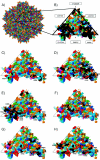
Structure of AAV-2 capsid and location of mutations. (A) Space-filling diagram of AAV-2 capsid. Amino acids are colored as follows: red, residues D and E; pink, W; brown, P; orange, S and T; yellow, C and M; dark blue, R and K; medium blue, N and Q; light blue, F and Y; light green, H; dark green, I, L, and V; gray, A; and white, G. The black triangle defines the boundary of one asymmetric structural unit. There are 60 asymmetric structural units per capsid. Approximately 145 amino acids out of 735 amino acids in VP1 are on the surface. (B) Positions
FIG. 2.
Heparin binding. (A) Heparin binding by capsids with mutations in or near the heparin binding site. Wild-type AAV-2 and wild-type AAV-8 were used as positive and negative controls, respectively. (B) Heparin binding by mutants with mutations in the dead zone. These mutants have <10% of the wild type's transduction in vitro. The R588A mutant and wild-type AAV-2 were used as negative and positive controls, respectively. B, bound to heparin; UB, unbound. Only VP3 is shown.
FIG. 3.
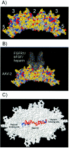
Model of AAV-2 capsid binding to a fibroblast growth factor receptor 1/basic fibroblast growth factor/heparin complex. (A) Two trimers of the AAV-2 capsid monomer. Axes of symmetry (2, 3, and 5) are indicated. The AAV-2 capsid is shown in space-filling format and colored as follows: red, acidic amino acid; blue, basic; yellow, polar; and gray, hydrophobic. The dimple is centered over the twofold axis of symmetry and is located at the top of the figure. (B) Docking of a fibroblast growth factor receptor 1/basic fibroblast growth factor/heparin complex into the dimple. The receptor complex is shown in the wire frame format. (C) Location of heparin in the model. Two trimers of the AAV-2 capsid monomer are shown in white and rotated 90° toward the reader relative to their positions in Fig. 3A. The heparin is shown in Corey, Pauling, and Koltun colors. The five basic amino acids that comprise the AAV-2 heparin binding site are colored blue.
Similar articles
- Genetic modifications of the adeno-associated virus type 2 capsid reduce the affinity and the neutralizing effects of human serum antibodies.
Huttner NA, Girod A, Perabo L, Edbauer D, Kleinschmidt JA, Büning H, Hallek M. Huttner NA, et al. Gene Ther. 2003 Dec;10(26):2139-47. doi: 10.1038/sj.gt.3302123. Gene Ther. 2003. PMID: 14625569 - Mapping an Adeno-associated Virus 9-Specific Neutralizing Epitope To Develop Next-Generation Gene Delivery Vectors.
Giles AR, Govindasamy L, Somanathan S, Wilson JM. Giles AR, et al. J Virol. 2018 Sep 26;92(20):e01011-18. doi: 10.1128/JVI.01011-18. Print 2018 Oct 15. J Virol. 2018. PMID: 30089698 Free PMC article. - Monoclonal antibodies against the adeno-associated virus type 2 (AAV-2) capsid: epitope mapping and identification of capsid domains involved in AAV-2-cell interaction and neutralization of AAV-2 infection.
Wobus CE, Hügle-Dörr B, Girod A, Petersen G, Hallek M, Kleinschmidt JA. Wobus CE, et al. J Virol. 2000 Oct;74(19):9281-93. doi: 10.1128/jvi.74.19.9281-9293.2000. J Virol. 2000. PMID: 10982375 Free PMC article. - Characterization of the Adeno-Associated Virus 1 and 6 Sialic Acid Binding Site.
Huang LY, Patel A, Ng R, Miller EB, Halder S, McKenna R, Asokan A, Agbandje-McKenna M. Huang LY, et al. J Virol. 2016 May 12;90(11):5219-5230. doi: 10.1128/JVI.00161-16. Print 2016 Jun 1. J Virol. 2016. PMID: 26962225 Free PMC article. - Capsid antibodies to different adeno-associated virus serotypes bind common regions.
Gurda BL, DiMattia MA, Miller EB, Bennett A, McKenna R, Weichert WS, Nelson CD, Chen WJ, Muzyczka N, Olson NH, Sinkovits RS, Chiorini JA, Zolotutkhin S, Kozyreva OG, Samulski RJ, Baker TS, Parrish CR, Agbandje-McKenna M. Gurda BL, et al. J Virol. 2013 Aug;87(16):9111-24. doi: 10.1128/JVI.00622-13. Epub 2013 Jun 12. J Virol. 2013. PMID: 23760240 Free PMC article.
Cited by
- Immunogenicity assessment of AAV-based gene therapies: An IQ consortium industry white paper.
Yang TY, Braun M, Lembke W, McBlane F, Kamerud J, DeWall S, Tarcsa E, Fang X, Hofer L, Kavita U, Upreti VV, Gupta S, Loo L, Johnson AJ, Chandode RK, Stubenrauch KG, Vinzing M, Xia CQ, Jawa V. Yang TY, et al. Mol Ther Methods Clin Dev. 2022 Aug 2;26:471-494. doi: 10.1016/j.omtm.2022.07.018. eCollection 2022 Sep 8. Mol Ther Methods Clin Dev. 2022. PMID: 36092368 Free PMC article. Review. - Cryo-electron Microscopy Reconstruction and Stability Studies of the Wild Type and the R432A Variant of Adeno-associated Virus Type 2 Reveal that Capsid Structural Stability Is a Major Factor in Genome Packaging.
Drouin LM, Lins B, Janssen M, Bennett A, Chipman P, McKenna R, Chen W, Muzyczka N, Cardone G, Baker TS, Agbandje-McKenna M. Drouin LM, et al. J Virol. 2016 Sep 12;90(19):8542-51. doi: 10.1128/JVI.00575-16. Print 2016 Oct 1. J Virol. 2016. PMID: 27440903 Free PMC article. - Mutants at the 2-Fold Interface of Adeno-associated Virus Type 2 (AAV2) Structural Proteins Suggest a Role in Viral Transcription for AAV Capsids.
Aydemir F, Salganik M, Resztak J, Singh J, Bennett A, Agbandje-McKenna M, Muzyczka N. Aydemir F, et al. J Virol. 2016 Jul 27;90(16):7196-7204. doi: 10.1128/JVI.00493-16. Print 2016 Aug 15. J Virol. 2016. PMID: 27252527 Free PMC article. - Evidence for pH-dependent protease activity in the adeno-associated virus capsid.
Salganik M, Venkatakrishnan B, Bennett A, Lins B, Yarbrough J, Muzyczka N, Agbandje-McKenna M, McKenna R. Salganik M, et al. J Virol. 2012 Nov;86(21):11877-85. doi: 10.1128/JVI.01717-12. Epub 2012 Aug 22. J Virol. 2012. PMID: 22915820 Free PMC article. - In vitro and in vivo gene therapy vector evolution via multispecies interbreeding and retargeting of adeno-associated viruses.
Grimm D, Lee JS, Wang L, Desai T, Akache B, Storm TA, Kay MA. Grimm D, et al. J Virol. 2008 Jun;82(12):5887-911. doi: 10.1128/JVI.00254-08. Epub 2008 Apr 9. J Virol. 2008. PMID: 18400866 Free PMC article.
References
- Arbetman, A. E., M Lochrie, S. Zhou, J. Wellman, C. Scallan, M. M. Doroudchi, B. Randlev, S. Patarroyo-White, T. Liu, P. Smith, H. Lehmkuhl, L. A. Hobbs, G. F. Pierce, and P. Colosi. 2005. A novel caprine adeno-associated virus (AAV) capsid (AAV-Go.1) is closely related to the primate AAV-5 and has unique tropism and neutralization properties. J. Virol. 779:15238-15245. - PMC - PubMed
- Blacklow, N. R., M. D. Hoggan, and W. P. Rowe. 1968. Serologic evidence for human infection with adenovirus-associated viruses. J. Natl. Cancer Inst. 40:319-327. - PubMed
- Buning, H., M. Braun-Falco, and M. Hallek. 2004. Progress in the use of adeno-associated viral vectors for gene therapy. Cells Tissues Organs 177:139-150. - PubMed
Publication types
MeSH terms
Substances
LinkOut - more resources
Full Text Sources
Other Literature Sources


