Cocaine-induced dendritic spine formation in D1 and D2 dopamine receptor-containing medium spiny neurons in nucleus accumbens - PubMed (original) (raw)
Cocaine-induced dendritic spine formation in D1 and D2 dopamine receptor-containing medium spiny neurons in nucleus accumbens
Ko-Woon Lee et al. Proc Natl Acad Sci U S A. 2006.
Abstract
Psychostimulant-induced alteration of dendritic spines on dopaminoceptive neurons in nucleus accumbens (NAcc) has been hypothesized as an adaptive neuronal response that is linked to long-lasting addictive behaviors. NAcc is largely composed of two distinct subpopulations of medium-sized spiny neurons expressing high levels of either dopamine D1 or D2 receptors. In the present study, we analyzed dendritic spine density after chronic cocaine treatment in distinct D1 or D2 receptor-containing medium-sized spiny neurons in NAcc. These studies made use of transgenic mice that expressed EGFP under the control of either the D1 or D2 receptor promoter (Drd1-EGFP or Drd2-EGFP). After 28 days of cocaine treatment and 2 days of withdrawal, spine density increased in both Drd1-EGFP- and Drd2-EGFP-positive neurons. However, the increase in spine density was maintained only in Drd1-EGFP-positive neurons 30 days after drug withdrawal. Notably, increased DeltaFosB expression also was observed in Drd1-EGFP- and Drd2-EGFP-positive neurons after 2 days of drug withdrawal but only in Drd1-EGFP-positive neurons after 30 days of drug withdrawal. These results suggest that the increased spine density observed after chronic cocaine treatment is stable only in D1-receptor-containing neurons and that DeltaFosB expression is associated with the formation and/or the maintenance of dendritic spines in D1 as well as D2 receptor-containing neurons in NAcc.
Conflict of interest statement
Conflict of interest statement: No conflicts declared.
Figures
Fig. 1.
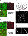
Analysis of MSNs in Drd1-EGFP and Drd2-EGFP mice. (a and b) Fixed brain slices from NAcc of Drd1-EGFP (a) or Drd2-EGFP (b) BAC transgenic mice were immunostained for GFP and NeuN (as a general neuronal marker). The merged images show, in yellow, colocalization of GFP and NeuN. Of neurons in NAcc of Drd1-EGFP mice, 58% expressed GFP, whereas 48% of neurons in NAcc of Drd2-EGFP mice expressed GFP. The numbers of GFP-positive and NeuN-positive neurons were counted independently; all GFP-positive neurons were also NeuN-positive. (c) Visualization of DiI in a single MSN in NAcc. The region of NAcc analyzed in Lower is indicated in the schematic in Upper (red circle) (Bregma, 1.34 mm). Twenty pictures were taken at different z levels (0.5- to 1-μm intervals); the images were then stacked and flattened. (d) Examples of distal dendrites visualized with DiI and used for analysis of spines. (Scale bars: a_–_c, 50 μm; d, 10 μm.)
Fig. 2.
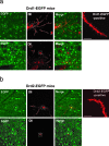
Analysis of dendritic spines in Drd1-EGFP and Drd2-EGFP mice. Neurons in NAcc of either Drd1-EGFP mice (a) or Drd2-EGFP mice (b) were first labeled with DiI (red) and then subjected to immunohistochemistry using an anti-GFP antibody (EGFP, green). Only MSNs were labeled with DiI. Images of DiI staining and GFP staining were overlaid (Merge). (a Upper) The dashed circles show an example of double labeling of a DiI-positive and Drd1-EGFP-positive cell. (a Lower) The dashed squares show a lack of double labeling of cells in slices from Drd1-EGFP mice. (b Upper) The dashed circles show an example of double labeling of a DiI-positive and Drd2-EGFP-positive cell. (b Lower) The dashed squares show a lack of double labeling of cells in slices from Drd2-EGFP mice. The rightmost images in a Upper and b Upper show examples of DiI-stained distal dendrites in Drd1-EGFP or Drd2-EGFP mice, respectively. (Scale bars: 10 μm.) The examples shown were from saline-treated mice.
Fig. 3.
Chronic cocaine-induced increases in spine density in Drd1-EGFP- or Drd2-EGFP-positive MSNs in NAcc. (a and b) Drd1-EGFP (a) or Drd2-EGFP (b) mice were treated with saline (Sal) or cocaine (Coc, 30 mg/kg) for 4 weeks. Mouse brains 2WD or 30WD were processed for DiI labeling and immunohistochemistry as shown in Fig. 2. All spine-like protrusions on distal dendrites were included in the analysis. Data are expressed as the number of spines per 10 μm of dendritic length (mean ± SEM); ∗∗∗, P < 0.001 versus saline treated group, Kolmogorov–Smirnov test. (c and d) Cumulative frequency plots showing the distribution of spine density from individual neurons that were analyzed in a and b. One to two dendrites from one neuron in 10–15 animals per group were analyzed. The total numbers of dendrites analyzed were as follows. For D1: saline, 49 (2WD) and 38 (30WD); cocaine, 74 (2WD) and 75 (30WD). For D2: saline, 50 (2WD) and 43 (30WD); cocaine, 79 (2WD) and 89 (30WD).
Fig. 4.
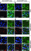
Chronic cocaine induces ΔFosB expression in Drd1-EGFP- or Drd2-EGFP-positive MSNs in NAcc. Drd1-EGFP (a, c, and e) or Drd2-EGFP (b, d, and f) mice were treated with saline or chronic cocaine as described in Fig. 3. 2WD (c and d) or 30WD (e and f), the expression of GFP (green) for the identification of Drd1-EGFP- or Drd2-EGFP-positive MSNs and ΔFosB (red) was analyzed by immunohistochemistry. The localization of NeuN (purple) was also analyzed to show the position of neuronal nuclei. NeuN, GFP, and ΔFosB images were merged to examine their coexpression (Merge). Photomicrographs are representative of results from multiple brain sections obtained from three to four animals in each treatment group. Quantitative analysis is shown in Table 1.
Similar articles
- Acute binge pattern cocaine administration induces region-specific effects in D1-r- and D2-r-expressing cells in eGFP transgenic mice.
Lawhorn C, Edusei E, Zhou Y, Ho A, Kreek MJ. Lawhorn C, et al. Neuroscience. 2013 Dec 3;253:123-31. doi: 10.1016/j.neuroscience.2013.08.032. Epub 2013 Aug 31. Neuroscience. 2013. PMID: 24001687 - Methylphenidate-induced dendritic spine formation and DeltaFosB expression in nucleus accumbens.
Kim Y, Teylan MA, Baron M, Sands A, Nairn AC, Greengard P. Kim Y, et al. Proc Natl Acad Sci U S A. 2009 Feb 24;106(8):2915-20. doi: 10.1073/pnas.0813179106. Epub 2009 Feb 6. Proc Natl Acad Sci U S A. 2009. PMID: 19202072 Free PMC article. - Stress and Cocaine Trigger Divergent and Cell Type-Specific Regulation of Synaptic Transmission at Single Spines in Nucleus Accumbens.
Khibnik LA, Beaumont M, Doyle M, Heshmati M, Slesinger PA, Nestler EJ, Russo SJ. Khibnik LA, et al. Biol Psychiatry. 2016 Jun 1;79(11):898-905. doi: 10.1016/j.biopsych.2015.05.022. Epub 2015 Jun 6. Biol Psychiatry. 2016. PMID: 26164802 Free PMC article. - Licking microstructure in response to novel rewards, reward devaluation and dopamine antagonists: Possible role of D1 and D2 medium spiny neurons in the nucleus accumbens.
D'Aquila PS. D'Aquila PS. Neurosci Biobehav Rev. 2024 Oct;165:105861. doi: 10.1016/j.neubiorev.2024.105861. Epub 2024 Aug 17. Neurosci Biobehav Rev. 2024. PMID: 39159734 Review. - Dyadic social interaction inhibits cocaine-conditioned place preference and the associated activation of the accumbens corridor.
Zernig G, Pinheiro BS. Zernig G, et al. Behav Pharmacol. 2015 Sep;26(6):580-94. doi: 10.1097/FBP.0000000000000167. Behav Pharmacol. 2015. PMID: 26221832 Free PMC article. Review.
Cited by
- Cell type-specific epigenetic priming of gene expression in nucleus accumbens by cocaine.
Mews P, Van der Zee Y, Gurung A, Estill M, Futamura R, Kronman H, Ramakrishnan A, Ryan M, Reyes AA, Garcia BA, Browne CJ, Sidoli S, Shen L, Nestler EJ. Mews P, et al. Sci Adv. 2024 Oct 4;10(40):eado3514. doi: 10.1126/sciadv.ado3514. Epub 2024 Oct 4. Sci Adv. 2024. PMID: 39365860 Free PMC article. - Changes in nucleus accumbens core translatome accompanying incubation of cocaine craving.
Kawa AB, Hashimoto JG, Beutler MM, Guizzetti M, Wolf ME. Kawa AB, et al. bioRxiv [Preprint]. 2024 Sep 16:2024.09.15.613147. doi: 10.1101/2024.09.15.613147. bioRxiv. 2024. PMID: 39345421 Free PMC article. Preprint. - Chronic Cocaine Use and Parkinson's Disease: An Interpretative Model.
Carbone MG, Maremmani I. Carbone MG, et al. Int J Environ Res Public Health. 2024 Aug 21;21(8):1105. doi: 10.3390/ijerph21081105. Int J Environ Res Public Health. 2024. PMID: 39200714 Free PMC article. Review. - Conditional knockout of Shank3 in the ventral CA1 by quantitative in vivo genome-editing impairs social memory in mice.
Chung M, Imanaka K, Huang Z, Watarai A, Wang MY, Tao K, Ejima H, Aida T, Feng G, Okuyama T. Chung M, et al. Nat Commun. 2024 Jun 12;15(1):4531. doi: 10.1038/s41467-024-48430-x. Nat Commun. 2024. PMID: 38866749 Free PMC article. - Post-transcriptional regulation and subcellular localization of G-protein γ7 subunit: implications for striatal function and behavioral responses to cocaine.
Pelletier OB, Brunori G, Wang Y, Robishaw JD. Pelletier OB, et al. Front Neuroanat. 2024 May 2;18:1394659. doi: 10.3389/fnana.2024.1394659. eCollection 2024. Front Neuroanat. 2024. PMID: 38764487 Free PMC article.
References
- Totterdell S., Smith A. D. J. Chem. Neuroanat. 1989;2:285–298. - PubMed
- Smith Y., Bevan M. D., Shink E., Bolam J. P. Neuroscience. 1998;86:353–387. - PubMed
- Heikkila R. E., Orlansky H., Cohen G. Biochem. Pharmacol. 1975;24:847–852. - PubMed
- Ritz M. C., Lamb R. J., Goldberg S. R., Kuhar M. J. Science. 1987;237:1219–1223. - PubMed
- Nestler E. J. Trends Pharmacol. Sci. 2004;25:210–218. - PubMed
Publication types
MeSH terms
Substances
LinkOut - more resources
Full Text Sources
Other Literature Sources
Molecular Biology Databases
Miscellaneous


