The role of Frizzled3 and Frizzled6 in neural tube closure and in the planar polarity of inner-ear sensory hair cells - PubMed (original) (raw)
The role of Frizzled3 and Frizzled6 in neural tube closure and in the planar polarity of inner-ear sensory hair cells
Yanshu Wang et al. J Neurosci. 2006.
Abstract
In the mouse, Frizzled3 (Fz3) and Frizzled6 (Fz6) have been shown previously to control axonal growth and guidance in the CNS and hair patterning in the skin, respectively. Here, we report that Fz3 and Fz6 redundantly control neural tube closure and the planar orientation of hair bundles on a subset of auditory and vestibular sensory cells. In the inner ear, Fz3 and Fz6 proteins are localized to the lateral faces of sensory and supporting cells in all sensory epithelia in a pattern that correlates with the axis of planar polarity. Interestingly, the polarity of Fz6 localization with respect to the asymmetric position of the kinocilium is reversed between vestibular hair cells in the cristae of the semicircular canals and auditory hair cells in the organ of Corti. Vangl2, one of two mammalian homologs of the Drosophila planar cell polarity (PCP) gene van Gogh/Strabismus, is also required for correct hair bundle orientation on a subset of auditory sensory cells and on all vestibular sensory cells. In the inner ear of a Vangl2 mutant (Looptail; Lp), Fz3 and Fz6 proteins accumulate to normal levels but do not localize correctly at the cell surface. These results support the view that vertebrates and invertebrates use similar molecular mechanisms to control a wide variety of PCP-dependent developmental processes. This study also establishes the vestibular sensory epithelium as a tractable tissue for analyzing PCP, and it introduces the use of genetic mosaics for determining the absolute orientation of PCP proteins in mammals.
Figures
External morphology of Fz3−/−;Fz6−/− mice at E18. A, B, D, E, A fully open neural tube (craniorachischisis) is observed in nearly all Fz3−/−;Fz6−/− embryos. The neural tube in Fz3+/+;Fz6−/− and Fz3+/−;Fz6−/− littermates is indistinguishable from WT. C, F, Cross sections through the eye, stained with phalloidin. The lens is at center-left, with the cornea immediately to the right of the lens. The arrowheads indicate the point of fusion in the closed eyelids (C) or the edges of the open eyelids (F). The eyelids remain open in ∼10% of Fz3−/−;Fz6−/− late-gestation embryos. Eyelid closure occurs normally in Fz3+/+;Fz6−/− and Fz3+/−;Fz6−/− littermates.
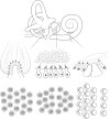
Diagram of the mammalian inner ear and the orientation of sensory hair bundles. Top, Three-dimensional arrangement of the mammalian inner-ear ducts and the locations of sensory epithelia. Center, Cross-sectional views through the principal types of sensory epithelia. Bottom, En face views of these sensory epithelia showing the apical face of the sensory hair cells and the arrangement of hair bundles. The crista (semicircular canal), utricle, and organ of Corti (cochlea) are shown left to right. The saccule closely resembles the utricle, but in the saccule, the hair bundles face away from each other rather toward each other across the equator (Denman-Johnson and Forge, 1999). In the en face view, each kinocilium (•) resides adjacent to a cluster of stereocila. In this view, the stereocilia form a “V” in cochlear hair cells and a disc in hair cells from the utricle, saccule, and cristae.
Comparison of hair bundle orientation defects in the organ of Corti in WT, Fz3−/−;Fz6−/−, and Lp/Lp mice at E18. A, Top, Stereocilia labeled with phalloidin (red) and kinocilia labeled with anti-acetylated tubulin (green), near the base of the organ of Corti at E18. Bottom, Diagrams showing the scoring of hair bundle orientation for the images above. OHC1, Inner row of outer hair cells; OHC2, central row of outer hair cells; OHC3, outer row of outer hair cells. The genotype is indicated above each column of panels. B, IHC, OHC1, OHC2, and OHC3 orientations for each of the three genotypes indicated above. The convention for angular measurements is shown in the inset of the top left panel. Using Fisher’s exact test, the p values for the three pairwise comparisons of the distributions of hair bundle angles between genotypes are listed below in the following order: Lp/Lp versus WT, WT versus Fz3−/−;Fz6−/−, Fz3−/−;Fz6−/− versus Lp/Lp. IHC: 5 × 10⁻⁵, <2 × 10⁻¹⁶, <2 × 10⁻¹⁶; OHC1: 1 × 10⁻⁴, 2 × 10⁻⁵, 0.2; OHC2: 3 × 10⁻⁵, 2 × 10⁻⁴, 1 × 10⁻³; OHC3: 1 × 10⁻⁷, 4 × 10⁻⁶, 2 × 10⁻⁷.
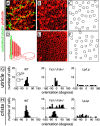
Comparison of defects in hair bundle orientation in WT, Fz3−/−;Fz6−/−, and Lp/Lp utricles and cristae at E18. A–C, Flat-mounted Lp/Lp crista. A, Stereocilia labeled with phalloidin (red) and kinocilia labeled with anti-acetylated tubulin (green) in an optical section immediately above the apical surface of the epithelium. B, The same crista, showing only the phalloidin stain with the optical plane just below the apical surface of the epithelium. Within the cuticular plate of each hair cell, a small zone that lacks phalloidin staining marks the site from which the kinocilium projects (compare A, B). C, Scoring of hair bundle orientations in A and B. D–F, The effect of mechanically removing the hair bundle. D, Schematic of the intact hair bundle inserted into the cuticular plate (left) and the cuticular plate after the hair bundle has been mechanically removed (right). E, Flat-mounted WT utricle labeled with phalloidin after hair bundle removal. F, Scoring of hair bundle orientations in E. G, H, Histograms showing the hair bundle orientations for the indicated genotypes. The convention for angular measurements of hair bundles is shown in the histogram for the WT utricle. For the utricle, the dissected sensory epithelium is without anatomic landmarks, and therefore the angle values have been arbitrarily rotated to center the peak at 0°. For cristae, 0° was set parallel to the direction of fluid flow in the semicircular canal. Histograms for two Fz3−/−;Fz6−/− cristae are shown to illustrate the phenotypic variability seen in hair bundle orientation. Using Fisher’s exact test, the p values for the pairwise comparisons of the distributions of hair bundle angles between genotypes are listed below. Utricle: Lp/Lp versus WT, 8 × 10⁻⁶; WT versus Fz3−/−;Fz6−/−, 0.2; Fz3−/−;Fz6−/− versus Lp/Lp, 4 × 10_−5_. Cristae: Lp/Lp versus WT, 1 × 10_−5_; WT versus upper Fz3−/−;Fz6−/−, 2 × 10_−7_; WT versus lower Fz3−/−;Fz6−/−, 3 × 10_−5_; Lp/Lp versus upper Fz3−/−;Fz6−/−, 0.2; Lp/Lp versus lower Fz3−/−;Fz6−/−, 0.1.
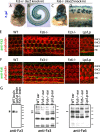
Asymmetric localization of Fz3 and Fz6 proteins in the organ of Corti and the effect of the Lp mutation. A–D, X-gal staining of the intact inner ear (A, C) and the dissected organ of Corti (B, D) from Fz3+/−(lacZ knock-in) mice at E18 (A, B) or Fz6−/−(nuclear lacZ knock-in) mice at postnatal day 5 (P5) (C, D). In the images of the intact inner ear, the cochlea is at the top. For both genotypes, strong X-gal staining is seen in all of the sensory epithelia as well as in some of the adjacent nonsensory epthelia, including a thin band of cells running along each semicircular canal. As seen in D, the nlacZ knock-in at the Fz6 locus shows variagated expression in the four rows of hair cells in the organ of Corti. E, F, Flat mounts of the organ of Corti showing immunolocalization of Fz3 and Fz6 in E18 WT, Fz3−/−, and Lp/Lp samples; the Fz6−/− sample is from P2. (Control experiments show identical Fz6 immunostaining patterns in WT samples at E18 and P2 but a greater Fz6 intensity at P2.) The specificities of the Fz3 and Fz6 antibody staining are demonstrated by their absence in Fz3−/− and Fz6−/− tissue, respectively. Relative to WT tissue, Fz6−/− tissue shows increased Fz3 immunostaining intensity, and Fz3−/− tissue shows increased Fz6 immunostaining intensity. In Lp/Lp tissue, localized Fz3 and Fz6 signals are absent or greatly reduced. G, Immunoblots of postnuclear supernatant proteins from E18 mouse brain (left) or E18 mouse inner ear (right) probed with anti-Fz3 or anti-Fz6 antibodies as indicated. The Fz3 and Fz6 protein bands are indicated by arrowheads; the Fz3 protein appears as a doublet. In the left panel, the Fz3 protein is seen in WT and Lp/Lp brains and is absent, as expected, from the Fz3−/− brain. We note that the lower level of Fz3 protein in the Lp/Lp brain may be secondary to the gross anatomic disruption that arises from the open neural tube, rather than reflecting a specific affect of Vangl2 on Fz3. In the two inner-ear immunoblots, several nonspecific bands are present and serve as internal controls for protein loading. Each Fz band is missing from the extract prepared from the corresponding homozygous mutant and is unaltered in the extract prepared from the Lp/Lp mutant. Molecular mass markers from top to bottom are as follows: left, 220, 130, 90, 70, 60, 40, 30, and 20 kDa; right, 130, 90, 70, 60, and 40 kDa.
Asymmetric localization of Fz3 and Fz6 proteins in the E18 utricle and crista. A, B, Flat mounts of utricles and cristae showing phalloidin staining (red) and Fz3 (A) or Fz6 (B) immunolocalization (green). In each row, the rightmost pair of flat-mount images shows phalloidin plus anti-Fz immunostaining (left member of the pair) and the corresponding anti-Fz signal alone (right member of each pair). WT tissues show strong Fz3 immunostaining (A, right panels), but Fz6 immunostaining is substantially stronger in Fz3−/− tissues compared with WT (B, right panels; and data not shown). The Fz3 and Fz6 proteins are preferentially localized to those sides of the polygonal epithelial cells that lie approximately at right angles to the hair bundle orientation, as quantitated in the histograms that accompany the WT and Fz3−/− flat mounts. Left, In Lp/Lp tissue, localized Fz3 and Fz6 immunostaining signals are absent or greatly reduced.
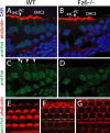
Apical localization of Fz proteins in the organ of Corti at postnatal day 0. A–D, Immunolocalization of Fz6 in frozen sections of the organ of Corti. WT (A, C) and Fz6−/− (B, D) control tissues stained for Fz6 (green) and labeled with phalloidin (red) and DAPI (4′,6-diamidino-2-phenylindole; blue). C, D, The anti-Fz6 signal alone. The Fz6 protein is concentrated at the borders of pillar cells and hair cells and is enriched near the apical face of the cells. PC, Pillar cell; OHC3, third row of outer hair cells. E–G, Successive optical sections through the WT organ of Corti immunostained for Fz3: hair bundles (E), apical region of the hair cell bodies (F), and nuclear region of the hair cell bodies (G). IHCs are at the bottom of the image, and OHC3s are at the top. In F and G, the cuboidal pillar cells can be seen sandwiched between the IHCs and OHC1s. Fz3 is concentrated in the apical region (F) of the hair cells and pillar cells.
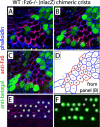
Absolute orientation of Fz6 accumulation determined using WT:Fz6−/−(nlacZ) chimeric sensory epithelia at postnatal day 2 (P2) to P3. A–C, Successive optical sections of a chimeric crista stained with anti-β-galactosidase (green; to visualize nuclear β-galactosidase in Fz6−/− cells), anti-Fz6 (red; produced only by WT cells), and phalloidin (blue). D, Schematic of Fz6 and phalloidin labeling. The cell boundaries are derived from B; the assignment of the presence (red) or absence (blue) of Fz6 labeling has been made with reference to all three optical sections (A–C) because the zones of most intense phalloidin or Fz6 labeling do not always reside within a single optical plane. Cells are scored as + for WT and − for Fz6−/−. The orientations of cell borders that display Fz6 labeling are approximately along a line from top right to bottom left. In regions where a Fz6−/− cell abuts a WT cell, Fz6 protein is found on the top left side but not on the bottom right side of the Fz6−/− cell, as seen for example, with the two adjacent Fz6−/− cells in the center of image. E, F, Analogous flat mount of the organ of Corti in a WT:Fz6−/−(nlacZ) chimera. The asterisks denote the locations of Fz6−/−(nlacZ) cell bodies, two of which exhibit the variegated absence of β-galactosidase expression illustrated in Figure 5_D_. The four rows of hair cell nuclei are several micrometers below the apical face of the epithelium (see Fig. 7), and they occupy a somewhat broader region when viewed as a flat mount (compare E, F). In the organ of Corti, Fz6 synthesized within each WT sensory cell localizes to only one lateral face of that cell; as expected, Fz6−/− cells lack Fz6 immunoreactivity.
Similar articles
- Asymmetric distribution of prickle-like 2 reveals an early underlying polarization of vestibular sensory epithelia in the inner ear.
Deans MR, Antic D, Suyama K, Scott MP, Axelrod JD, Goodrich LV. Deans MR, et al. J Neurosci. 2007 Mar 21;27(12):3139-47. doi: 10.1523/JNEUROSCI.5151-06.2007. J Neurosci. 2007. PMID: 17376975 Free PMC article. - Partial interchangeability of Fz3 and Fz6 in tissue polarity signaling for epithelial orientation and axon growth and guidance.
Hua ZL, Chang H, Wang Y, Smallwood PM, Nathans J. Hua ZL, et al. Development. 2014 Oct;141(20):3944-54. doi: 10.1242/dev.110189. Development. 2014. PMID: 25294940 Free PMC article. - PTK7 regulates myosin II activity to orient planar polarity in the mammalian auditory epithelium.
Lee J, Andreeva A, Sipe CW, Liu L, Cheng A, Lu X. Lee J, et al. Curr Biol. 2012 Jun 5;22(11):956-66. doi: 10.1016/j.cub.2012.03.068. Epub 2012 May 3. Curr Biol. 2012. PMID: 22560610 Free PMC article. - Conserved and Divergent Principles of Planar Polarity Revealed by Hair Cell Development and Function.
Deans MR. Deans MR. Front Neurosci. 2021 Oct 18;15:742391. doi: 10.3389/fnins.2021.742391. eCollection 2021. Front Neurosci. 2021. PMID: 34733133 Free PMC article. Review. - Current concepts of hair cell differentiation and planar cell polarity in inner ear sensory organs.
Sienknecht UJ. Sienknecht UJ. Cell Tissue Res. 2015 Jul;361(1):25-32. doi: 10.1007/s00441-015-2200-1. Epub 2015 May 12. Cell Tissue Res. 2015. PMID: 25959294 Review.
Cited by
- Planar cell polarity genes, Celsr1-3, in neural development.
Feng J, Han Q, Zhou L. Feng J, et al. Neurosci Bull. 2012 Jun;28(3):309-15. doi: 10.1007/s12264-012-1232-8. Neurosci Bull. 2012. PMID: 22622831 Free PMC article. Review. - Wnt/PCP proteins regulate stereotyped axon branch extension in Drosophila.
Ng J. Ng J. Development. 2012 Jan;139(1):165-77. doi: 10.1242/dev.068668. Development. 2012. PMID: 22147954 Free PMC article. - Requirement for Dlgh-1 in planar cell polarity and skeletogenesis during vertebrate development.
Rivera C, Simonson SJ, Yamben IF, Shatadal S, Nguyen MM, Beurg M, Lambert PF, Griep AE. Rivera C, et al. PLoS One. 2013;8(1):e54410. doi: 10.1371/journal.pone.0054410. Epub 2013 Jan 22. PLoS One. 2013. PMID: 23349879 Free PMC article. - The Wnt receptor Ryk plays a role in mammalian planar cell polarity signaling.
Macheda ML, Sun WW, Kugathasan K, Hogan BM, Bower NI, Halford MM, Zhang YF, Jacques BE, Lieschke GJ, Dabdoub A, Stacker SA. Macheda ML, et al. J Biol Chem. 2012 Aug 24;287(35):29312-23. doi: 10.1074/jbc.M112.362681. Epub 2012 Jul 6. J Biol Chem. 2012. PMID: 22773843 Free PMC article. - Planar cell polarity: one or two pathways?
Lawrence PA, Struhl G, Casal J. Lawrence PA, et al. Nat Rev Genet. 2007 Jul;8(7):555-63. doi: 10.1038/nrg2125. Epub 2007 Jun 12. Nat Rev Genet. 2007. PMID: 17563758 Free PMC article. Review.
References
- Adler PN (2002). Planar signaling and morphogenesis in Drosophila. Dev Cell 2:525–535. - PubMed
- Beall SA, Boekelheide K, Johnson KJ (2005). Hybrid GPCR/cadherin (Celsr) proteins in rat testis are expressed with cell type specificity and exhibit differential Sertoli cell-germ cell adhesion activity. J Androl 26:529–538. - PubMed
- Bhanot P, Brink M, Samos CH, Hsieh JC, Wang Y, Macke JP, Andrew D, Nathans J, Nusse R (1996). A new member of the frizzled family from Drosophila functions as a Wingless receptor. Nature 382:225–230. - PubMed
- Bhanot P, Fish M, Jemison JA, Nusse R, Nathans J, Cadigan KM (1999). Frizzled and Dfrizzled-2 function as redundant receptors for Wingless during Drosophila embryonic development. Development 126:4175–4186. - PubMed
Publication types
MeSH terms
Substances
LinkOut - more resources
Full Text Sources
Other Literature Sources
Molecular Biology Databases
Miscellaneous

