Local morphology predicts functional organization of the dorsal premotor region in the human brain - PubMed (original) (raw)
Comparative Study
Local morphology predicts functional organization of the dorsal premotor region in the human brain
Céline Amiez et al. J Neurosci. 2006.
Abstract
A confusing picture of the functional organization of the dorsal premotor region of the human brain emerged when functional neuroimaging studies that either examined visuomotor hand conditional activity or attempted to localize the human frontal eye field reported activity increases at the same general location, namely the junction of the superior precentral sulcus with the superior frontal sulcus. The present functional magnetic resonance imaging study examined visuomotor hand conditional activity and the locus of the frontal eye field as defined by a standard task, on a subject-by-subject basis, to clarify their location and reveal relationships between the pattern of local morphology and functional activity. The results demonstrate that visuomotor hand conditional activity and the frontal eye field lie within distinct parts of the superior precentral sulcus, revealing an organization of the human premotor cortex consistent with that observed in experimental studies in the monkey.
Figures
Figure 1.
Behavioral tasks. a, Visuomotor hand conditional task and motor control task. In both the visuomotor hand conditional and the motor control trials, one of four different colors was presented in a pseudo-random order in successive trials. The color occupied the entire screen. The subjects were instructed to look at the cross in the center of the colored screen during all trials. A written sentence at the beginning of each block of 16 trials indicated to the subjects the type of trial to be performed during that block. The sentence “Do the appropriate movement for each color” instructed the subjects that a block of 16 visuomotor hand conditional trials would follow. During these trials, the subjects had to press the appropriate one of four buttons in response to the presentation of one of the four colors. The arrows indicate the correct button to press depending on the presented color. The subjects were asked to press the top, left, right, and bottom buttons with their middle, index, and ring fingers and thumb, respectively. The sentence “Do the same movement for each color” instructed the subjects that a block of 16 motor control trials would follow. During these trials, the subjects had to press the same mouse button (with their index finger) for all of the colors presented. The duration of the intertrial interval was 1 s. b, Saccadic eye movement task and ocular fixation control task. The sentences “Follow the dot” and “Fixate on the dot” instructed the subject to perform saccadic eye movements or to fixate, respectively. During the saccadic eye movement trials, a dot was presented in one of three possible locations on the screen (i.e., left, center or right) for 750 ms in each location for a total of 22.5 s. The subjects had to perform a saccade to follow the dot to its current location on the screen. During the ocular fixation trials, the subjects had to fixate on the dot presented in the center of the screen for 22.5 s.
Figure 2.
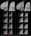
The location of the premotor hand region for conditional motor responses is in blue, the location of the saccadic eye movement region (i.e., the FEF) is in red, and the location of the primary motor cortex hand representation is in green in subjects 1 and 2. The foci of activity illustrated result from the subtractions reported in Tables 1–3. These foci are shown on each subject’s left hemisphere: lateral view (top left diagram) and top view (top right diagram). The green arrow indicates the point of the central sulcus in the depth of which the primary hand motor representation is located (i.e., precentral knob). For each subject, horizontal sections at different levels (z coordinate) in standard stereotaxic space are shown. The left horizontal sections are anatomical MRIs, and the right sections are the same ones with the premotor hand conditional (blue) and saccadic eye movement (red) foci displayed. In both subjects, the primary hand motor region is also displayed in green. In subject 1, the blue arrow marks the point of intersection of the SPSd with the caudal end of the SFS, and the yellow arrow marks the point of intersection of the SPSv with the SFS. In subject 2, the red arrow indicates the common point of intersection of the SPSd, the SPSv, and the caudal end of the SFS. Ant, Anterior part of the brain; CS, central sulcus; SF, Sylvian fissure.
Figure 3.
The location of the premotor hand region for conditional motor responses is in blue, the location of the saccadic eye movement region (i.e., the FEF) is in red, and the location of the primary motor cortex hand representation is in green in subjects 3–6. The foci of activity illustrated result from the subtractions reported in Tables 1–3. These foci are shown on each subject’s left hemisphere: lateral view (top left diagram) and top view (top right diagram). The green arrow indicates the point of the central sulcus in the depth of which the primary hand motor representation is located (i.e., Broca’s pli de passage moyen). For each subject, horizontal sections at different levels (z coordinate) in standard stereotaxic space are shown. The left horizontal sections are anatomical MRIs, and the right sections are the same ones with the premotor hand conditional (blue) and saccadic eye movement (red) foci displayed. In subjects 3 and 5, the primary hand motor region is also displayed in green. In subjects 4–6, the yellow dotted line indicates the level of the sagittal section (i.e., the x coordinate) illustrated within the yellow dotted box. Note that, in subject 5, the precentral gyrus has receded and therefore the SPSv and the central sulcus blend in certain locations. This can be appreciated by careful inspection of the horizontal section indicated by the orange line and box. Ant, Anterior part of the brain; CS, central sulcus; SF, Sylvian fissure.
Figure 4.
The location of the premotor hand region for conditional motor responses is in blue, the location of the saccadic eye movement region (i.e., the FEF) is in red, and the location of the primary motor cortex hand representation is in green in subjects 7 and 8. The foci of activity illustrated result from the subtractions reported in Tables 1–3. These foci are shown on each subject’s left hemisphere: lateral view (top left diagram) and top view (top right diagram). The green arrow indicates the point of the central sulcus in the depth of which the primary hand motor representation is located (i.e., Broca’s pli de passage moyen). For each subject, horizontal sections at different levels (z coordinate) in standard stereotaxic space are shown. The left horizontal sections are anatomical MRIs, and the right sections are the same ones with the premotor hand conditional (blue) and saccadic eye movement (red) foci displayed. In both subjects, the primary hand motor region is also displayed in green. In both subjects, the yellow dotted line indicates the level of the sagittal section (i.e., the x coordinate) illustrated within the yellow dotted box. Note that (1) on the lateral and top views of subject 8, the red arrow points to the location of the SPSv in the depth of which the saccadic eye movement region is located; (2) on the lateral and top views of subject 7, the blue arrow points to the location of the SPSd in the depth of which the premotor hand region for motor conditional responses is located; and (3) in the top view of subject 7, the asterisk indicates the narrow gyral passage between the SPSd and the SPSv. Ant, Anterior part of the brain; CS, central sulcus; SF, Sylvian fissure.
Figure 5.
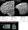
The premotor hand region, the saccadic eye movement region (i.e., the FEF), and the primary motor cortex hand representation in the human and monkey frontal cortex.a, The premotor cortex hand region for conditional responses (blue), the saccadic eye movement region (red), and the primary motor cortex hand representation (green) are shown on the cortical surface of the three-dimensional rendering of the left hemisphere of one human brain in standard stereotaxic space. The crosses correspond to the mean locations of activities reported in Tables 1–3. The blue, red, and green regions correspond to an estimate of the size of these regions based on the subject-by-subject analysis. b, On the left hemisphere of a macaque monkey brain, the following are indicated: the location of the dorsal premotor region involved in the performance of hand conditional responses (blue); the saccadic eye movement region (i.e., the classical FEF) in the concavity of the arcuate sulcus (red); and the hand representation of the primary motor cortex (green). c, Schematic representation of the sulcal patterns in the dorsal premotor region of the human brain. Type 1, The SPSd and SPSv approach each other at the caudal end of the SFS, giving the impression of one continuous sulcus; type 2a, the SPSv joins the SFS at a more anterior location than the SPSd; type 2b, the SPSv approaches but does not join the SFS.d, The hand representation of the primary motor cortex (area 4) in the human brain lies within the central sulcus at the level indicated in green in a. Within the central sulcus, the hand representation occupies a distinct morphological feature, a fold known as the precentral knob. A horizontal (left) and a sagittal (right) section through this part of the central sulcus illustrate this distinct morphological feature. CS, Central sulcus; AS, arcuate sulcus; PS, principalis sulcus; S, spur; SPdimple, superior precentral dimple.
Similar articles
- Human anterior intraparietal and ventral premotor cortices support representations of grasping with the hand or a novel tool.
Jacobs S, Danielmeier C, Frey SH. Jacobs S, et al. J Cogn Neurosci. 2010 Nov;22(11):2594-608. doi: 10.1162/jocn.2009.21372. J Cogn Neurosci. 2010. PMID: 19925200 - Spatial and effector processing in the human parietofrontal network for reaches and saccades.
Beurze SM, de Lange FP, Toni I, Medendorp WP. Beurze SM, et al. J Neurophysiol. 2009 Jun;101(6):3053-62. doi: 10.1152/jn.91194.2008. Epub 2009 Mar 25. J Neurophysiol. 2009. PMID: 19321636 - Dorsal premotor cortex and conditional movement selection: A PET functional mapping study.
Grafton ST, Fagg AH, Arbib MA. Grafton ST, et al. J Neurophysiol. 1998 Feb;79(2):1092-7. doi: 10.1152/jn.1998.79.2.1092. J Neurophysiol. 1998. PMID: 9463464 - The primary motor and premotor areas of the human cerebral cortex.
Chouinard PA, Paus T. Chouinard PA, et al. Neuroscientist. 2006 Apr;12(2):143-52. doi: 10.1177/1073858405284255. Neuroscientist. 2006. PMID: 16514011 Review. - Premotor and parietal cortex: corticocortical connectivity and combinatorial computations.
Wise SP, Boussaoud D, Johnson PB, Caminiti R. Wise SP, et al. Annu Rev Neurosci. 1997;20:25-42. doi: 10.1146/annurev.neuro.20.1.25. Annu Rev Neurosci. 1997. PMID: 9056706 Review.
Cited by
- Morphological patterns and spatial probability maps of the inferior frontal sulcus in the human brain.
Nolan E, Loh KK, Petrides M. Nolan E, et al. Hum Brain Mapp. 2024 Jul 15;45(10):e26759. doi: 10.1002/hbm.26759. Hum Brain Mapp. 2024. PMID: 38989632 Free PMC article. - Functional connectivity profile of the human inferior frontal junction: involvement in a cognitive control network.
Sundermann B, Pfleiderer B. Sundermann B, et al. BMC Neurosci. 2012 Oct 3;13:119. doi: 10.1186/1471-2202-13-119. BMC Neurosci. 2012. PMID: 23033990 Free PMC article. - Diffusion-weighted imaging tractography-based parcellation of the human lateral premotor cortex identifies dorsal and ventral subregions with anatomical and functional specializations.
Tomassini V, Jbabdi S, Klein JC, Behrens TE, Pozzilli C, Matthews PM, Rushworth MF, Johansen-Berg H. Tomassini V, et al. J Neurosci. 2007 Sep 19;27(38):10259-69. doi: 10.1523/JNEUROSCI.2144-07.2007. J Neurosci. 2007. PMID: 17881532 Free PMC article. - The role of contralesional dorsal premotor cortex after stroke as studied with concurrent TMS-fMRI.
Bestmann S, Swayne O, Blankenburg F, Ruff CC, Teo J, Weiskopf N, Driver J, Rothwell JC, Ward NS. Bestmann S, et al. J Neurosci. 2010 Sep 8;30(36):11926-37. doi: 10.1523/JNEUROSCI.5642-09.2010. J Neurosci. 2010. PMID: 20826657 Free PMC article. - Diffusion MRI data, sulcal anatomy, and tractography for eight species from the Primate Brain Bank.
Bryant KL, Ardesch DJ, Roumazeilles L, Scholtens LH, Khrapitchev AA, Tendler BC, Wu W, Miller KL, Sallet J, van den Heuvel MP, Mars RB. Bryant KL, et al. Brain Struct Funct. 2021 Nov;226(8):2497-2509. doi: 10.1007/s00429-021-02268-x. Epub 2021 Jul 15. Brain Struct Funct. 2021. PMID: 34264391 Free PMC article.
References
- Blanke O, Spinelli L, Thut G, Michel CM, Perrig S, Landis T, Seeck M (2000). Location of the human frontal eye field as defined by electrical cortical stimulation: anatomical, functional and electrophysiological characteristics. NeuroReport 11:1907–1913. - PubMed
- Boling W, Olivier A, Bittar RG, Reutens D (1999). Localization of hand motor activation in Broca’s pli de passage moyen. J Neurosurg 91:903–910. - PubMed
- Boussaoud D, Wise SP (1993a). Primate frontal cortex: neuronal activity following attentional versus intentional cues. Exp Brain Res 95:15–27. - PubMed
Publication types
MeSH terms
Substances
LinkOut - more resources
Full Text Sources


