Expression of the interleukin-7 receptor alpha chain (CD127) on virus-specific CD8+ T cells identifies functionally and phenotypically defined memory T cells during acute resolving hepatitis B virus infection - PubMed (original) (raw)
Comparative Study
Expression of the interleukin-7 receptor alpha chain (CD127) on virus-specific CD8+ T cells identifies functionally and phenotypically defined memory T cells during acute resolving hepatitis B virus infection
Tobias Boettler et al. J Virol. 2006 Apr.
Abstract
Virus-specific CD8+ T cells play a central role in the outcome of several viral infections, including hepatitis B virus (HBV) infection. A key feature of virus-specific CD8+ T cells is the development of memory. The mechanisms resulting in the establishment of T-cell memory are still only poorly understood. It has been suggested that T-cell memory may depend on the survival of virus-specific CD8+ T cells in the contraction phase. Indeed, a population of effector cells that express high levels of the interleukin-7 receptor alpha chain (CD127) as the precursors of memory CD8+ T cells has recently been identified in mice. However, very little information is currently available about the kinetics of CD127 expression in an acute resolving viral infection in humans and its association with disease pathogenesis, viral load, and functional and phenotypical T-cell characteristics. To address these important issues, we analyzed the HBV-specific CD8+ T-cell response longitudinally in a cohort of six patients with acute HBV infection who spontaneously cleared the virus. We observed the emergence of CD127 expression on antigen-specific CD8+ memory T cells during the course of infection. Importantly, the up-regulation of CD127 correlated phenotypically with a loss of CD38 and PD-1 expression and acquisition of CCR7 expression: functionally with an enhanced proliferative capacity and clinically with the decline in serum alanine aminotransferase levels and viral clearance. These results suggest that the expression of CD127 is a marker for the development of functionally and phenotypically defined antigen-specific CD8+ memory T cells in cleared human viral infections.
Figures
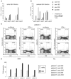
FIG. 1.
Multispecific CD8+ T-cell responses during acute resolving HBV infection. (A and B) PBMC of six patients with acute HBV infection were analyzed for CD8+ T-cell responses to four immunodominant HBV epitopes in the early phase (A) and after resolved infection (B) with HLA-A2 multimers. Multimer data are shown as the percentages of multimer-positive cells among all CD8+ T cells. Early and late time points for each patient are defined in Table 1. (C) Representative density plots of each patient showing multimer-positive CD8+ T-cell responses.
FIG. 2.
Phenotypic characterization of HBV-specific CD8+ T cells. For each patient, surface staining of HBV-specific CD8+ T cells was performed using an HBV multimer with a strong ex vivo response (Table 1) and antibodies to CD127 (A), CD38 (B), CCR7 (C), or CD27 (D). The phenotype of HBV-specific CD8+ T cells was analyzed in the early phase (gray bars) and after resolved infection (black bars). Data are shown as the percentages of all multimer-positive CD8+ T cells. Representative density plots of each surface staining from patients 1 and 3 during acute (early) and after resolved infection (late) gated on CD8+multimer-positive cells are shown on the right.
FIG. 3.
Courses of acute resolving HBV infection. Clinical and virological data are shown for three patients at several time points during and after acute HBV infection. (A, B, and C) Viral load (VL), ALT levels, and dominant multimer response. GE, genome equivalents; U/l, units per liter. (D, E, and F) Kinetics of CD38 expression of HBV-multimer-positive CD8+ T cells. (G, H, and I) Kinetics of CCR7 expression of HBV-multimer-positive CD8+ T cells. (J, K, and L) Kinetics of CD127 expression of HBV-multimer-positive CD8+ T cells. Data are shown as the percentages of multimer-positive CD8+ T cells specific for the respective surface marker. (M, N, and O) Expansion in the proliferative capacity of multimer-specific CD8+ T cells. Calculation of the expansion was performed as described in Materials and Methods. “n.d.,” not done.
FIG. 4.
Functional capacities of HBV-specific CD8+ T cells. (A and B) PBMC of five patients with acute HBV infection were analyzed for HBV-specific CD8+ T-cell IFN-γ production after stimulation with four immunodominant HBV epitopes in the early phase (A) and after resolved infection (B). HBV-specific IFN-γ production of CD8+ T cells was examined by intracellular cytokine staining. Data are shown as the percentages of peptide-specific IFN-γ-producing CD8+ T cells in relation to the frequency of the respective multimer-positive CD8+ T cells. (C) Representative plots of multimer and IFN-γ staining for patient 2 performed at the clinical onset of infection (early) and after resolution (late). The displayed numbers indicate the percentages of multimer-positive or IFN-γ-producing CD8+ T cells. The negative control results were <0.03% in all cases and are not shown. (D) The proliferative capacity of HBV-specific CD8+ T cells was studied in the early phase of infection (gray bars) and after resolved infection (black bars). PBMC were stimulated using the dominant epitope from each patient as shown in Table 1 and were cultured for 14 days prior to staining with the corresponding multimer. Cells were gated on multimer-positive CD8+ cells. Calculation of the expansion was performed as described in Materials and Methods.
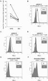
FIG. 5.
PD-1 expression on HBV- and Flu-specific CD8+ T cells. Virus-specific CD8+ T cells were detected via multimer staining and analyzed for PD-1 expression. (A) PD-1 expression on HBV-specific CD8+ T cells was analyzed at clinical presentation of acute hepatitis B (early) and after resolved infection (late). Data are shown as the mean PD-1 fluorescence results (⋄, patient 1; ▪, patient 2; ▴, patient 3; ×, patient 4; ○, patient 6). Expression of PD-1 on HBV-specific CD8+ T cells (B), whole CD8+ T cells (C), and influenza virus-specific CD8+ T cells (D) of patient 2 is demonstrated by overlay histograms of both time points. The expression of PD-1 on unstimulated (E) and stimulated (F) PBMC from one young, healthy donor served as a positive control.
Similar articles
- Dynamic decrease in PD-1 expression correlates with HBV-specific memory CD8 T-cell development in acute self-limited hepatitis B patients.
Zhang Z, Jin B, Zhang JY, Xu B, Wang H, Shi M, Wherry EJ, Lau GK, Wang FS. Zhang Z, et al. J Hepatol. 2009 Jun;50(6):1163-73. doi: 10.1016/j.jhep.2009.01.026. Epub 2009 Mar 29. J Hepatol. 2009. PMID: 19395117 - Loss of IL-7 receptor alpha-chain (CD127) expression in acute HCV infection associated with viral persistence.
Golden-Mason L, Burton JR Jr, Castelblanco N, Klarquist J, Benlloch S, Wang C, Rosen HR. Golden-Mason L, et al. Hepatology. 2006 Nov;44(5):1098-109. doi: 10.1002/hep.21365. Hepatology. 2006. PMID: 17058243 - Dynamic programmed death 1 expression by virus-specific CD8 T cells correlates with the outcome of acute hepatitis B.
Zhang Z, Zhang JY, Wherry EJ, Jin B, Xu B, Zou ZS, Zhang SY, Li BS, Wang HF, Wu H, Lau GK, Fu YX, Wang FS. Zhang Z, et al. Gastroenterology. 2008 Jun;134(7):1938-49, 1949.e1-3. doi: 10.1053/j.gastro.2008.03.037. Epub 2008 Mar 22. Gastroenterology. 2008. PMID: 18455515 - Establishment and recall of CD8+ T-cell memory in a model of localized transient infection.
Kedzierska K, La Gruta NL, Turner SJ, Doherty PC. Kedzierska K, et al. Immunol Rev. 2006 Jun;211:133-45. doi: 10.1111/j.0105-2896.2006.00386.x. Immunol Rev. 2006. PMID: 16824123 Review. - Expression of γ-chain cytokine receptors on CD8+ T cells in HIV infection with a focus on IL-7Rα (CD127).
Crawley AM, Angel JB. Crawley AM, et al. Immunol Cell Biol. 2012 Apr;90(4):379-87. doi: 10.1038/icb.2011.66. Epub 2011 Aug 23. Immunol Cell Biol. 2012. PMID: 21863001 Review.
Cited by
- Immune profiling of responses to influenza vaccination in patients with myeloproliferative neoplasms.
Bachanová P, How J, Dzeng R, Mukherjee S, Pavlovic M, Lombardi J, Hobbs G, Reeves PM. Bachanová P, et al. EJHaem. 2024 Apr 11;5(3):573-577. doi: 10.1002/jha2.868. eCollection 2024 Jun. EJHaem. 2024. PMID: 38895092 Free PMC article. - Multifactoral immune modulation potentiates durable remission in multiple models of aggressive malignancy.
Halpert MM, Burns BA, Rosario SR, Withers HG, Trivedi AJ, Hofferek CJ, Gephart BD, Wang H, Vazquez-Perez J, Amanya SB, Hyslop ST, Yang J, Kemnade JO, Sandulache VC, Konduri V, Decker WK. Halpert MM, et al. FASEB J. 2024 May 31;38(10):e23644. doi: 10.1096/fj.202302675R. FASEB J. 2024. PMID: 38738472 - IL-7 producing immunotherapy improves ex vivo T cell functions of immunosenescent patients, especially post hip fracture.
Marton C, Minaud A, Coupet CA, Chauvin M, Dhiab J, Vallet H, Boddaert J, Kehrer N, Bastien B, Inchauspe G, Barraud L, Sauce D. Marton C, et al. Hum Vaccin Immunother. 2023 Aug 1;19(2):2232247. doi: 10.1080/21645515.2023.2232247. Hum Vaccin Immunother. 2023. PMID: 37417353 Free PMC article. - Upregulation of PD-1 Expression and High sPD-L1 Levels Associated with COVID-19 Severity.
Beserra DR, Alberca RW, Branco ACCC, de Mendonça Oliveira L, de Souza Andrade MM, Gozzi-Silva SC, Teixeira FME, Yendo TM, da Silva Duarte AJ, Sato MN. Beserra DR, et al. J Immunol Res. 2022 Aug 1;2022:9764002. doi: 10.1155/2022/9764002. eCollection 2022. J Immunol Res. 2022. PMID: 35971391 Free PMC article. - Lifelong cytomegalovirus and early-LIFE irradiation synergistically potentiate age-related defects in response to vaccination and infection.
Pugh JL, Coplen CP, Sukhina AS, Uhrlaub JL, Padilla-Torres J, Hayashi T, Nikolich-Žugich J. Pugh JL, et al. Aging Cell. 2022 Jul;21(7):e13648. doi: 10.1111/acel.13648. Epub 2022 Jun 3. Aging Cell. 2022. PMID: 35657768 Free PMC article.
References
- Appay, V., P. R. Dunbar, M. Callan, P. Klenerman, G. M. Gillespie, L. Papagno, G. S. Ogg, A. King, F. Lechner, C. A. Spina, S. Little, D. V. Havlir, D. D. Richman, N. Gruener, G. Pape, A. Waters, P. Easterbrook, M. Salio, V. Cerundolo, A. J. McMichael, and S. L. Rowland-Jones. 2002. Memory CD8+ T cells vary in differentiation phenotype in different persistent virus infections. Nat. Med. 8:379-385. - PubMed
- Appay, V., and S. L. Rowland-Jones. 2004. Lessons from the study of T-cell differentiation in persistent human virus infection. Semin. Immunol. 16:205-212. - PubMed
- Bachmann, M. F., P. Wolint, K. Schwarz, P. Jager, and A. Oxenius. 2005. Functional properties and lineage relationship of CD8+ T cell subsets identified by expression of IL-7 receptor α and CD62L. J. Immunol. 175:4686-4696. - PubMed
- Chemnitz, J. M., R. V. Parry, K. E. Nichols, C. H. June, and J. L. Riley. 2004. SHP-1 and SHP-2 associate with immunoreceptor tyrosine-based switch motif of programmed death 1 upon primary human T cell stimulation, but only receptor ligation prevents T cell activation. J. Immunol. 173:945-954. - PubMed
- Fuller, M. J., D. A. Hildeman, S. Sabbaj, D. E. Gaddis, A. E. Tebo, L. Shang, P. A. Goepfert, and A. J. Zajac. 2005. Cutting edge: emergence of CD127 high functionally competent memory T cells is compromised by high viral loads and inadequate T cell help. J. Immunol. 174:5926-5930. - PubMed
Publication types
MeSH terms
Substances
LinkOut - more resources
Full Text Sources
Other Literature Sources
Medical
Molecular Biology Databases
Research Materials


