Hepatocyte nuclear factor 4alpha is essential for embryonic development of the mouse colon - PubMed (original) (raw)
Hepatocyte nuclear factor 4alpha is essential for embryonic development of the mouse colon
Wendy D Garrison et al. Gastroenterology. 2006 Apr.
Abstract
Background & aims: Hepatocyte nuclear factor 4 alpha (HNF4alpha) is a transcription factor that has been shown to be required for hepatocyte differentiation and development of the liver. It has also been implicated in regulating expression of genes that act in the epithelium of the lower gastrointestinal tract. This implied that HNF4alpha might be required for development of the gut.
Methods: Mouse embryos were generated in which Hnf4a was ablated in the epithelial cells of the fetal colon by using Cre-loxP technology. Embryos were examined by using a combination of histology, immunohistochemistry, DNA microarray, reverse-transcription polymerase chain reaction, electrophoretic mobility shift assays, and chromatin immunoprecipitation analyses to define the consequences of loss of HNF4alpha on colon development.
Results: Embryos were recovered at E18.5 that lacked HNF4alpha in their colons. Although early stages of colonic development occurred, HNF4alpha-null colons failed to form normal crypts. In addition, goblet-cell maturation was perturbed and expression of an array of genes that encode proteins with diverse roles in colon function was disrupted. Several genes whose expression in the colon was dependent on HNF4alpha contained HNF4alpha-binding sites within putative transcriptional regulatory regions and a subset of these sites were occupied by HNF4alpha in vivo.
Conclusions: HNF4alpha is a transcription factor that is essential for development of the mammalian colon, regulates goblet-cell maturation, and is required for expression of genes that control normal colon function and epithelial cell differentiation.
Figures
Figure 1
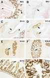
HNF4α is expressed in the epithelium of the lower gastrointestinal tract during embryogenesis. Embryos were collected at E9.5 (A and B), E11.5 (C and D), E14.5 (E), E15.5 (F), and E17.5 (G and H) and transverse sections processed for immunohistochemistry using an anti-HNF4α antibody. At E9.5, HNF4α (brown nuclear staining) was identified in the hepatoblasts (A, arrowhead) as well as in the epithelium of the hindgut and midgut (B, arrow) but not in the foregut (A, *). There was a sharp rostral boundary of expression of HNF4α within the stomach (C, ^). Caudal to this boundary, HNF4α was restricted to the epithelium (C–H, arrows) of the gut throughout the remainder of development, including that of the small intestine (D–G) and colon (H).
Figure 2
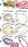
Loss of HNF4α in the epithelial cells disrupts development of the colon. (A and B) Foxa3cre mice were bred to a line of transgenic mice, Gt(ROSA)26SortmlSho, that expresses β-galactosidase only after Cre-mediated deletion of _lox_P-flanked DNA sequences and resulting E9.5 embryos were stained for β-galactosidase expression. Expression of β-galactosidase was detected in the colon (arrowhead) of whole embryos (A) and sections (B) through these embryos revealed the expression to be restricted to the epithelial cells of the colon (arrowhead). (C–F) Micrographs of immunohistochemistry experiments using an anti-HNF4α antibody. HNF4α was identified in the nuclei (open arrowheads) of control (C, Hnf4aloxP/+Foxa3Cre) but was undetectable in a subset of experimental (D, Hnf4aloxP/loxPFoxa3Cre; F, Hnf4aloxP/−Foxa3Cre) E18.5 colons. Some Hnf4aloxP/loxPFoxa3Cre colons displayed a chimeric expression of HNF4α (E). (G and H) H&E–stained sections of E18.5 embryos revealed the formation of crypts (G, arrow) and the presence of mature goblets cells (G, arrowhead) in control E18.5 colons, whereas crypt formation and goblet cell maturation was disrupted in colons lacking HNF4α (H). Boxed areas are shown at a higher resolution in the inset.
Figure 3
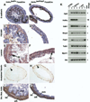
HNF4α is necessary for multiple aspects of colon development. (A–J) Micrographs showing results of immunohistochemistry (brown staining) performed on sections of control (A, C, E, G, I; Hnf4aloxP/+Foxa3Cre) and mutant (B, D, F, H, J; Hnf4aloxP/loxPFoxa3Cre) E18.5 embryos using antibodies that recognize E-cadherin (A and B), PECAM1 (C and D), laminin (E and F), neural acetylated tubulin (G and H,) and α smooth-muscle actin (I and J). (K) RT-PCR analyses of mRNA isolated from Hnf4aloxP/+Foxa3Cre (control, lanes 2–4) and Hnf4aloxP/loxPFoxa3Cre (null, lanes 5–7) E18.5 colons using oligonucleotide pairs that amplified Hnf4a, Cdh1, Bmp4, Foxl1, Tcf21, Myh11, and Ihh mRNAs. Amplification of Hprt mRNA was used as a loading control and reactions lacking input DNA (0 DNA, lane 1) confirmed that amplicons originated from input RNA. Levels of amplicons were determined by phosphoimager analyses and were presented as an average fold difference (fold change) between control and mutant samples after normalizing to levels of the Hprt amplicon.
Figure 4
Epithelial cell proliferation is reduced in colons lacking HNF4α. (A) Epithelial cells were counted in 3 H&E-stained colon sections from 2 Hnf4aloxP/+Foxa3Cre (Con.1 and Con. 2, shaded box) or 2 Hnf4aloxP/loxPFoxa3Cre (Null 1, Null 2, open box) E18.5 embryos, and results are presented graphically. Statistical significance (**) was calculated by using ANOVA. (B and C) Proliferating cells were identified by using immunohistochemistry with anti–Ki-67 antibodies (brown staining, arrowhead) on control (B) and mutant (C) E18.5 colons. A high-resolution image of the boxed area is contained within inset. (D) Counts of proliferating cells were collected from 3 independent sections and are presented as in A. (E) RT-PCR analyses of mRNA isolated from Hnf4aloxP/+Foxa3Cre (control, lanes 2–4) and Hnf4aloxP/loxPFoxa3Cre (null, lanes 5–7) E18.5 colons using oligonucleotide pairs that amplified Hprt as a loading control, Myc and Foxm1 mRNAs. 0 DNA (lane 1) was included to exclude the possibility of contaminating DNA. Average fold changes in amplicon levels between control and mutant colons are shown.
Figure 5
Goblet-cell maturation is blocked by the loss of HNF4α. (A and B) Alcian blue histochemistry identified goblet cells in both control (Hnf4aloxP/+Foxa3Cre) and null (Hnf4aloxP/loxPFoxa3Cre) E18.5 colons (blue-stained cells). Insets contain high-resolution image of boxed regions showing that mature goblet cells (arrow) predominate in control colons, whereas immature goblet cells (arrowhead) are most abundant in HNF4α-null colons. (C) The number of mature or immature goblet cells were counted in 3 sections from each of 2 Hnf4aloxP/+Foxa3Cre (control 1, control 2; shaded boxes) and Hnf4aloxP/loxPFoxa3Cre (null 1, null 2; open boxes) E18.5 colons and data are presented as a bar graph. Statistical significance (**) was calculated by using ANOVA. (D) RT-PCR analyses of mRNA isolated from Hnf4aloxP/+Foxa3Cre (control, lanes 2–4) and Hnf4aloxP/loxPFoxa3Cre (null, lanes 5–7) E18.5 colons using oligonucleotide pairs that amplified Hprt as a loading control; mucins-1, -2, -3, and -4; and Klf4. 0 DNA (lane 1) showed the absence of contaminating DNA, and average fold changes in amplicon levels between control and mutant colons are shown.
Figure 6
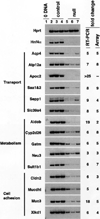
Loss of HNF4α disrupts expression of multiple genes encoding proteins that contribute to colon function. RT-PCR analyses of mRNA isolated from Hnf4aloxP/+Foxa3Cre (control, lanes 2–4) and Hnf4aloxP/loxPFoxa3Cre (null, lanes 5–7) E18.5 colons using oligonucleotide pairs that amplified Hprt as a loading control; Aqp4, Atp12a, Apoc2, Saa1 and 2, Sepp1, and Slc39a4, which encode proteins with a variety roles in transport; Aldob, Cyp2d26, Gatm, Neu3, and Sult1b1 that encode proteins having diverse roles in metabolism; and Cldn2, Mucdhl, Muc3, and Xlkd1 that encode proteins with various contributions to cell adhesion. 0 DNA (lane 1) confirmed the absence of contaminating DNA and average fold changes in mRNA levels between control and mutant colons calculated from RT-PCR analysis or Affymetrix array analysis are shown.
Figure 7
HNF4α-binding sites are present in genes whose expression is dependent on HNF4α. (A) Table showing the position and sequence of putative HNF4α-binding sites predicted to be present within genes whose expression is HNF4α dependent. HNF4α-binding site numbers (H4 sites) were assigned by using the criteria established by Yang et al. (B) EMSA was performed by using an HNF4α-binding site from the Apoc3 promoter and extracts from Cos-7 cells transfected with a control plasmid (mock, lane 1) or a plasmid expressing exogenous HNF4α (lanes 2–17). A specific shift caused by binding of HNF4α to the Apoc3 HNF4α-binding site is indicated with an arrowhead. Inclusion of 150-fold molar excess of unlabeled oligonucleotides corresponding to HNF4α-binding sites in the either the Apoc3 promoter (H4.21, lane 3) or binding sites predicted through computer modeling in Aldob (lane 4), Apoc2 (lanes 6–7), Aqp4 (lane 8), Cldn2 (lane 9), Gatm (lane 10), Muc3 (lane 11), Mucdh1 (lane 12), Neu3 (lane 13), Saa1 and 2 (lane 14), and Slc39a4 (lanes 15 and 16) all competed for binding of HNF4α to the labeled Apoc3 HNF4α-binding site. One binding site (lane 5) in the Apoc2 gene failed to compete, as did a negative control (N.C.) binding site for the transcription factor Foxa (lane 17).
Figure 8
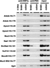
Genomic sequences within several genes whose expression is dependent on HNF4α are occupied by HNF4α within the colon. Chromatin immunoprecipitation was performed on 2 independent colons and a brain sample, a negative control tissue that does not express HNF4α, by using antibodies that precipitated either HNF4α or an unrelated protein, PES1. DNA sequences that coprecipitated with these proteins were identified by PCR by using primers that flanked the HNF4α-binding sites within Aldob, Apoc2, Muc3, Saa1, Slc39a4, Sult1b, and Mucdhl. PCR of the Hprt gene or the HNF4α-binding site 2 of the Muc3 gene (Muc3 H4.77) failed to show any enrichment in the colon, confirming that DNA sequences that were precipitated with anti-HNF4α were specific and reflected a bona fide interaction with HNF4α in the colon.
Similar articles
- Hepatocyte nuclear factor-4alpha promotes gut neoplasia in mice and protects against the production of reactive oxygen species.
Darsigny M, Babeu JP, Seidman EG, Gendron FP, Levy E, Carrier J, Perreault N, Boudreau F. Darsigny M, et al. Cancer Res. 2010 Nov 15;70(22):9423-33. doi: 10.1158/0008-5472.CAN-10-1697. Epub 2010 Nov 9. Cancer Res. 2010. PMID: 21062980 - The stable repression of mesenchymal program is required for hepatocyte identity: a novel role for hepatocyte nuclear factor 4α.
Santangelo L, Marchetti A, Cicchini C, Conigliaro A, Conti B, Mancone C, Bonzo JA, Gonzalez FJ, Alonzi T, Amicone L, Tripodi M. Santangelo L, et al. Hepatology. 2011 Jun;53(6):2063-74. doi: 10.1002/hep.24280. Hepatology. 2011. PMID: 21384409 Free PMC article. - Hepatocyte nuclear factor 4alpha is implicated in endoplasmic reticulum stress-induced acute phase response by regulating expression of cyclic adenosine monophosphate responsive element binding protein H.
Luebke-Wheeler J, Zhang K, Battle M, Si-Tayeb K, Garrison W, Chhinder S, Li J, Kaufman RJ, Duncan SA. Luebke-Wheeler J, et al. Hepatology. 2008 Oct;48(4):1242-50. doi: 10.1002/hep.22439. Hepatology. 2008. PMID: 18704925 Free PMC article. - Inverse regulation of claudin-2 and -7 expression by p53 and hepatocyte nuclear factor 4α in colonic MCE301 cells.
Hirota C, Takashina Y, Ikumi N, Ishizuka N, Hayashi H, Tabuchi Y, Yoshino Y, Matsunaga T, Ikari A. Hirota C, et al. Tissue Barriers. 2021 Jan 2;9(1):1860409. doi: 10.1080/21688370.2020.1860409. Epub 2020 Dec 23. Tissue Barriers. 2021. PMID: 33356822 Free PMC article. Review. - Role of hepatocyte nuclear factor 4α (HNF4α) in cell proliferation and cancer.
Walesky C, Apte U. Walesky C, et al. Gene Expr. 2015;16(3):101-8. doi: 10.3727/105221615X14181438356292. Gene Expr. 2015. PMID: 25700366 Free PMC article. Review.
Cited by
- Hepatocyte-specific deletion of hepatocyte nuclear factor-4α in adult mice results in increased hepatocyte proliferation.
Walesky C, Gunewardena S, Terwilliger EF, Edwards G, Borude P, Apte U. Walesky C, et al. Am J Physiol Gastrointest Liver Physiol. 2013 Jan 1;304(1):G26-37. doi: 10.1152/ajpgi.00064.2012. Epub 2012 Oct 25. Am J Physiol Gastrointest Liver Physiol. 2013. PMID: 23104559 Free PMC article. - Association between genetic variants in the HNF4A gene and childhood-onset Crohn's disease.
Marcil V, Sinnett D, Seidman E, Boudreau F, Gendron FP, Beaulieu JF, Menard D, Lambert M, Bitton A, Sanchez R, Amre D, Levy E. Marcil V, et al. Genes Immun. 2012 Oct;13(7):556-65. doi: 10.1038/gene.2012.37. Epub 2012 Aug 23. Genes Immun. 2012. PMID: 22914433 Free PMC article. - Effect of electroacupuncture at Sibai on the gastric myoelectric activities of denervated rats.
Chang XR, Yan J, Zhao YL, Li JS, Liu JH, He JF. Chang XR, et al. World J Gastroenterol. 2006 Sep 28;12(36):5897-901. doi: 10.3748/wjg.v12.i36.5897. World J Gastroenterol. 2006. PMID: 17007061 Free PMC article. - Identification of an endogenous ligand bound to a native orphan nuclear receptor.
Yuan X, Ta TC, Lin M, Evans JR, Dong Y, Bolotin E, Sherman MA, Forman BM, Sladek FM. Yuan X, et al. PLoS One. 2009;4(5):e5609. doi: 10.1371/journal.pone.0005609. Epub 2009 May 19. PLoS One. 2009. PMID: 19440305 Free PMC article. - Hepatocyte nuclear factor 4α transactivates the mitochondrial alanine aminotransferase gene in the kidney of Sparus aurata.
Salgado MC, Metón I, Anemaet IG, González JD, Fernández F, Baanante IV. Salgado MC, et al. Mar Biotechnol (NY). 2012 Feb;14(1):46-62. doi: 10.1007/s10126-011-9386-3. Epub 2011 May 24. Mar Biotechnol (NY). 2012. PMID: 21607544
References
- Radtke F, Clevers H. Self-renewal and cancer of the gut: two sides of a coin. Science. 2005;307:1904–1909. - PubMed
- Stainier DY. No organ left behind: tales of gut development and evolution. Science. 2005;307:1902–1904. - PubMed
- Badman MK, Flier JS. The gut and energy balance: visceral allies in the obesity wars. Science. 2005;307:1909–1914. - PubMed
- Pennisi E. The dynamic gut. Science. 2005;307:1896–1899. - PubMed
- Sanderson IR, Walker WA. Development of the gastrointestinal tract. Hamilton, Ontario, Canada: B.C. Decker; 2000.
Publication types
MeSH terms
Substances
Grants and funding
- R01 DK053892/DK/NIDDK NIH HHS/United States
- F32 DK067808-02/DK/NIDDK NIH HHS/United States
- F32 DK067808/DK/NIDDK NIH HHS/United States
- F32 DK067808-01/DK/NIDDK NIH HHS/United States
- F32 DK067808-03/DK/NIDDK NIH HHS/United States
LinkOut - more resources
Full Text Sources
Other Literature Sources
Molecular Biology Databases
