Neural correlates of processing valence and arousal in affective words - PubMed (original) (raw)
Neural correlates of processing valence and arousal in affective words
P A Lewis et al. Cereb Cortex. 2007 Mar.
Abstract
Psychological frameworks conceptualize emotion along 2 dimensions, "valence" and "arousal." Arousal invokes a single axis of intensity increasing from neutral to maximally arousing. Valence can be described variously as a bipolar continuum, as independent positive and negative dimensions, or as hedonic value (distance from neutral). In this study, we used functional magnetic resonance imaging to characterize neural activity correlating with arousal and with distinct models of valence during presentation of affective word stimuli. Our results extend observations in the chemosensory domain suggesting a double dissociation in which subregions of orbitofrontal cortex process valence, whereas amygdala preferentially processes arousal. In addition, our data support the physiological validity of descriptions of valence along independent axes or as absolute distance from neutral but fail to support the validity of descriptions of valence along a bipolar continuum.
Figures
Figure 1
Three unidimensional models of emotional valence. In the bipolar distribution (A), activation increases from most negative to most positive. In the independent model (Wundt 1924; Lang and others 1993; Barrett and Russell 1998) (B), activation increases in an independent way for positive and negative valences (Watson and Tellegen 1988; Cacioppo and Berntson 1994). In the U-shaped distribution (C), valence increases from most neutral to most intense regardless of whether positive or negative (Winston and others 2003, 2005; Cunningham and others 2004). In the psychological models upon which these constructs are based (e.g., Wundt 1924; Watson and Tellegen 1988; Lang and others 1993; Cacioppo and Berntson 1994; Barrett and Russell 1998), “activation” was intended as an unspecified psychological response. In the context of brain imaging, however, this can easily be translated as neural activity or even blood oxygen level–dependent response.
Figure 2
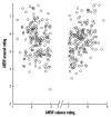
Distribution of our word stimuli in terms of valence and arousal. Normative valence, as taken from the ANEW (Bradley and Lang 1999) database, is shown on the x axis, and normative arousal is shown on the y axis.
Figure 3
Valence: Areas of activity significantly modulated by changing valence are rendered onto the SPM canonical brain at P < 0.001. Activity associated with increasingly positive valence is shown in red, activity associated with increasingly negative valence in blue, activity associated with the conjunction of these 2 (U-shaped model) in purple, and activity associated with the interaction of increasingly negative valence and arousal in cyan. Parameter estimates are shown for lateral orbitofrontal cortex (C), anterior cingulate (D top), and subgenual cingulate (D bottom). Note that responses in these regions followed the U-shaped model, that is, activity was modulated by increasingly intense valence in both positive and negative words. Parameter estimates represent (from left to right) valence, arousal, and the interaction of valence and arousal in negative (left) and positive (right) words.
Figure 4
Arousal: Areas of activity significantly modulated by arousal are rendered onto the SPM canonical brain at P < 0.001. The head of putamen, which is significantly modulated by increasing arousal in positive words, is shown in red (A), regions modulated by increasing arousal in negative words, including brain stem (A), pallidum (B), and amygdala (C), are shown in blue. Regions of activity shared between increasing arousal in positive and negative words (independent of valence), including ventral striatum (A, B), anterior insula (B), pallidum (B), and amygdala (C), are shown in purple. Insets in (C) show amygdala responses to both negative words (C1, 2, blue) and U-shaped model (C2,3, purple). Regions modulated by the interaction of increasingly negative valence and arousal are shown in cyan (C). Parameter estimates are shown for the activities shown in purple (correlating with increasing arousal in both positive and negative words) in ventral striatum (A), anterior insula (upper B), and pallidum (lower B), and amygdala (C). Parameter estimates represent (from left to right) valence, arousal, and the interaction of valence and arousal in negative (left) and positive (right) words. Activity in the amygdala is shown in (C), with insets showing the same at a visualization threshold of P < 0.005, sagittal view (C1,3) and coronal view (C2).
Similar articles
- Emotional valence and arousal affect reading in an interactive way: neuroimaging evidence for an approach-withdrawal framework.
Citron FM, Gray MA, Critchley HD, Weekes BS, Ferstl EC. Citron FM, et al. Neuropsychologia. 2014 Apr;56(100):79-89. doi: 10.1016/j.neuropsychologia.2014.01.002. Epub 2014 Jan 15. Neuropsychologia. 2014. PMID: 24440410 Free PMC article. - The neurophysiological bases of emotion: An fMRI study of the affective circumplex using emotion-denoting words.
Posner J, Russell JA, Gerber A, Gorman D, Colibazzi T, Yu S, Wang Z, Kangarlu A, Zhu H, Peterson BS. Posner J, et al. Hum Brain Mapp. 2009 Mar;30(3):883-95. doi: 10.1002/hbm.20553. Hum Brain Mapp. 2009. PMID: 18344175 Free PMC article. - Amygdala responses to Valence and its interaction by arousal revealed by MEG.
Styliadis C, Ioannides AA, Bamidis PD, Papadelis C. Styliadis C, et al. Int J Psychophysiol. 2014 Jul;93(1):121-33. doi: 10.1016/j.ijpsycho.2013.05.006. Epub 2013 May 18. Int J Psychophysiol. 2014. PMID: 23688672 - Remembering emotional experiences: the contribution of valence and arousal.
Kensinger EA. Kensinger EA. Rev Neurosci. 2004;15(4):241-51. doi: 10.1515/revneuro.2004.15.4.241. Rev Neurosci. 2004. PMID: 15526549 Review. - Multiple scales of valence processing in the brain.
Man V, Cunningham WA. Man V, et al. Soc Neurosci. 2021 Feb;16(1):57-67. doi: 10.1080/17470919.2019.1692068. Epub 2019 Nov 21. Soc Neurosci. 2021. PMID: 31711368 Review.
Cited by
- The role of the somatosensory system in the feeling of emotions: a neurostimulation study.
Giraud M, Javadi AH, Lenatti C, Allen J, Tamè L, Nava E. Giraud M, et al. Soc Cogn Affect Neurosci. 2024 Oct 18;19(1):nsae062. doi: 10.1093/scan/nsae062. Soc Cogn Affect Neurosci. 2024. PMID: 39275796 Free PMC article. - On the brain struggles to recognize basic facial emotions with face masks: an fMRI study.
Abutalebi J, Gallo F, Fedeli D, Houdayer E, Zangrillo F, Emedoli D, Spina A, Bellini C, Del Maschio N, Iannaccone S, Alemanno F. Abutalebi J, et al. Front Psychol. 2024 Jan 26;15:1339592. doi: 10.3389/fpsyg.2024.1339592. eCollection 2024. Front Psychol. 2024. PMID: 38344280 Free PMC article. - Tactile estimation of hedonic and sensory properties during active touch: An electroencephalography study.
Henderson J, Mari T, Hewitt D, Newton-Fenner A, Hopkinson A, Giesbrecht T, Marshall A, Stancak A, Fallon N. Henderson J, et al. Eur J Neurosci. 2023 Sep;58(6):3412-3431. doi: 10.1111/ejn.16101. Epub 2023 Jul 30. Eur J Neurosci. 2023. PMID: 37518981 Free PMC article. - Neural correlates of emotional valence for faces and words.
Ballotta D, Maramotti R, Borelli E, Lui F, Pagnoni G. Ballotta D, et al. Front Psychol. 2023 Feb 23;14:1055054. doi: 10.3389/fpsyg.2023.1055054. eCollection 2023. Front Psychol. 2023. PMID: 36910761 Free PMC article. - Short-Term Head-Out Whole-Body Cold-Water Immersion Facilitates Positive Affect and Increases Interaction between Large-Scale Brain Networks.
Yankouskaya A, Williamson R, Stacey C, Totman JJ, Massey H. Yankouskaya A, et al. Biology (Basel). 2023 Jan 29;12(2):211. doi: 10.3390/biology12020211. Biology (Basel). 2023. PMID: 36829490 Free PMC article.
References
- Anderson AK, Christoff K, Stappen I, Panitz D, Ghahremani DG, Glover G, Gabrieli JD, Sobel N. Dissociated neural representations of intensity and valence in human olfaction. Nat Neurosci. 2003;6(2):196–202. - PubMed
- Barrett L, Russell J. Independence and bipolarity in the structure of current affect. J Pers Soc Psychol. 1998;74:967–984.
- Baxter MG, Murray EA. The amygdala and reward. Nat Rev Neurosci. 2002;3(7):563–573. - PubMed
- Bensafi M, Rouby C, Farget V, Bertrand B, Vigouroux M, Holley A. Autonomic nervous system responses to odours: the role of pleasantness and arousal. Chem Senses. 2002;27(8):703–709. - PubMed
Publication types
MeSH terms
LinkOut - more resources
Full Text Sources
Medical


