PAR1b promotes cell-cell adhesion and inhibits dishevelled-mediated transformation of Madin-Darby canine kidney cells - PubMed (original) (raw)
PAR1b promotes cell-cell adhesion and inhibits dishevelled-mediated transformation of Madin-Darby canine kidney cells
Maya Elbert et al. Mol Biol Cell. 2006 Aug.
Abstract
Mammalian Par1 is a family of serine/threonine kinases comprised of four homologous isoforms that have been associated with tumor suppression and differentiation of epithelial and neuronal cells, yet little is known about their cellular functions. In polarizing kidney epithelial (Madin-Darby canine kidney [MDCK]) cells, the Par1 isoform Par1b/MARK2/EMK1 promotes the E-cadherin-dependent compaction, columnarization, and cytoskeletal organization characteristic of differentiated columnar epithelia. Here, we identify two functions of Par1b that likely contribute to its role as a tumor suppressor in epithelial cells. 1) The kinase promotes cell-cell adhesion and resistance of E-cadherin to extraction by nonionic detergents, a measure for the association of the E-cadherin cytoplasmic domain with the actin cytoskeleton, which is critical for E-cadherin function. 2) Par1b attenuates the effect of Dishevelled (Dvl) expression, an inducer of wnt signaling that causes transformation of epithelial cells. Although Dvl is a known Par1 substrate in vitro, we determined, after mapping the PAR1b-phosphorylation sites in Dvl, that PAR1b did not antagonize Dvl signaling by phosphorylating the wnt-signaling molecule. Instead, our data suggest that both proteins function antagonistically to regulate the assembly of functional E-cadherin-dependent adhesion complexes.
Figures
Figure 1.
Par1b regulates cell–cell adhesion. (A) Doxycycline-inducible Par1b-KO cell lines. Parental TET-ON cells and two Par1b-KO clones (cl.12 and cl.A) were cultured in the presence (+dox) or absence (−dox) of doxycycline as described in Materials and Methods. An antibody that recognizes all Par1 isoforms was used to probe for Par1 expression in equal amounts of SDS lysates. Par1b-specific bands are indicated by asterisks. (B) Cell–cell adhesion in hanging drop assays. Parental MDCK-TET-ON cells and Par1b-KO cells cl.12 and A (top) or Par1b-MDCK-TET-OFF cells cl.12 and 24 (bottom) were cultured in the presence or absence of dox, Cell–cell adhesion upon suspension in hanging drops was determined from 4× wide-field images (right) as described in Materials and Methods. The areas of cell clusters in six random fields taken from five different hanging drops were determined for each culture condition and classified as indicated. An area of 105 μm2 corresponds to ∼600 cells, 2 × 104 μm2 corresponds to ∼100 cells, 104 μm2 corresponds to ∼55 cells, and 3.5 × 103 μm2 corresponds to 20 cells. Presented are the average clusters per field for each classifier from one representative experiment out of two (Par1b-MDCK) or three (Par1b-KO) independent experiments. Bars, 300 μm.
Figure 2.
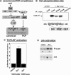
Dvl1 promotes wnt signaling in MDCK cells and is phosphorylated by Par1b on two sites in vitro. (A) Models for Dvl in the canonical and planar polarity wnt pathways. The wnt signal is transduced via the membrane receptor Frizzled and its coreceptor LRP to Dvl, a soluble protein with three functional domains. Canonical pathway: Dvl inhibits GSK3β. GSK3β functions in a destruction complex with Axin and APC to phosphorylate β-catenin and targets it for degradation. β-catenin that accumulates in the nucleus acts as cotranscription factor for TCF/LEF; requires all three domains of Dvl. Planar polarity pathway: Dvl activates JNK, presumably via rho- GTPases, which leads to c-Jun–dependent gene transcription and to cytoskeletal rearrangements; requires the Dvl-DEP domain. (B) PAR1b phosphorylates Dvl1 on Ser 236 and Ser 243 in vitro. Immobilized immune complexes containing MDCK-derived PAR1b (+PAR1b-myc) or IgG (control) were incubated with purified recombinant DvlM proteins (amino acids 173–395) and [γ32P]ATP as described in Materials and Methods. Evolutionary conserved Ser/Thr residues in the target region (amino acids 213–248) were sequentially substituted to alanine by site-directed mutagenesis. Substitution of both Ser 236 and Ser 243 to alanine abolished PAR1b-dependent phosphorylation. (C) Dvl promotes TCF/LEF activation. TOPFLASH or FOPFLASH control assays of MDCK cells cotransfected with either 12 μg of vector DNA or WT-Dvl plasmids. Normalized luciferase activity is expressed as -fold increase over the baseline activity in vector-transfected cells. (D) Dvl promotes JNK activation Cells were transfected with either vector DNA or WT-Dvl cDNA as described in C and analyzed for JNK activity with c-Jun as substrate as described in Materials and Methods. Duplicate samples are presented. Top, phospho-c-Jun. Bottom, JNK immunoprecipitation.
Figure 3.
PAR1b and Dvl function antagonistically to regulate MDCK polarization. (A) Dvl and Dvl S236,243A expression reduce cell–cell adhesion, block lumen formation, compaction, and MT reorganization. Control MDCK cells transiently transfected with vector DNA, Dvl wild type, or DvlS236,243S were subjected to cell–cell adhesion assays (top), collagen overlay (middle), or Ca2+-switch assays (bottom). Cells were labeled for the indicated markers. Presented are 4× wide-field images in the top panel, confocal x-z views in the two middle panels, and a confocal x-y section taken through the center of the cells in the MT panels (bottom). Note the cross section through vertical MTs in the control cells that is absent in the Dvl samples. (B) PAR1b blocks the Dvl and DvlS236,243A phenotypes. PAR1b-MDCK cells were transfected, cultured, and processed as the control cells described in A. Bars, 300 μm (cell–cell adhesion panels) and 10 μm (all other panels). Table shows results from a hanging drop assay performed as described in Materials and Methods and in Figure 1. Data are representative of three (MDCK control) and two (MDCK-Par1b) independent experiments.
Figure 4.
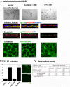
Dvl regulates MDCK polarity independent of canonical wnt signaling. Control cells were either mock transfected (vector) or transfected with 12 μg of cDNA encoding DvlΔDEP or myc-tagged β-cateninΔN90 in the presence (B) or absence (A and C) of the TOPFLASH reporter plasmids. (A) DvlΔDEP but not β-cateninΔN90 mimic the Dvl phenotype. MDCK cells were subjected to cell–cell adhesion assays, collagen overlay, or Ca2+-switch and analyzed for tubulogenesis (collagen overlay, x-z view), lumen formation, cell compaction, and shape (middle, x-z view) and MT organization (bottom, x-y view) as described in Figure 3. Graph shows -fold stimulation of TOPFLASH-dependent luciferase activity by Dvl, DvlΔDEP, and β-cateninΔN90. The value for the Dvl-bar is derived from Figure 2C. Image shows myc-labeling of vector and β-cateninΔN90 transfected cells; x-y view through the cell center indicates homogenous expression of recombinant β-catenin within the cell population. Bars, 300 μm (cell–cell adhesion panels) and 10 μm (all other panels). (C) Hanging drop assay. Assays were performed as described in Figure 1 and Materials and Methods. Data are representative of three independent experiments.
Figure 5.
PAR1b and Dvl regulate the association of E-cadherin with microfilaments. (A) Total E-cadherin levels. MDCK-TET-OFF cells transiently expressing the indicated recombinant proteins (lanes 1–5) or parental TET-ON (control), Par1b-KO cl12, and Par1b-KO cl.A cells induced for 2 d with dox (lanes 6–8) were plated at confluence for 24 h, and equal amounts of SDS lysates were probed for E-cadherin in immunoblots. (B) Tx100-resistent E-cadherin. Total SDS lysates (1 and 2) and SDS lysates of Tx100-extracted cells (3 and 4) were probed for the presence of E-cadherin in quantitative immunoblots. Two times as much lysate from samples 3 and 4 was loaded as from 1 and 2; Graphs express the ratio of Tx100-resistant E-cadherin (3 and 4) to total E-cadherin (1 and 2) for each cell culture condition from three independent experiments with SEs. Note that the total protein levels are not adjusted between the different cell culture conditions. Top, parental TET-ON cells or Par1b-KO clones 12 and A were either induced (+dox) or remained uninduced (−dox). The small decrease in Tx100-resistant E-cadherin in the uninduced Par1b-KO clones (−dox) compared with the parental cells is likely due to leakiness of the expression system, because both Par1-KO clones express slightly less Par1 than the parental cells even before doxycycline induction. Bottom, parental TET-OFF cells (control MDCK) transfected with either vector, Dvl, or βcateninΔN90 cDNA and Par1b-MDCK cells transfected with either vector or Dvl cDNA were cultured under inducible conditions (−dox). Note that Par1b-KO and Dvl expression do not decrease the total Tx100-resistant actin pool (Supplemental Figure S3).
Similar articles
- Par1b promotes hepatic-type lumen polarity in Madin Darby canine kidney cells via myosin II- and E-cadherin-dependent signaling.
Cohen D, Tian Y, Müsch A. Cohen D, et al. Mol Biol Cell. 2007 Jun;18(6):2203-15. doi: 10.1091/mbc.e07-02-0095. Epub 2007 Apr 4. Mol Biol Cell. 2007. PMID: 17409351 Free PMC article. - Dishevelled-induced phosphorylation regulates membrane localization of Par1b.
Terabayashi T, Funato Y, Miki H. Terabayashi T, et al. Biochem Biophys Res Commun. 2008 Oct 31;375(4):660-5. doi: 10.1016/j.bbrc.2008.08.098. Epub 2008 Aug 28. Biochem Biophys Res Commun. 2008. PMID: 18760999 - The serine/threonine kinase Par1b regulates epithelial lumen polarity via IRSp53-mediated cell-ECM signaling.
Cohen D, Fernandez D, Lázaro-Diéguez F, Müsch A. Cohen D, et al. J Cell Biol. 2011 Feb 7;192(3):525-40. doi: 10.1083/jcb.201007002. Epub 2011 Jan 31. J Cell Biol. 2011. PMID: 21282462 Free PMC article. - Dishevelled: The hub of Wnt signaling.
Gao C, Chen YG. Gao C, et al. Cell Signal. 2010 May;22(5):717-27. doi: 10.1016/j.cellsig.2009.11.021. Epub 2009 Dec 13. Cell Signal. 2010. PMID: 20006983 Review. - Spatial integration of E-cadherin adhesion, signalling and the epithelial cytoskeleton.
Braga V. Braga V. Curr Opin Cell Biol. 2016 Oct;42:138-145. doi: 10.1016/j.ceb.2016.07.006. Epub 2016 Aug 6. Curr Opin Cell Biol. 2016. PMID: 27504601 Review.
Cited by
- Rac1 acts in conjunction with Nedd4 and dishevelled-1 to promote maturation of cell-cell contacts.
Nethe M, de Kreuk BJ, Tauriello DV, Anthony EC, Snoek B, Stumpel T, Salinas PC, Maurice MM, Geerts D, Deelder AM, Hensbergen PJ, Hordijk PL. Nethe M, et al. J Cell Sci. 2012 Jul 15;125(Pt 14):3430-42. doi: 10.1242/jcs.100925. Epub 2012 Mar 30. J Cell Sci. 2012. PMID: 22467858 Free PMC article. - Par1b promotes hepatic-type lumen polarity in Madin Darby canine kidney cells via myosin II- and E-cadherin-dependent signaling.
Cohen D, Tian Y, Müsch A. Cohen D, et al. Mol Biol Cell. 2007 Jun;18(6):2203-15. doi: 10.1091/mbc.e07-02-0095. Epub 2007 Apr 4. Mol Biol Cell. 2007. PMID: 17409351 Free PMC article. - Inhibition of the miR-192/215-Rab11-FIP2 axis suppresses human gastric cancer progression.
Zhang X, Peng Y, Huang Y, Deng S, Feng X, Hou G, Lin H, Wang J, Yan R, Zhao Y, Fan X, Meltzer SJ, Li S, Jin Z. Zhang X, et al. Cell Death Dis. 2018 Jul 13;9(7):778. doi: 10.1038/s41419-018-0785-5. Cell Death Dis. 2018. PMID: 30006518 Free PMC article. - Establishment of epithelial polarity--GEF who's minding the GAP?
Ngok SP, Lin WH, Anastasiadis PZ. Ngok SP, et al. J Cell Sci. 2014 Aug 1;127(Pt 15):3205-15. doi: 10.1242/jcs.153197. Epub 2014 Jul 2. J Cell Sci. 2014. PMID: 24994932 Free PMC article. Review. - Par1b links lumen polarity with LGN-NuMA positioning for distinct epithelial cell division phenotypes.
Lázaro-Diéguez F, Cohen D, Fernandez D, Hodgson L, van Ijzendoorn SC, Müsch A. Lázaro-Diéguez F, et al. J Cell Biol. 2013 Oct 28;203(2):251-64. doi: 10.1083/jcb.201303013. J Cell Biol. 2013. PMID: 24165937 Free PMC article.
References
- Beghini A. The neural progenitor-restricted isoform of the MARK4 gene in 19q13.2 is upregulated in human gliomas and overexpressed in a subset of glioblastoma cell lines. Oncogene. 2003;22:2581–2591. - PubMed
- Bershadsky A. Magic touch: how does cell-cell adhesion trigger actin assembly? Trends Cell Biol. 2004;14:589–593. - PubMed
- Bohm H., Brinkmann V., Drab M., Henske A., Kurzchalia T. V. Mammalian homologues of C. elegans PAR-1 are asymmetrically localized in epithelial cells and may influence their polarity. Curr. Biol. 1997;7:603–606. - PubMed
Publication types
MeSH terms
Substances
Grants and funding
- R01 GM034107/GM/NIGMS NIH HHS/United States
- GM-34107/GM/NIGMS NIH HHS/United States
- R01 DK064842/DK/NIDDK NIH HHS/United States
- EY07138/EY/NEI NIH HHS/United States
- T32 EY007138/EY/NEI NIH HHS/United States
LinkOut - more resources
Full Text Sources
Other Literature Sources


