Context fear learning in the absence of the hippocampus - PubMed (original) (raw)
Context fear learning in the absence of the hippocampus
Brian J Wiltgen et al. J Neurosci. 2006.
Abstract
Lesions of the rodent hippocampus invariably abolish context fear memories formed in the recent past but do not always prevent new learning. To better understand this discrepancy, we thoroughly examined the acquisition of context fear in rats with pretraining excitotoxic lesions of the dorsal hippocampus. In the first experiment, animals received a shock immediately after placement in the context or after variable delays. Immediate shock produced no context fear learning in lesioned rats or controls. In contrast, delayed shock produced robust context fear learning in both groups. The absence of fear with immediate shock occurs because animals need time to form a representation of the context before shock is presented. The fact that it occurs in both sham and lesioned rats suggests that they learn about the context in a similar manner. However, despite learning about the context in the delay condition, lesioned rats did not acquire as much fear as controls. The second experiment showed that this lesion-induced deficit could be overcome by increasing the number of conditioning trials. Lesioned animals learned normally after multiple shocks, regardless of freezing level or trial spacing. The last experiment showed that animals with complete hippocampus lesions could also learn about the context, although the same lesions produced devastating retrograde amnesia. These results demonstrate that alternative systems can acquire context fear but do so less efficiently than the hippocampus.
Figures
Figure 1.
Photomicrographs showing thionin-stained coronal brain sections after excitotoxic lesions of the dorsal hippocampus. From top to bottom, the sections are −0.80, −1.60, −2.60, −3.60, −4.60, and −6.0 mm posterior to bregma.
Figure 2.
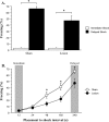
Mean ± SEM percentage freezing during the context test for sham and DH-lesioned animals. A, Freezing levels for animals that received an immediate or delayed shock. B, Freezing levels for animals that received shock 12, 24, 48, 192, or 340 s after placement in the context during training. *p < 0.05, significant group difference.
Figure 3.
Mean ± SEM percentage freezing during the context test for sham and DH-lesioned animals. Separate groups of animals received one or three shocks during a 48 or 340 s training session. *p < 0.05, significant group difference.
Figure 4.
Mean ± SEM percentage freezing during each minute of the context test for sham and DH-lesioned animals. A, Freezing time course for animals receiving a single shock during training. B, Freezing time course for animals receiving three shocks during training.
Figure 5.
Mean ± SEM velocity (in centimeters per second) for sham and DH-lesioned animals during the 2 s shock and an equivalent baseline period. A, Velocity for animals receiving a single shock. B, Velocity for animals receiving three shocks.
Figure 6.
Photomicrographs showing thionin-stained coronal brain sections after excitotoxic lesions of the dorsal and ventral hippocampus (left) or sham surgery (right). From top to bottom, the sections are −0.30, −1.80, −3.0, −4.0, −4.50, and −4.60 mm posterior to bregma.
Figure 7.
Mean ± SEM percentage freezing during the context test after posttraining (Retro) or pretraining (Antero) lesions of the hippocampus. Lesions made 1 d after intense training produced robust retrograde amnesia for context fear. Anterograde amnesia was not observed after moderate retraining. *p < 0.05, significant group difference.
Similar articles
- Retrograde amnesia following hippocampal lesions in the shock-probe conditioning test.
Lehmann H, Lecluse V, Houle A, Mumby DG. Lehmann H, et al. Hippocampus. 2006;16(4):379-87. doi: 10.1002/hipo.20159. Hippocampus. 2006. PMID: 16411184 - Posttraining but not pretraining lesions of the hippocampus interfere with feature-negative discrimination of fear-potentiated startle.
Heldt SA, Coover GD, Falls WA. Heldt SA, et al. Hippocampus. 2002;12(6):774-86. doi: 10.1002/hipo.10033. Hippocampus. 2002. PMID: 12542229 - Absence of systems consolidation of fear memories after dorsal, ventral, or complete hippocampal damage.
Sutherland RJ, O'Brien J, Lehmann H. Sutherland RJ, et al. Hippocampus. 2008;18(7):710-8. doi: 10.1002/hipo.20431. Hippocampus. 2008. PMID: 18446823 - Hippocampus and contextual fear conditioning: recent controversies and advances.
Anagnostaras SG, Gale GD, Fanselow MS. Anagnostaras SG, et al. Hippocampus. 2001;11(1):8-17. doi: 10.1002/1098-1063(2001)11:1<8::AID-HIPO1015>3.0.CO;2-7. Hippocampus. 2001. PMID: 11261775 Review. - A complex associative structure formed in the mammalian brain during acquisition of a simple visual discrimination task: dorsolateral striatum, amygdala, and hippocampus.
McDonald RJ, King AL, Wasiak TD, Zelinski EL, Hong NS. McDonald RJ, et al. Hippocampus. 2007;17(9):759-74. doi: 10.1002/hipo.20333. Hippocampus. 2007. PMID: 17623852 Review.
Cited by
- Know thy SEFL: Fear sensitization and its relevance to stressor-related disorders.
Nishimura KJ, Poulos AM, Drew MR, Rajbhandari AK. Nishimura KJ, et al. Neurosci Biobehav Rev. 2022 Nov;142:104884. doi: 10.1016/j.neubiorev.2022.104884. Epub 2022 Sep 26. Neurosci Biobehav Rev. 2022. PMID: 36174795 Free PMC article. Review. - Network supporting contextual fear learning after dorsal hippocampal damage has increased dependence on retrosplenial cortex.
Coelho CAO, Ferreira TL, Kramer-Soares JC, Sato JR, Oliveira MGM. Coelho CAO, et al. PLoS Comput Biol. 2018 Aug 7;14(8):e1006207. doi: 10.1371/journal.pcbi.1006207. eCollection 2018 Aug. PLoS Comput Biol. 2018. PMID: 30086129 Free PMC article. - Memory consolidation in both trace and delay fear conditioning is disrupted by intra-amygdala infusion of the protein synthesis inhibitor anisomycin.
Kwapis JL, Jarome TJ, Schiff JC, Helmstetter FJ. Kwapis JL, et al. Learn Mem. 2011 Oct 25;18(11):728-32. doi: 10.1101/lm.023945.111. Print 2011 Nov. Learn Mem. 2011. PMID: 22028394 Free PMC article. - Hippocampal Engrams and Contextual Memory.
Vasudevan K, Hassell JE Jr, Maren S. Vasudevan K, et al. Adv Neurobiol. 2024;38:45-66. doi: 10.1007/978-3-031-62983-9_4. Adv Neurobiol. 2024. PMID: 39008010 Free PMC article. Review. - Neural and cellular mechanisms of fear and extinction memory formation.
Orsini CA, Maren S. Orsini CA, et al. Neurosci Biobehav Rev. 2012 Aug;36(7):1773-802. doi: 10.1016/j.neubiorev.2011.12.014. Epub 2012 Jan 2. Neurosci Biobehav Rev. 2012. PMID: 22230704 Free PMC article. Review.
References
- Anagnostaras SG, Gale GD, Fanselow MS (2001). Hippocampus and contextual fear conditioning: recent controversies and advances. Hippocampus 11:8–17. - PubMed
- Cho YH, Friedman E, Silva AJ (1999). Ibotenate lesions of the hippocampus impair spatial learning but not contextual fear conditioning in mice. Behav Brain Res 98:77–87. - PubMed
- de Hoz L, Moser EI, Morris RG (2005). Spatial learning with unilateral and bilateral hippocampal networks. Eur J Neurosci 22:745–754. - PubMed
- Fanselow MS (1984). Opiate modulation of the active and inactive components of the postshock reaction: parallels between naloxone pretreatment and shock intensity. Behav Neurosci 98:269–277. - PubMed
Publication types
MeSH terms
LinkOut - more resources
Full Text Sources
Medical
Miscellaneous


