Protein disulfide isomerase-like proteins play opposing roles during retrotranslocation - PubMed (original) (raw)
Protein disulfide isomerase-like proteins play opposing roles during retrotranslocation
Michele L Forster et al. J Cell Biol. 2006.
Abstract
Misfolded proteins in the endoplasmic reticulum (ER) are retained in the organelle or retrotranslocated to the cytosol for proteasomal degradation. ER chaperones that guide these opposing processes are largely unknown. We developed a semipermeabilized cell system to study the retrotranslocation of cholera toxin (CT), a toxic agent that crosses the ER membrane to reach the cytosol during intoxication. We found that protein disulfide isomerase (PDI) facilitates CT retrotranslocation, whereas ERp72, a PDI-like protein, mediates its ER retention. In vitro analysis revealed that PDI and ERp72 alter CT's conformation in a manner consistent with their roles in retrotranslocation and ER retention. Moreover, we found that PDI's and ERp72's opposing functions operate on endogenous ER misfolded proteins. Thus, our data identify PDI family proteins that play opposing roles in ER quality control and establish an assay to further delineate the mechanism of CT retrotranslocation.
Figures
Figure 1.
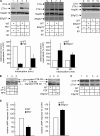
Retrotranslocation of the CTA1 subunit. (A) HeLa cells were incubated with CT for 90 min at 4 or 37°C with or without BFA or NEM. Cells were permeabilized, centrifuged, and the supernatant and pellet fractions were separated, subjected to nonreducing SDS-PAGE, and immunoblotted with the indicated antibodies. CTA, CTA1, and CTB are 28, 22, and 11 kD, respectively. (B) As in A except a CT mutant, R192H, was used. PDI is 58 kD. (C) As in A except where indicated, cells were treated with MG132. Polyubiquitinated proteins range from 100 to 300 kD. White lines indicate that intervening lanes have been spliced out.
Figure 2.
Down-regulation of PDI-like proteins. (A) Structural organization of PDI, ERp72, and ERp57. (B) PDI, ERp72, ERp57, and ERp29 protein levels were examined in wild-type (WT), PDI−, ERp72−, and ERp57− cells. (C) Wild-type, PDI−, ERp72−, and ERp57− cells were incubated with 2 μg/ml tunicamycin (TM) for 8 h, and BiP expression was assessed by SDS-PAGE and immunoblot analysis. BiP is 78 kD.
Figure 3.
PDI and ERp72 exert opposing effects on CT retrotranslocation. (A, top) Wild-type (WT), PDI−, ERp72−, and ERp57− cells were incubated with CT for 45 or 90 min, permeabilized, and the supernatant fraction was analyzed as in Fig. 1. (bottom) The intensities of the CTA and CTA1 bands in the supernatant were quantified. Graphs show the mean ± SD (error bars) of two to four experiments. (B) Wild-type, PDI−, and ERp72− cells were incubated with CT-GS, and the cell lysate was subjected to SDS-PAGE followed by immunoblotting with an anti-CTB antibody. Nonglycosylated and glycosylated CTB are 17 and 21 kD, respectively. (C) Whole cell lysates from CT-intoxicated wild-type, PDI−, and ERp72− cells were prepared in the presence of NEM and subjected to immunoblotting with CTA antibody. (D) Wild-type, PDI−, and ERp72− cells were incubated with CT for 45 or 90 min, and the cAMP level was measured by a cAMP Biotrak Enzyme Immunoassay System (GE Healthcare). Means ± SD of two to four experiments are shown. White lines indicate that intervening lanes have been spliced out.
Figure 4.
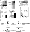
Opposing effects of PDI and ERp72 on the conformation of CT. (A) His-tagged ERp72 protein was purified from bacteria and analyzed by SDS-PAGE followed by Coomassie staining or immunoblotting with an antibody against ERp72. (B) CTA was incubated with BSA, PDI, or ERp72 followed by the addition of trypsin. Samples were subjected to SDS-PAGE followed by immunoblotting with an anti-CTA antibody.
Figure 5.
A general role of PDI and ERp72 in the retrotranslocation of ER misfolded proteins. (A, top) Wild-type (WT), PDI−, or ERp72− cells were treated with MG132, and the cell lysate was subjected to SDS-PAGE followed by immunoblotting with the indicated antibodies. (bottom) The total ubiquitin signal intensity was measured. Graphs show the mean ± SD (error bars) of three to five experiments. (B) Wild-type, PDI−, or ERp72− cells were treated with TNFα, and the lysate was subjected to SDS-PAGE followed by immunoblotting with an antibody against IκBα. IκBα is 39 kD. (C) CHO cells stably expressing cogTg (CHO-P) and PDI (CHO-PDI) or ERp72 (CHO-ERp72) were pulse labeled with [35S]methionine and chased for the times indicated. CogTg was immunoprecipitated from the cell lysate with anti-Tg antibody and analyzed by SDS-PAGE. Data show quantification of the radioactive cogTg band intensity. (D) Diagram depicting the similarity between the quality control system in the ER and cytosol. See last paragraph of Results and discussion. White lines indicate that intervening lanes have been spliced out.
Similar articles
- Unfolded cholera toxin is transferred to the ER membrane and released from protein disulfide isomerase upon oxidation by Ero1.
Tsai B, Rapoport TA. Tsai B, et al. J Cell Biol. 2002 Oct 28;159(2):207-16. doi: 10.1083/jcb.200207120. Epub 2002 Oct 28. J Cell Biol. 2002. PMID: 12403808 Free PMC article. - ERdj5 is required as a disulfide reductase for degradation of misfolded proteins in the ER.
Ushioda R, Hoseki J, Araki K, Jansen G, Thomas DY, Nagata K. Ushioda R, et al. Science. 2008 Jul 25;321(5888):569-72. doi: 10.1126/science.1159293. Science. 2008. PMID: 18653895 - Structure of the catalytic a(0)a fragment of the protein disulfide isomerase ERp72.
Kozlov G, Azeroual S, Rosenauer A, Määttänen P, Denisov AY, Thomas DY, Gehring K. Kozlov G, et al. J Mol Biol. 2010 Aug 27;401(4):618-25. doi: 10.1016/j.jmb.2010.06.045. Epub 2010 Jun 26. J Mol Biol. 2010. PMID: 20600112 - Proteins of the PDI family: unpredicted non-ER locations and functions.
Turano C, Coppari S, Altieri F, Ferraro A. Turano C, et al. J Cell Physiol. 2002 Nov;193(2):154-63. doi: 10.1002/jcp.10172. J Cell Physiol. 2002. PMID: 12384992 Review. - Protein folding includes oligomerization - examples from the endoplasmic reticulum and cytosol.
Christis C, Lubsen NH, Braakman I. Christis C, et al. FEBS J. 2008 Oct;275(19):4700-27. doi: 10.1111/j.1742-4658.2008.06590.x. Epub 2008 Aug 1. FEBS J. 2008. PMID: 18680510 Review.
Cited by
- Functional relationship between protein disulfide isomerase family members during the oxidative folding of human secretory proteins.
Rutkevich LA, Cohen-Doyle MF, Brockmeier U, Williams DB. Rutkevich LA, et al. Mol Biol Cell. 2010 Sep 15;21(18):3093-105. doi: 10.1091/mbc.E10-04-0356. Epub 2010 Jul 21. Mol Biol Cell. 2010. PMID: 20660153 Free PMC article. - Transient covalent interactions of newly synthesized thyroglobulin with oxidoreductases of the endoplasmic reticulum.
Di Jeso B, Morishita Y, Treglia AS, Lofrumento DD, Nicolardi G, Beguinot F, Kellogg AP, Arvan P. Di Jeso B, et al. J Biol Chem. 2014 Apr 18;289(16):11488-11496. doi: 10.1074/jbc.M113.520767. Epub 2014 Mar 5. J Biol Chem. 2014. PMID: 24599957 Free PMC article. - The recognition and retrotranslocation of misfolded proteins from the endoplasmic reticulum.
Nakatsukasa K, Brodsky JL. Nakatsukasa K, et al. Traffic. 2008 Jun;9(6):861-70. doi: 10.1111/j.1600-0854.2008.00729.x. Epub 2008 Feb 24. Traffic. 2008. PMID: 18315532 Free PMC article. Review. - Protein Quality Control in the Endoplasmic Reticulum.
Adams BM, Oster ME, Hebert DN. Adams BM, et al. Protein J. 2019 Jun;38(3):317-329. doi: 10.1007/s10930-019-09831-w. Protein J. 2019. PMID: 31004255 Free PMC article. Review. - A large and intact viral particle penetrates the endoplasmic reticulum membrane to reach the cytosol.
Inoue T, Tsai B. Inoue T, et al. PLoS Pathog. 2011 May;7(5):e1002037. doi: 10.1371/journal.ppat.1002037. Epub 2011 May 12. PLoS Pathog. 2011. PMID: 21589906 Free PMC article.
References
- Ellgaard, L., and A. Helenius. 2003. Quality control in the endoplasmic reticulum. Nat. Rev. Mol. Cell Biol. 4:181–191. - PubMed
- Fujinaga, Y., A.A. Wolf, C. Rodighiero, H. Wheeler, B. Tsai, L. Allen, M.G. Jobling, T. Rapoport, R.K. Holmes, and W.I. Lencer. 2003. Gangliosides that associate with lipid rafts mediate transport of cholera and related toxins from the plasma membrane to endoplasmic reticulum. Mol. Biol. Cell. 14:4783–4793. - PMC - PubMed
- Karin, M., and Y. Ben-Neriah. 2000. Phosphorylation meets ubiquitination: the control of NF-κB activity. Annu. Rev. Immunol. 18:621–663. - PubMed
MeSH terms
Substances
LinkOut - more resources
Full Text Sources
Other Literature Sources
Research Materials
Miscellaneous


