Spontaneous neuronal activity distinguishes human dorsal and ventral attention systems - PubMed (original) (raw)
Spontaneous neuronal activity distinguishes human dorsal and ventral attention systems
Michael D Fox et al. Proc Natl Acad Sci U S A. 2006.
Erratum in
- Proc Natl Acad Sci U S A. 2006 Sep 5;103(36):13560
Abstract
On the basis of task-related imaging studies in normal human subjects, it has been suggested that two attention systems exist in the human brain: a bilateral dorsal attention system involved in top-down orienting of attention and a right-lateralized ventral attention system involved in reorienting attention in response to salient sensory stimuli. An important question is whether this functional organization emerges only in response to external attentional demands or is represented more fundamentally in the internal dynamics of brain activity. To address this question, we examine correlations in spontaneous fluctuations of the functional MRI blood oxygen level-dependent signal in the absence of task, stimuli, or explicit attentional demands. We identify a bilateral dorsal attention system and a right-lateralized ventral attention system solely on the basis of spontaneous activity. Further, we observe regions in the prefrontal cortex correlated with both systems, a potential mechanism for mediating the functional interaction between systems. These findings demonstrate that the neuroanatomical substrates of human attention persist in the absence of external events, reflected in the correlation structure of spontaneous activity.
Conflict of interest statement
Conflict of interest statement: No conflicts declared.
Figures
Fig. 1.
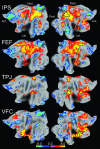
Z score maps showing voxels significantly correlated or anticorrelated with seed regions in the IPS, FEF, TPJ, and VFC during resting fixation (P < 0.01, random effects). Data are displayed on the flattened left hemisphere (Left) and the flattened right hemisphere (Right) of an average human brain with anterior (Ant.), posterior (Post.), medial (Med.), and lateral (Lat.) directions noted. The seed regions are outlined in black. The IPS and FEF correlation maps are similar to each other and largely bilateral, whereas the TPJ and VFC correlation maps are similar to each other and right lateralized. Prominent anticorrelations also are present, especially in the IPS and FEF maps, and have been discussed in ref. .
Fig. 2.
Seed regions can be partitioned into an IPS/FEF system and a TPJ/VFC system on the basis of spontaneous activity. (A) Temporal correlation coefficient between the regional time courses from each pair of regions. (B) Spatial correlation coefficient between each pair of resting state correlation maps. (C) Percent overlap of significant positively correlated voxels for each pair of resting-state correlation maps. Displayed values are averaged across subjects and resting state conditions. P values represent the least significant pairwise comparison (two-tailed paired t test) between the IPS/FEF or TPJ/VFC and all other columns.
Fig. 3.
Resting-state correlation maps associated with the IPS and FEF regions are significantly more bilateral than those associated with the TPJ and VFC regions. (A) Spatial correlation coefficient between each resting state correlation map and that same map flipped about the midline. (B) Fractional overlap of significant positively correlated voxels between each resting state correlation map and that same map flipped about the midline. Displayed values are averaged across subjects and resting state conditions. P values show the least significant pairwise comparison (two-tailed paired t test) between the IPS or FEF and the TPJ or VFC.
Fig. 4.
Conjunction of thresholded correlation maps across fixation, eyes-open, and eyes-closed resting-state conditions. (A) Voxels significantly correlated (P < 0.01) with the IPS (green), the FEF (cyan), and both the IPS and FEF (defined as the dorsal attention system, blue) in all three resting-state conditions. (B) Voxels significantly correlated (P < 0.01) with the TPJ (orange), the VFC (dark yellow), and both the TPJ and VFC (defined as the ventral attention system, red) in all three resting-state conditions. (C) The IPS/FEF dorsal attention system from A (blue), the TPJ/VFC ventral attention system from B (red), and the overlap between them (yellow).
Fig. 5.
Intrinsically defined dorsal and ventral attention systems and the overlap between them. Voxels in the dorsal system (blue scale) were significantly correlated (P < 0.01) with both the IPS and FEF regions in all three resting state conditions (fixation, eyes open, and eyes closed). Voxels in the ventral system (red scale) were significantly correlated with both the TPJ and VFC regions in all three resting-state conditions. Voxels significantly correlated with all four regions in all three conditions are shown in yellow. Data are displayed on the lateral and medial surfaces of the left hemisphere (Left), the dorsal surface (Center), and the lateral and medial surfaces of the right hemisphere (Right).
Similar articles
- Spatiotemporal dynamics of attentional orienting and reorienting revealed by fast optical imaging in occipital and parietal cortices.
Parisi G, Mazzi C, Colombari E, Chiarelli AM, Metzger BA, Marzi CA, Savazzi S. Parisi G, et al. Neuroimage. 2020 Nov 15;222:117244. doi: 10.1016/j.neuroimage.2020.117244. Epub 2020 Aug 14. Neuroimage. 2020. PMID: 32798674 - Concurrent TMS-fMRI Reveals Interactions between Dorsal and Ventral Attentional Systems.
Leitão J, Thielscher A, Tünnerhoff J, Noppeney U. Leitão J, et al. J Neurosci. 2015 Aug 12;35(32):11445-57. doi: 10.1523/JNEUROSCI.0939-15.2015. J Neurosci. 2015. PMID: 26269649 Free PMC article. - Trial history effects in the ventral attentional network.
Scalf PE, Ahn J, Beck DM, Lleras A. Scalf PE, et al. J Cogn Neurosci. 2014 Dec;26(12):2789-97. doi: 10.1162/jocn_a_00678. Epub 2014 Jun 24. J Cogn Neurosci. 2014. PMID: 24960047 - Functional imaging of brain responses to pain. A review and meta-analysis (2000).
Peyron R, Laurent B, García-Larrea L. Peyron R, et al. Neurophysiol Clin. 2000 Oct;30(5):263-88. doi: 10.1016/s0987-7053(00)00227-6. Neurophysiol Clin. 2000. PMID: 11126640 Review. - Visual attention: Linking prefrontal sources to neuronal and behavioral correlates.
Clark K, Squire RF, Merrikhi Y, Noudoost B. Clark K, et al. Prog Neurobiol. 2015 Sep;132:59-80. doi: 10.1016/j.pneurobio.2015.06.006. Epub 2015 Jul 6. Prog Neurobiol. 2015. PMID: 26159708 Review.
Cited by
- EEG Evidence of Acute Stress Enhancing Inhibition Control by Increasing Attention.
Yan B, Wang Y, Yang Y, Wu D, Sun K, Xiao W. Yan B, et al. Brain Sci. 2024 Oct 10;14(10):1013. doi: 10.3390/brainsci14101013. Brain Sci. 2024. PMID: 39452026 Free PMC article. - Cortical signatures of dyslexia and remediation: an intrinsic functional connectivity approach.
Koyama MS, Di Martino A, Kelly C, Jutagir DR, Sunshine J, Schwartz SJ, Castellanos FX, Milham MP. Koyama MS, et al. PLoS One. 2013;8(2):e55454. doi: 10.1371/journal.pone.0055454. Epub 2013 Feb 11. PLoS One. 2013. PMID: 23408984 Free PMC article. - Consistency of network modules in resting-state FMRI connectome data.
Moussa MN, Steen MR, Laurienti PJ, Hayasaka S. Moussa MN, et al. PLoS One. 2012;7(8):e44428. doi: 10.1371/journal.pone.0044428. Epub 2012 Aug 31. PLoS One. 2012. PMID: 22952978 Free PMC article. - Mapping the functional and structural connectivity of the scene network.
Watson DM, Andrews TJ. Watson DM, et al. Hum Brain Mapp. 2024 Feb 15;45(3):e26628. doi: 10.1002/hbm.26628. Hum Brain Mapp. 2024. PMID: 38376190 Free PMC article. - Mixed Effects Models for Resampled Network Statistics Improves Statistical Power to Find Differences in Multi-Subject Functional Connectivity.
Narayan M, Allen GI. Narayan M, et al. Front Neurosci. 2016 Apr 12;10:108. doi: 10.3389/fnins.2016.00108. eCollection 2016. Front Neurosci. 2016. PMID: 27147940 Free PMC article.
References
- Pashler H. E. Cambridge, MA: MIT Press; 1998. The Psychology of Attention.
- Alport A. The Foundations of Cognitive Science. In: Posner M. I., editor. Cambridge, MA: MIT Press; 1990.
- Posner M. I., Petersen S. E. Annu. Rev. Neurosci. 1990;13:25–42. - PubMed
- Corbetta M., Shulman G. L. Nat. Rev. Neurosci. 2002;3:201–215. - PubMed
- Corbetta M., Kincade J. M., Ollinger J. M., McAvoy M. P., Shulman G. L. Nat. Neurosci. 2000;3:292–297. - PubMed
Publication types
MeSH terms
Substances
Grants and funding
- NS 06833/NS/NINDS NIH HHS/United States
- P50 NS006833/NS/NINDS NIH HHS/United States
- F30 NS054398/NS/NINDS NIH HHS/United States
- NS 048013/NS/NINDS NIH HHS/United States
- MH 7192-06/MH/NIMH NIH HHS/United States
- R01 NS048013/NS/NINDS NIH HHS/United States
- F30 NS 054398-01/NS/NINDS NIH HHS/United States
- R01 MH071920/MH/NIMH NIH HHS/United States
LinkOut - more resources
Full Text Sources
Other Literature Sources


