Inner retinal photoreception independent of the visual retinoid cycle - PubMed (original) (raw)
Inner retinal photoreception independent of the visual retinoid cycle
Daniel C Tu et al. Proc Natl Acad Sci U S A. 2006.
Abstract
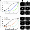
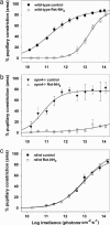
Mice lacking the visual cycle enzymes RPE65 or lecithin-retinol acyl transferase (Lrat) have pupillary light responses (PLR) that are less sensitive than those of mice with outer retinal degeneration (rd/rd or rdta). Inner retinal photoresponses are mediated by melanopsin-expressing, intrinsically photosensitive retinal ganglion cells (ipRGCs), suggesting that the melanopsin-dependent photocycle utilizes RPE65 and Lrat. To test this hypothesis, we generated rpe65(-/-); rdta and lrat(-/-); rd/rd mutant mice. Unexpectedly, both rpe65(-/-); rdta and lrat(-/-); rd/rd mice demonstrate paradoxically increased PLR photosensitivity compared with mice mutant in visual cycle enzymes alone. Acute pharmacologic inhibition of the visual cycle of melanopsin-deficient mice with all-trans-retinylamine results in a near-total loss of PLR sensitivity, whereas treatment of rd/rd mice has no effect, demonstrating that the inner retina does not require the visual cycle. Treatment of rpe65(-/-); rdta with 9-cis-retinal partially restores PLR sensitivity. Photic sensitivity in P8 rpe65(-/-) and lrat(-/-) ipRGCs is intact as measured by ex vivo multielectrode array recording. These results demonstrate that the melanopsin-dependent ipRGC photocycle is independent of the visual retinoid cycle.
Conflict of interest statement
Conflict of interest statement: No conflicts declared.
Figures
Fig. 1.
PLRs. Irradiance–response relations for PLRs of rdta, rpe65_−/−, rpe65_−/−;rdta (a) and rd/rd, lrat_−/−, lrat_−/−;rd/rd (b) mice stimulated by 470-nm narrow bandpass filtered light (n = 6–9, mean ± SEM). Pupillary constriction (c_–_n) was stimulated by 470 nm light (3.98 × 10¹⁴ photons·sec−1·cm−2). An rdta pupil before (c) and after (d) 30 s of light exposure is shown. An rpe65_−/− pupil before (e) and after (f) 30 s of light exposure is shown. An rpe65_−/−; rdta pupil before (g) and after (h) 30 s of light exposure is shown. An rd/rd pupil before (i) and after (j) 30 s of light exposure is shown. An lrat_−/− pupil before (k) and after (l) 30 s of light exposure is shown. An lrat_−/−; rd/rd pupil before (m) and after (n) 30 s of light exposure is shown.
Fig. 2.
_All_-_trans_-retinylamine inhibits outer but not inner retinal photosensitivity. Irradiance–response relations for PLRs of C57BL/6 (a), _opn4_−/− (b), and rd/rd (c) mice before (control) and 24 h after (Ret-NH2) oral gavage of 1 mg Ret-NH2. n = 3, 3, and 5 for C57BL/6, _opn4_−/−, and rd/rd, respectively; mean ± SEM.
Fig. 3.
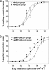
9-_cis_-retinal partially rescues PLR sensitivity of rpe65_−/−;rdta mice. Irradiance–response relations for PLRs of rd/rd (a) and rpe65_−/−;rdta (b) mice before and 24 h after oral gavage of 1-mg 9-cis_-retinal. n = 5 for rpe65_−/−;rdta, n = 6 for rd/rd; mean ± SEM; asterisks indicate significance P < 0.05 by Wilcoxon ranked sums test.
Fig. 4.
ipRGC light responses of _rpe65_−/− and _lrat_−/− mice. Light-induced action potentials recorded from individual ipRGCs from C57BL/6 (a), _rpe65_−/− (b), and _lrat_−/− (c) P8 mouse retinas via multielectrode array. Timing of light stimulus (60 s, 480 nm, 4.11 × 10¹³ photons·sec−1·cm−2) is indicated by step in horizontal line below recordings. Zero voltage is indicated by dashed line. Cumulative average time course of type I ipRGC activity from C57BL/6 (d), _rpe65_−/− (e), and _lrat_−/− (f) retinas in response to subsaturating and saturating light stimulation (60 s, 480 nm light pulse is indicated by step in horizontal line below histograms). n = 27, 45, and 43 cells for d, e, and f, respectively; mean, 1-s bins.
Fig. 5.
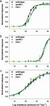
Irradiance–response relations of _rpe65_−/− and _lrat_−/− ipRGCs. Irradiance–response relationships for P8 type I (a), II (b), and III (c) ipRGCs from C57BL/6, _rpe65_−/−, and _lrat_−/− mice. For C57BL/6, _rpe65_−/−, and _lrat_−/−, respectively: n = 27, 45, and 43 cells in a, n = 6, 3, and 8 cells in b, and n = 4, 5, and 8 cells in c; mean ± SEM.
Comment in
- Chromophore regeneration: melanopsin does its own thing.
Lucas RJ. Lucas RJ. Proc Natl Acad Sci U S A. 2006 Jul 5;103(27):10153-10154. doi: 10.1073/pnas.0603955103. Epub 2006 Jun 26. Proc Natl Acad Sci U S A. 2006. PMID: 16801536 Free PMC article. No abstract available.
Similar articles
- Prolonged Melanopsin-based Photoresponses Depend in Part on RPE65 and Cellular Retinaldehyde-binding Protein (CRALBP).
Harrison KR, Reifler AN, Chervenak AP, Wong KY. Harrison KR, et al. Curr Eye Res. 2021 Apr;46(4):515-523. doi: 10.1080/02713683.2020.1815793. Epub 2020 Sep 7. Curr Eye Res. 2021. PMID: 32841098 Free PMC article. - Intrinsically photosensitive retinal ganglion cells detect light with a vitamin A-based photopigment, melanopsin.
Fu Y, Zhong H, Wang MH, Luo DG, Liao HW, Maeda H, Hattar S, Frishman LJ, Yau KW. Fu Y, et al. Proc Natl Acad Sci U S A. 2005 Jul 19;102(29):10339-44. doi: 10.1073/pnas.0501866102. Epub 2005 Jul 12. Proc Natl Acad Sci U S A. 2005. PMID: 16014418 Free PMC article. - Nonvisual light responses in the Rpe65 knockout mouse: rod loss restores sensitivity to the melanopsin system.
Doyle SE, Castrucci AM, McCall M, Provencio I, Menaker M. Doyle SE, et al. Proc Natl Acad Sci U S A. 2006 Jul 5;103(27):10432-10437. doi: 10.1073/pnas.0600934103. Epub 2006 Jun 20. Proc Natl Acad Sci U S A. 2006. PMID: 16788070 Free PMC article. - Function of the protein RPE65 in the visual cycle.
Wolf G. Wolf G. Nutr Rev. 2005 Mar;63(3):97-100. doi: 10.1111/j.1753-4887.2005.tb00127.x. Nutr Rev. 2005. PMID: 15825812 Review. - Key enzymes of the retinoid (visual) cycle in vertebrate retina.
Kiser PD, Golczak M, Maeda A, Palczewski K. Kiser PD, et al. Biochim Biophys Acta. 2012 Jan;1821(1):137-51. doi: 10.1016/j.bbalip.2011.03.005. Epub 2011 Apr 5. Biochim Biophys Acta. 2012. PMID: 21447403 Free PMC article. Review.
Cited by
- Melanopsin-dependent nonvisual responses: evidence for photopigment bistability in vivo.
Mure LS, Rieux C, Hattar S, Cooper HM. Mure LS, et al. J Biol Rhythms. 2007 Oct;22(5):411-24. doi: 10.1177/0748730407306043. J Biol Rhythms. 2007. PMID: 17876062 Free PMC article. - Light aversion in mice depends on nonimage-forming irradiance detection.
Thompson S, Recober A, Vogel TW, Kuburas A, Owens JA, Sheffield VC, Russo AF, Stone EM. Thompson S, et al. Behav Neurosci. 2010 Dec;124(6):821-7. doi: 10.1037/a0021568. Behav Neurosci. 2010. PMID: 21038932 Free PMC article. - Intrinsically photosensitive retinal ganglion cells: many subtypes, diverse functions.
Schmidt TM, Chen SK, Hattar S. Schmidt TM, et al. Trends Neurosci. 2011 Nov;34(11):572-80. doi: 10.1016/j.tins.2011.07.001. Epub 2011 Aug 3. Trends Neurosci. 2011. PMID: 21816493 Free PMC article. Review. - Melanopsin and mechanisms of non-visual ocular photoreception.
Sexton T, Buhr E, Van Gelder RN. Sexton T, et al. J Biol Chem. 2012 Jan 13;287(3):1649-56. doi: 10.1074/jbc.R111.301226. Epub 2011 Nov 10. J Biol Chem. 2012. PMID: 22074930 Free PMC article. Review. - Protein Phosphatase 2A and Clathrin-Mediated Endocytosis Facilitate Robust Melanopsin Light Responses and Resensitization.
Valdez-Lopez JC, Gebreegziabher M, Bailey RJ, Flores J, Awotunde O, Burnett T, Robinson PR. Valdez-Lopez JC, et al. Invest Ophthalmol Vis Sci. 2020 Oct 1;61(12):10. doi: 10.1167/iovs.61.12.10. Invest Ophthalmol Vis Sci. 2020. PMID: 33049058 Free PMC article.
References
- Ebihara S., Tsuji K. Physiol. Behav. 1980;24:523–527. - PubMed
- Freedman M. S., Lucas R. J., Soni B., von Schantz M., Munoz M., David-Gray Z., Foster R. Science. 1999;284:502–504. - PubMed
- Lucas R. J., Freedman M. S., Munoz M., Garcia-Fernandez J. M., Foster R. G. Science. 1999;284:505–507. - PubMed
- Lucas R. J., Douglas R. H., Foster R. G. Nat. Neurosci. 2001;4:621–626. - PubMed
Publication types
MeSH terms
Substances
Grants and funding
- R01 EY014988/EY/NEI NIH HHS/United States
- R01 EY009339/EY/NEI NIH HHS/United States
- P30 EY011373/EY/NEI NIH HHS/United States
- P30 EY002687/EY/NEI NIH HHS/United States
- EY14988/EY/NEI NIH HHS/United States
- EY09339/EY/NEI NIH HHS/United States
- P30 EY11373/EY/NEI NIH HHS/United States
- P30 EY 02687/EY/NEI NIH HHS/United States
LinkOut - more resources
Full Text Sources
Molecular Biology Databases
Miscellaneous


