Ca2+/calmodulin-dependent protein kinase II is a modulator of CARMA1-mediated NF-kappaB activation - PubMed (original) (raw)
. 2006 Jul;26(14):5497-508.
doi: 10.1128/MCB.02469-05.
Todd Green, Joseph Rapley, Heather Wachtel, Cosmas Giallourakis, Aimee Landry, Zhifang Cao, Naifang Lu, Ando Takafumi, Hidemi Goto, Mark J Daly, Ramnik J Xavier
Affiliations
- PMID: 16809782
- PMCID: PMC1592706
- DOI: 10.1128/MCB.02469-05
Ca2+/calmodulin-dependent protein kinase II is a modulator of CARMA1-mediated NF-kappaB activation
Kazuhiro Ishiguro et al. Mol Cell Biol. 2006 Jul.
Abstract
CARMA1 is a central regulator of NF-kappaB activation in lymphocytes. CARMA1 and Bcl10 functionally interact and control NF-kappaB signaling downstream of the T-cell receptor (TCR). Computational analysis of expression neighborhoods of CARMA1-Bcl10MALT 1 for enrichment in kinases identified calmodulin-dependent protein kinase II (CaMKII) as an important component of this pathway. Here we report that Ca(2+)/CaMKII is redistributed to the immune synapse following T-cell activation and that CaMKII is critical for NF-kappaB activation induced by TCR stimulation. Furthermore, CaMKII enhances CARMA1-induced NF-kappaB activation. Moreover, we have shown that CaMKII phosphorylates CARMA1 on Ser109 and that the phosphorylation facilitates the interaction between CARMA1 and Bcl10. These results provide a novel function for CaMKII in TCR signaling and CARMA1-induced NF-kappaB activation.
Figures
FIG. 1.
Schema for identification of CaMKII as a potential modulator of CARMA1-mediated NF-κB activation by use of kinase and pathway component expression signatures. (A) (i) Literature analysis of pathway components in TCR-mediated NF-κB activation identified CARMA1, Bcl10, and MALT1 as a functional module whose components exhibit tissue-restricted expression profiles. We hypothesized that an additional CARMA1 regulatory kinase (kinase X) would exhibit a coregulated expression signature with CARMA1, Bcl10, and MALT1. (ii) We derived a list of potential candidate kinases based on expression profiles. Three rank-ordered lists of kinase expression profiles with respect to CARMA1, Bcl10, and MALT1 gene sets were generated. For CARMA1, MALT1, and Bcl10, each of the probes representing the 460 kinases in the data set are ranked from most similar to each to least similar in expression profile across the GNF compendium based on pairwise Pearson's correlation coefficient. Subsequently, 13 kinases were identified among 460, including CaMKIIγ, which exhibited significant correlation with the target gene set of the CARMA1 signaling pathway. (iii) Motif Scan was employed to examine if CARMA1 had potential consensus CaMKII phosphorylation sites. (B) Expression level correlation between CaMKIIγ and the CARMA1-MALT1-Bcl10 gene set. A Z-score normalization (y axis) of microarray intensity values was performed and plotted for CaMKIIγ and the CARMA1-MALT1-Bcl10 target gene set in each of the tissue/cell types (x axis) in the GNF compendium data set (all 79 tissues/cells in tissue compendium are plotted, and a selected subset is labeled). Also indicated are the Pearson's pairwise correlation values (r) between CaMKIIγ and the individual gene set members as a quantification of expression similarity. DRG, dorsal root ganglion.
FIG. 2.
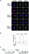
CaMKII redistributes to the immune synapse in an activation-dependent manner. (A) Raji cells were loaded with a fluorescent cell tracker chloromethylbenzoyl derivative of aminocoumarin (blue) and pulsed with SEE for 20 min. See Materials and Methods for details. Alexa Fluor 488 is shown in green, and phalloidin conjugated with Alexa Fluor 568 is shown in red. Representative results from one of three experiments are shown. (B) Quantification of CaMKII redistribution to the contact area in Jurkat cells. Conjugates displaying CaMKII redistribution to the submembranous area were counted under the fluorescence microscope at a magnification of ×100. Results are representative of three independent experiments. n, total number of conjugates examined for each experimental condition. (C) After incubation with KN93 or DMSO, Jurkat cells were stimulated with anti-CD3 antibody or TNF-α and then the nuclear extract was obtained as described in Materials and Methods. The nuclear extract (10 μg of total proteins) was subjected to SDS-PAGE, transferred to a polyvinylidene difluoride membrane, and blotted with anti-p65 antibody or antiretinoblastoma (Rb) antibody.
FIG. 3.
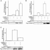
siRNAs of CaMKII interfere with NF-κB activation induced by TCR stimulation. (A and B) Jurkat cells were electroporated with 10 μg of siRNA construct (vector, D1424, D1488, G371, or G1660), NF-κB reporter, and pRL-TK construct. Three days later, the cells were incubated with anti-CD3 antibody (1 μg/ml) or TNF-α (1 ng/ml) for 6 h, and then luciferase activities were determined as described in the text. (C) Jurkat cells were electroporated with 10 μg of siRNA construct (vector, D1488, or G371) or 5 μg of D1488 and 5 μg of G371, NF-κB reporter, and pRL-TK. Three days later, the cells were incubated with anti-CD3 antibody (1 μg/ml) or anti-CD28 antibody (0.2 μg/ml) for 6 h, and then luciferase activity was determined. (A to C, lower panels) Equal amounts of protein from lysates used for luciferase assays were blotted for CaMKII and actin. (D) Jurkat cells were electroporated with siRNA construct (vector, D1488, or G371), SRE reporter, and pRL-TK construct. Three days later, the cells were incubated with anti-CD3 antibody (1 μg/ml) for 6 h, and then SRE reporter activity was determined. (E) Jurkat cells were electroporated with siRNA construct (vector or D1488), NFAT reporter, and pRL-TK construct. Three days later, the cells were incubated with phorbol myristate acetate (PMA) (50 ng/ml) plus ionomycin (1 μM) for 6 h, and then NFAT reporter activity was determined. For results in all panels, four independent experiments were performed. Bars represent standard errors.
FIG. 4.
Synergistic NF-κB activation of CARMA1 with CaMKII upstream of IKK. (A) Jurkat cells were electroporated with NF-κB reporter, pRL-TK construct, 2 μg of CaMKII1-290, and 2 μg of Flag-CARMA1 construct. One day later, luciferase activities were determined. (B) Jurkat cells were electroporated with NF-κB reporter, pRL-TK construct, 2 μg of the IKKβ dominant-negative (d/n) mutant (with S177A and S181A mutations), and CaMKII1-290 or Flag-CARMA1. One day later, luciferase activities were determined. (C) Jurkat cells were transfected as described for panel B except that CaMKII1-290 and CARMA1 were cotransfected. Western blot analysis using cell lysates was performed with rabbit anti-CaMKII antibody or monoclonal anti-Flag antibody (lower panels). Three independent experiments were performed in duplicate. Bars represent standard errors.
FIG. 5.
Synergistic NF-κB activation of CaMKII with CARMA1 but not with CARMA3. (A) Jurkat cells were electroporated with NF-κB-dependent reporter, pRL-TK construct, 2 μg of CaMKII1-290, and 50 ng of Myc-CARMA1 or Myc-CARMA3. The total amount of DNA was equalized with empty vector. One day later, luciferase activities were determined as described in the text. (B) The Motif Scan program Scansite predicted amino acid residues phosphorylated with CaMKII in the N-terminal regions of CARMA1 and CARMA3 (boldface type). The residues predicted specifically in CARMA1 are shaded. Asterisks indicate residues conserved between CARMA1 and CARMA3. The arrow indicates the P-3 position of CARMA1 Ser109 or CARMA3 Ser121, where CaMKII prefers Arg or Lys for substrates. (C) Jurkat cells were electroporated with NF-κB reporter, pRL-TK construct, 2 μg CaMKII1-290, and 50 ng of Myc-CARMA1 or its mutant with the S109A mutation. The total amount of DNA was equalized with empty vectors. One day later, luciferase activities were determined. (D and E) Results are from an experiment similar to that described for panel C, except with Myc-CARMA1 or its mutant with the (D) T110A or T160A mutation or the (E) S243A or S432A mutation. The total amount of DNA was equalized with empty vectors. One day later, luciferase activities were determined. (F) Jurkat cells were electroporated with NF-κB reporter, pRL-TK construct, and 100 ng of Myc-CARMA1 or its mutant with the S109D mutation. One day later, luciferase activities were determined. (G) Results are from an experiment similar to that described for panel F, except that 18 h later, the cells were incubated with anti-CD3 antibody (1 μg/ml) for a further 6 h, and then luciferase activities were determined. (Lower panels) Cell lysates were blotted with anti-Myc antibody or rabbit anti-CaMKII antibody. Four independent experiments were performed. Bars represent standard errors. WT, wild type.
FIG. 6.
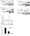
Ser109 phosphorylation of CARMA1 with CaMKII. (A) Purified CARMA1 and CARMA3 were phosphorylated in vitro by wild-type CaMKII. (B) Purified CARMA1 and the CARMA1 S109A mutant were phosphorylated in vitro in the presence and absence of a CaMKII inhibitor as indicated. The amount of CARMA1 was determined by Coomassie staining, and then the dried gels were exposed to a phosphorimager. The remainder of each sample was analyzed by Western blotting using an antibody against CaMKII (lower panel). (C) 293T cells were transfected with Myc-CARMA1 or its S109A mutant and CaMKII1-290. The cells were lysed 18 h later. Monoclonal anti-Myc antibody was used for blotting. (D) Jurkat cells were electroporated with siRNA construct (vector or D1488) and Myc-CARMA1 construct. Three days later, the cells were incubated with anti-CD3 antibody (5 μg/ml) and anti-CD28 antibody (1 μg/ml) for 10 min and then lysed. (E) Jurkat cells were electroporated with Myc-CARMA1 or its S109A mutant. One day later, the cells were incubated with anti-CD3 antibody and anti-CD28 antibody for 10 min and then lysed. Phosphoproteins were isolated via IMAC as described in the text. Lower panels show total lysates immunoblotted with anti-Myc antibody. The experiments were performed three times, and representative results are presented. WT, wild type.
FIG. 7.
Regulation of binding between CARMA1 and Bcl10 via Ser109 phosphorylation. (A and B) 293T cells were transfected with 2 μg of Flag-Bcl10 construct and 0.5 μg of Myc-CARMA1 or its S109A mutant (A) or its S109D mutant (B). The total amount of DNA was equalized with empty vectors. The cells were lysed 18 h later. Monoclonal anti-Flag antibody was used for immunoprecipitation (IP). Monoclonal anti-Myc antibody or anti-Flag antibody was used for blotting. Results are representative of three independent experiments. (C and D) Jurkat cells (C) or CARMA1 mutant Jurkat cells (D) were electroporated with NF-κB reporter, pRL-TK construct, and Myc-CARMA1 or its S109A mutant. Eighteen hours later, the cells were incubated with anti-CD3 antibody (1 μg/ml) for a further 6 h, and NF-κB luciferase activities were determined. Four independent experiments were performed. Bars represent standard errors. (E) 293T cells were transfected with 1 μg of Flag-Bcl10, 0.5 μg of Myc-CARMA1, and 2 μg of CaMKII1-290. The total amount of DNA was equalized with empty vectors. The cells were lysed 18 h later. Monoclonal anti-Flag antibody was used for IP. Monoclonal anti-Myc antibody or anti-Flag antibody was used for blotting. (F) Jurkat cells were electroporated with 10 μg of siRNA D1488 or G371 constructs or vector and 5 μg of Flag-CARMA1 construct. Three days later, the cells were incubated with anti-CD3 antibody (5 μg/ml) for 10 min, lysed, and immunoprecipitated with anti-Flag antibody. Total lysates were immunoblotted with anti-Flag or anti-Bcl10 antibody. Results are representative of three independent experiments. WT, wild type; Unstim., unstimulated; Stim., stimulated.
Similar articles
- Protein kinase C-δ negatively regulates T cell receptor-induced NF-κB activation by inhibiting the assembly of CARMA1 signalosome.
Liu Y, Song R, Gao Y, Li Y, Wang S, Liu HY, Wang Y, Hu YH, Shu HB. Liu Y, et al. J Biol Chem. 2012 Jun 8;287(24):20081-7. doi: 10.1074/jbc.M111.335463. Epub 2012 Apr 23. J Biol Chem. 2012. PMID: 22528498 Free PMC article. - Bcl10 is phosphorylated on Ser138 by Ca2+/calmodulin-dependent protein kinase II.
Ishiguro K, Ando T, Goto H, Xavier R. Ishiguro K, et al. Mol Immunol. 2007 Mar;44(8):2095-100. doi: 10.1016/j.molimm.2006.09.012. Epub 2006 Oct 18. Mol Immunol. 2007. PMID: 17052756 - The Ca2+-dependent phosphatase calcineurin controls the formation of the Carma1-Bcl10-Malt1 complex during T cell receptor-induced NF-kappaB activation.
Palkowitsch L, Marienfeld U, Brunner C, Eitelhuber A, Krappmann D, Marienfeld RB. Palkowitsch L, et al. J Biol Chem. 2011 Mar 4;286(9):7522-34. doi: 10.1074/jbc.M110.155895. Epub 2011 Jan 3. J Biol Chem. 2011. PMID: 21199863 Free PMC article. - Role of the CARMA1/BCL10/MALT1 complex in lymphoid malignancies.
Juilland M, Thome M. Juilland M, et al. Curr Opin Hematol. 2016 Jul;23(4):402-9. doi: 10.1097/MOH.0000000000000257. Curr Opin Hematol. 2016. PMID: 27135977 Free PMC article. Review. - Critical protein-protein interactions within the CARMA1-BCL10-MALT1 complex: Take-home points for the cell biologist.
Cheng J, Maurer LM, Kang H, Lucas PC, McAllister-Lucas LM. Cheng J, et al. Cell Immunol. 2020 Sep;355:104158. doi: 10.1016/j.cellimm.2020.104158. Epub 2020 Jul 7. Cell Immunol. 2020. PMID: 32721634 Review.
Cited by
- Protein kinase C-δ negatively regulates T cell receptor-induced NF-κB activation by inhibiting the assembly of CARMA1 signalosome.
Liu Y, Song R, Gao Y, Li Y, Wang S, Liu HY, Wang Y, Hu YH, Shu HB. Liu Y, et al. J Biol Chem. 2012 Jun 8;287(24):20081-7. doi: 10.1074/jbc.M111.335463. Epub 2012 Apr 23. J Biol Chem. 2012. PMID: 22528498 Free PMC article. - Nilotinib Improves Bioenergetic Profiling in Brain Astroglia in the 3xTg Mouse Model of Alzheimer's Disease.
Adlimoghaddam A, Odero GG, Glazner G, Turner RS, Albensi BC. Adlimoghaddam A, et al. Aging Dis. 2021 Apr 1;12(2):441-465. doi: 10.14336/AD.2020.0910. eCollection 2021 Apr. Aging Dis. 2021. PMID: 33815876 Free PMC article. - Structural variants of IFNα preferentially promote antiviral functions.
Vázquez N, Schmeisser H, Dolan MA, Bekisz J, Zoon KC, Wahl SM. Vázquez N, et al. Blood. 2011 Sep 1;118(9):2567-77. doi: 10.1182/blood-2010-12-325027. Epub 2011 Jul 14. Blood. 2011. PMID: 21757613 Free PMC article. - Cellular and molecular imaging of CAR-T cell-based immunotherapy.
Liu L, Yoon CW, Yuan Z, Guo T, Qu Y, He P, Yu X, Zhu Z, Limsakul P, Wang Y. Liu L, et al. Adv Drug Deliv Rev. 2023 Dec;203:115135. doi: 10.1016/j.addr.2023.115135. Epub 2023 Nov 4. Adv Drug Deliv Rev. 2023. PMID: 37931847 Free PMC article. Review. - WNT11 Promotes immune evasion and resistance to Anti-PD-1 therapy in liver metastasis.
Jiang W, Guan B, Sun H, Mi Y, Cai S, Wan R, Li X, Lian P, Li D, Zhao S. Jiang W, et al. Nat Commun. 2025 Feb 7;16(1):1429. doi: 10.1038/s41467-025-56714-z. Nat Commun. 2025. PMID: 39920102 Free PMC article.
References
- Andersson, L., and J. Porath. 1986. Isolation of phosphoproteins by immobilized metal (Fe3+) affinity chromatography. Anal. Biochem. 154:250-254. - PubMed
- Bauch, A., K. S. Campbell, and M. Reth. 1998. Interaction of the CD5 cytoplasmic domain with the Ca2+/calmodulin-dependent kinase IIdelta. Eur. J. Immunol. 28:2167-2177. - PubMed
- Bertin, J., L. Wang, Y. Guo, M. D. Jacobson, J. L. Poyet, S. M. Srinivasula, S. Merriam, P. S. DiStefano, and E. S. Alnemri. 2001. CARD11 and CARD14 are novel caspase recruitment domain (CARD)/membrane-associated guanylate kinase (MAGUK) family members that interact with BCL10 and activate NF-kappa B. J. Biol. Chem. 276:11877-11882. - PubMed
- Bui, J. D., S. Calbo, K. Hayden-Martinez, L. P. Kane, P. Gardner, and S. M. Hedrick. 2000. A role for CaMKII in T cell memory. Cell 100:457-467. - PubMed
Publication types
MeSH terms
Substances
LinkOut - more resources
Full Text Sources
Molecular Biology Databases
Research Materials
Miscellaneous


