Chlorella viruses - PubMed (original) (raw)
Review
Chlorella viruses
Takashi Yamada et al. Adv Virus Res. 2006.
Abstract
Chlorella viruses or chloroviruses are large, icosahedral, plaque-forming, double-stranded-DNA-containing viruses that replicate in certain strains of the unicellular green alga Chlorella. DNA sequence analysis of the 330-kbp genome of Paramecium bursaria chlorella virus 1 (PBCV-1), the prototype of this virus family (Phycodnaviridae), predict approximately 366 protein-encoding genes and 11 tRNA genes. The predicted gene products of approximately 50% of these genes resemble proteins of known function, including many that are completely unexpected for a virus. In addition, the chlorella viruses have several features and encode many gene products that distinguish them from most viruses. These products include: (1) multiple DNA methyltransferases and DNA site-specific endonucleases, (2) the enzymes required to glycosylate their proteins and synthesize polysaccharides such as hyaluronan and chitin, (3) a virus-encoded K(+) channel (called Kcv) located in the internal membrane of the virions, (4) a SET domain containing protein (referred to as vSET) that dimethylates Lys27 in histone 3, and (5) PBCV-1 has three types of introns; a self-splicing intron, a spliceosomal processed intron, and a small tRNA intron. Accumulating evidence indicates that the chlorella viruses have a very long evolutionary history. This review mainly deals with research on the virion structure, genome rearrangements, gene expression, cell wall degradation, polysaccharide synthesis, and evolution of PBCV-1 as well as other related viruses.
Figures
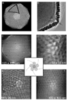
Fig. 1.
Three‐dimensional image reconstruction of chlorella virus PBCV‐1 from cryo‐electron micrographs and atomic force micrographs. (A) The virion capsid consists of 12 pentasymmetrons and 20 trisymmetrons. Five trisymmetrons are highlighted in the reconstruction (blue) and a single pentasymmetron is colored yellow. A pentavalent capsomer (white) lies at the center of each pentasymmetron. Each pentasymmeton consists of one pentamer plus 30 trimers. Eleven capsomers form the edge of each trisymmetron (black dots) and therefore each trisymmetron has 66 trimers (Yan et al., 2000). (B) Dense material (blue arrow) (cell wall digesting enzyme(s)?) is present at each vertex (red arrow) between the vertex and the membrane. (C–F) Atomic force microscopy images of the surfaces of PBCV‐1 virions showing the pentameric arrangements of proteins around the fivefold vertices, with a unique protein on the vertex (Kuznetsov et al., 2005). (See Color Insert.)
Fig. 2.
Comparison of the nucleotide sequences at the PBCV‐1 and CVK2 terminal hairpin ends. The most distal portions of the terminal inverted repeats (shown by arrows) are relatively similar between PBCV‐1 and CVK2 DNAs (unidentical bases are shown in red), whereas the sequences of the covalently closed hairpin loops (35 nucleotides for PBCV‐1 and 43 nucleotides for CVK2) are completely different. The hairpin loops exist in one of two forms (shown in blue and green); the two forms are complementary when the sequences are inverted (flip‐flop) (Zhang et al., 1994). (See Color Insert.)
Fig. 3.
Comparison of the location of 22 genes in four chlorovirus genomes: between PBCV‐1 and CVK2 (A), and PBCV‐1 and MT325 (Pbi‐virus) (B). Corresponding gene positions are connected by lines. PBCV‐1 genes used as landmarks are (1) A3R, (2) A35L, (3) A50L, (4) A100R, (5) A107L, (6) A125L, (7) A181R, (8) A185R, (9) A245R, (10) A292L, (11) tRNA genes, (12) A357L, (13) A413L, (14) A430L, (15) A448L, (16) A464R, (17) A533R, (18) A575L, (19) A577L, (20) A604L, (21) A646L, and (22) A666L. Some PBCV‐1 genes are duplicated or missing in the other virus genomes. Although a few minor rearrangements occur (ex. a large insertion between (2) and (3) in CVK2), in general, colinearity exists among the NC64A viruses (A). In contrast, there is almost no colinearity between the genomes of PBCV‐1 and MT325 (B). Unpublished sequence data for NY‐2A and MT325 (J. L. Van Etten and M. V. Graves ) are used in the comparison.
Fig. 4.
A diagram illustrating the genetic differences between HA and/or chitin synthesizing chloroviruses (Ali et al., 2005). Line 1 shows the location of three PBCV‐1 genes involved in the synthesis of HA. Viruses CVIK1 and CVHA1 that synthesize both HA and chitin (Line 2) have a chitin synthesizing gene inserted in the position of the PBCV‐1 A330R ORF. Viruses that only synthesize chitin have two chs genes. These viruses may still have a ugdh gene (viruses CVK2, NY‐2A, and AR158 in Line 3) or lose it (virus CVSA1 in Line 4). It is equally likely that the events depicted could occur in reverse order, that is, from Line 4 to Line 1. (See Color Insert.)
Similar articles
- Unusual life style of giant chlorella viruses.
Van Etten JL. Van Etten JL. Annu Rev Genet. 2003;37:153-95. doi: 10.1146/annurev.genet.37.110801.143915. Annu Rev Genet. 2003. PMID: 14616059 Review. - Giant viruses infecting algae.
Van Etten JL, Meints RH. Van Etten JL, et al. Annu Rev Microbiol. 1999;53:447-94. doi: 10.1146/annurev.micro.53.1.447. Annu Rev Microbiol. 1999. PMID: 10547698 Review. - Initial Events Associated with Virus PBCV-1 Infection of Chlorella NC64A.
Thiel G, Moroni A, Dunigan D, Van Etten JL. Thiel G, et al. Prog Bot. 2010 Jan 1;71(3):169-183. doi: 10.1007/978-3-642-02167-1_7. Prog Bot. 2010. PMID: 21152366 Free PMC article. - Identification of a Chlorovirus PBCV-1 Protein Involved in Degrading the Host Cell Wall during Virus Infection.
Agarkova IV, Lane LC, Dunigan DD, Quispe CF, Duncan GA, Milrot E, Minsky A, Esmael A, Ghosh JS, Van Etten JL. Agarkova IV, et al. Viruses. 2021 Apr 28;13(5):782. doi: 10.3390/v13050782. Viruses. 2021. PMID: 33924931 Free PMC article. - Virion-associated restriction endonucleases of chloroviruses.
Agarkova IV, Dunigan DD, Van Etten JL. Agarkova IV, et al. J Virol. 2006 Aug;80(16):8114-23. doi: 10.1128/JVI.00486-06. J Virol. 2006. PMID: 16873267 Free PMC article.
Cited by
- Chlorella virus ATCV-1 encodes a functional potassium channel of 82 amino acids.
Gazzarrini S, Kang M, Abenavoli A, Romani G, Olivari C, Gaslini D, Ferrara G, van Etten JL, Kreim M, Kast SM, Thiel G, Moroni A. Gazzarrini S, et al. Biochem J. 2009 May 13;420(2):295-303. doi: 10.1042/BJ20090095. Biochem J. 2009. PMID: 19267691 Free PMC article. - Mechanism of function of viral channel proteins and implications for drug development.
Fischer WB, Wang YT, Schindler C, Chen CP. Fischer WB, et al. Int Rev Cell Mol Biol. 2012;294:259-321. doi: 10.1016/B978-0-12-394305-7.00006-9. Int Rev Cell Mol Biol. 2012. PMID: 22364876 Free PMC article. Review. - Identification of GIG1, a GlcNAc-induced gene in Candida albicans needed for normal sensitivity to the chitin synthase inhibitor nikkomycin Z.
Gunasekera A, Alvarez FJ, Douglas LM, Wang HX, Rosebrock AP, Konopka JB. Gunasekera A, et al. Eukaryot Cell. 2010 Oct;9(10):1476-83. doi: 10.1128/EC.00178-10. Epub 2010 Jul 30. Eukaryot Cell. 2010. PMID: 20675577 Free PMC article. - Remarkable sequence similarity between the dinoflagellate-infecting marine girus and the terrestrial pathogen African swine fever virus.
Ogata H, Toyoda K, Tomaru Y, Nakayama N, Shirai Y, Claverie JM, Nagasaki K. Ogata H, et al. Virol J. 2009 Oct 27;6:178. doi: 10.1186/1743-422X-6-178. Virol J. 2009. PMID: 19860921 Free PMC article. - Evolutionary entanglement of mobile genetic elements and host defence systems: guns for hire.
Koonin EV, Makarova KS, Wolf YI, Krupovic M. Koonin EV, et al. Nat Rev Genet. 2020 Feb;21(2):119-131. doi: 10.1038/s41576-019-0172-9. Epub 2019 Oct 14. Nat Rev Genet. 2020. PMID: 31611667 Review.
References
- Ali MMA, Kawasaki T, Yamada T. Genetic rearrangements on the chlorovirus genome that switch between hyaluronan synthesis and chitin synthesis. Virology. 2005;342:102–110. - PubMed
- Becker B, Lesenmann DE, Reisser W. Ultrastructural studies on a chlorella virus from Germany. Arch. Virol. 1993;130:145–155. - PubMed
- Belfield GP, Tuite MF. Translation elongation factor 3: A fungus‐specific translation factor? Mol. Microbiol. 1993;9:411–418. - PubMed
- Bell PJ. Viral eukaryogenesis: Was the ancestor of the nucleus a complex DNA virus. J. Mol. Evol. 2001;53:251–256. - PubMed
- Benson SD, Bamford JKH, Bamford DH, Burnett RM. Does common architechture reveal a virus lineage spanning all three domains of life? Mol. Cell. 2004;16:673–685. - PubMed
Publication types
MeSH terms
Substances
LinkOut - more resources
Full Text Sources
Other Literature Sources


