Requirements for the assembly and release of Newcastle disease virus-like particles - PubMed (original) (raw)
Requirements for the assembly and release of Newcastle disease virus-like particles
Homer D Pantua et al. J Virol. 2006 Nov.
Erratum in
- J Virol. 2007 Feb;81(3):1537
Abstract
Paramyxoviruses, such as Newcastle disease virus (NDV), assemble in and bud from plasma membranes of infected cells. To explore the role of each of the NDV structural proteins in virion assembly and release, virus-like particles (VLPs) released from avian cells expressing all possible combinations of the nucleoprotein (NP), membrane or matrix protein (M), an uncleaved fusion protein (F-K115Q), and hemagglutinin-neuraminidase (HN) protein were characterized for densities, protein content, and efficiencies of release. Coexpression of all four proteins resulted in the release of VLPs with densities and efficiencies of release (1.18 to 1.16 g/cm(3) and 83.8% +/- 1.1%, respectively) similar to those of authentic virions. Expression of M protein alone, but not NP, F-K115Q, or HN protein individually, resulted in efficient VLP release, and expression of all different combinations of proteins in the absence of M protein did not result in particle release. Expression of any combination of proteins that included M protein yielded VLPs, although with different densities and efficiencies of release. To address the roles of NP, F, and HN proteins in VLP assembly, the interactions of proteins in VLPs formed with different combinations of viral proteins were characterized by coimmunoprecipitation. The colocalization of M protein with cell surface F and HN proteins in cells expressing all combinations of viral proteins was characterized. Taken together, the results show that M protein is necessary and sufficient for NDV budding. Furthermore, they suggest that M-HN and M-NP interactions are responsible for incorporation of HN and NP proteins into VLPs and that F protein is incorporated indirectly due to interactions with NP and HN protein.
Figures
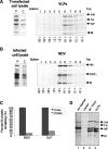
FIG. 1.
Coexpression of NP, F, HN, and M proteins resulted in VLP formation and release. Panel A: avian cells, cotransfected with pCAGGS-NP, -M, -F-K155Q, and -HN, were radioactively labeled with [35S]methionine and [35S]cysteine for 4 h (P) and then chased in nonradioactive medium for 8 h (C). Panel B: avian cells, infected with NDV, strain AV, at a multiplicity of infection of 5 for 5 h, were pulse-labeled for 30 min and chased in nonradioactive medium for 8 h. Radioactively labeled proteins in the extracts were immunoprecipitated with a cocktail of antibodies specific for all viral proteins, and the precipitated labeled proteins are shown on the left side of each panel. Particles in cell supernatants were purified as described in Materials and Methods. After flotation into sucrose gradients (right side of each panel), each gradient fraction was mixed with TNE containing 1% Triton X-100 and immunoprecipitated with the antibody cocktail. The density of each fraction (g/cm3) is shown at the bottom. Panel C shows the quantitation of efficiency of virion and VLP release as determined by the amount of M protein in the pulse and chase extracts and is the result of averaging results from three separate experiments. The standard deviation is shown. Panel D shows nonimmunoprecipitated radiolabeled particles released from NDV, strain B1, virus-infected avian cells and cells expressing NP, M, F-K115Q, and HN proteins. Particles were isolated by concentrating onto the 20% and 65% sucrose interface and then floated into a step gradient. Particles in the gradient fractions were then pelleted and resuspended in TNE buffer. HN, hemagglutinin-neuraminidase protein; F0, uncleaved fusion protein; NP, nucleocapsid protein; F1, cleaved fusion protein; M, membrane or matrix protein.
FIG. 2.
M protein is sufficient for particle release. Avian cells were transfected with pCAGGS-NP, -M, -F-K115Q, and -HN individually. Panel A shows radioactively labeled proteins in the extracts at time of pulse (left) or chase (right). Particles in the supernatants of avian cells, expressing NP, M, F, and HN individually, were concentrated and floated into sucrose gradients as described in the legend to Fig. 1. Panel B shows the distribution in the gradients of radioactively labeled proteins derived from each supernatant. Densities of gradient fractions are shown at bottom (g/cm3). Quantification of the amounts of proteins in particles present in all fractions is shown in panel C. The results of three separate experiments were averaged, and the standard deviation is shown.
FIG. 3.
M proteins are encased in membranous particles. Panel A: Avian cells were transfected with pCAGGS-M, and radioactively labeled VLPs were isolated and purified as described in Materials and Methods. Extract (upper panel) and VLPs, which were concentrated onto a 20% to 65% sucrose interface (middle panel), were treated with different concentrations (0.25, 0.5, 1, 5, 10, or 20 μg/ml; lanes 2 to 7, respectively) of proteinase K for 30 min on ice. In parallel, particles were incubated in 1% Triton X-100 prior to proteinase K treatment (bottom panel). After incubation with protease, reactions were stopped by adding 0.1 mM phenylmethylsulfonyl fluoride. M proteins were immunoprecipitated as described in Materials and Methods. Panel B: Electron microscopy of particles released from NDV-infected cells (B1), M protein-expressing cells (M), or cells expressing NP, M, F-K115Q, and HN proteins (VLP). Particles were purified as described in Materials and Methods. Middle and left panels show two representative particles from each preparation. Right panels show enlargements of the edges of typical particles.
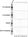
FIG. 4.
M protein is required for particle release. Avian cells were transfected with all possible combinations of cDNAs in the pCAGGS vector encoding NP, F, and HN proteins in the absence of M cDNA (F-K115Q+HN, F-K115Q+NP, HN+NP, NP+F-K115Q+HN). Particles in cell supernatants were purified as described in the legend to Fig. 1. Panels show proteins present in each gradient fraction. Radioactively labeled infected-cell extract was used as marker. Densities of fractions are shown at the bottom (g/cm3). The control for this experiment is shown in Fig. 1, panel A.
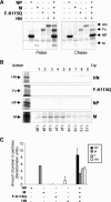
FIG. 5.
Effect of expressing NP, F, or HN protein with M protein on particle release. Avian cells, transfected with cDNAs encoding F-K115Q plus M, HN plus M, or NP plus M structural protein genes, as well as all four cDNAs, were labeled by using a pulse-chase protocol as described in the legend to Fig. 1. Particles present in the supernatants were concentrated and then floated into sucrose gradients as described in Materials and Methods. Panel A shows labeled proteins in cell extracts at end of pulse (top) or chase (bottom). Panel B shows the proteins present in each gradient fraction after immunoprecipitation of each fraction with an antibody cocktail. Densities (g/cm3) of the fractions are shown at the bottom. Panel C shows the quantification of each protein in particles in all fractions. Autoradiographs of gradient fractions from F-K115Q plus NP-, F-K115Q plus HN-, or HN plus NP-expressing cells did not contain any protein and are shown with gels in Fig. 4. Results are the averages for three experiments, and the standard deviation is shown.
FIG. 6.
Effect of expressing all combinations of three viral proteins on particle release. Avian cells, transfected with all possible combinations of three NDV structural protein genes including M cDNA, were labeled in a pulse-chase protocol, and particles in the supernatant were concentrated and floated into a sucrose gradient as for Fig. 1. The proteins in the cell extracts were immunoprecipitated with the antibody cocktail. Panel A shows labeled proteins in cell extracts at end of pulse (top) or chase (bottom). Panel B shows the proteins present in each gradient fraction after immunoprecipitation of each fraction with an antibody cocktail. Densities (g/cm3) of the fractions are shown at the bottom. Panel C shows quantification of the amounts of each protein in particles in all fractions. Autoradiograph of particles released from NP-, F-, and HN- protein-expressing cells is shown in Fig. 4.
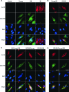
FIG. 7.
Colocalization of M protein with F and HN proteins. The cell surface localization of NDV F and HN proteins and the cellular localization of M proteins were analyzed by immunofluorescence microscopy. Avian cells were transfected either individually (A) or with F-K115Q plus M or HN plus M (B), with NP plus M plus F-K115Q, with NP plus M plus HN, with M plus F-K115Q plus HN (C), or with all 4 cDNAs (D). Nuclei were stained with 4′,6′-diamidino-2-phenylindole (blue) 40 h posttransfection. Intact transfected cells were stained with rabbit anti-F protein antibodies or anti-HN protein antibodies as indicated in the panels. Cells were permeabilized with 0.05% Triton X-100 prior to incubation with anti-M protein antibody. Secondary antibodies were anti-rabbit Alexa 488 conjugate (green) and anti-mouse Alexa 568 conjugate (red). Images were taken at magnification ×60 and were merged using Adobe Photoshop. Results shown are representative of all cells in the slide.
FIG. 8.
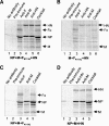
Coimmunoprecipitation of viral proteins in particles. Radioactively labeled particles released from cells expressing NP plus M plus F-K115Q plus HN (A), M plus F-K115Q plus HN (B), NP plus M plus F-K115Q (C), or NP plus M plus HN (D) were purified into a 20%- to 65%-sucrose interface. Particles were solubilized in TNE buffer containing 1% Triton X-100 at 4°C for 15 min. Solubilized particles were then incubated with excess amounts of cocktail of anti-F-protein antibodies (anti-HR1, anti-HR2, anti-Ftail, anti-F2-96, and monoclonal anti-F [G5]), anti-HN protein antibodies (mix of monoclonal antibodies), anti-M protein monoclonal antibody, or a cocktail of NDV-specific antibodies overnight at 4°C. No antibody as well as preimmune sera were used as negative controls. Immune complexes were precipitated with prewashed Pansorbin A for at least 2 h at 4°C with constant mixing. Samples were washed three times in cold TNE with 0.5% Triton X-100. All steps of coimmunoprecipitation were accomplished at 4°C. Proteins were resolved by sodium dodecyl sulfate-polyacrylamide gel electrophoresis as described in Materials and Methods. Results show one of three independent experiments, all with identical results.
FIG. 9.
Protein-protein interactions in particles. Inset shows viral protein-protein interactions detected by coimmunoprecipitation of proteins in particles. Also shown are interactions proposed to result in assembly of particles formed by coexpression of all combinations of NP, F, and HN proteins with M protein.
References
- Ali, A., and D. P. Nayak. 2000. Assembly of Sendai virus: M protein interacts with F and HN proteins and with the cytoplasmic tail and transmembrane domain of F protein. Virology 276:289-303. - PubMed
- Bieniasz, P. D. 2006. Late budding domains and host proteins in enveloped virus release. Virology 344:55-63. - PubMed
Publication types
MeSH terms
Substances
LinkOut - more resources
Full Text Sources
Other Literature Sources
Miscellaneous
