Interdigital webbing retention in bat wings illustrates genetic changes underlying amniote limb diversification - PubMed (original) (raw)
Comparative Study
. 2006 Oct 10;103(41):15103-7.
doi: 10.1073/pnas.0604934103. Epub 2006 Oct 2.
Affiliations
- PMID: 17015842
- PMCID: PMC1622783
- DOI: 10.1073/pnas.0604934103
Comparative Study
Interdigital webbing retention in bat wings illustrates genetic changes underlying amniote limb diversification
Scott D Weatherbee et al. Proc Natl Acad Sci U S A. 2006.
Abstract
Developmentally regulated programmed cell death sculpts the limbs and other embryonic organs in vertebrates. One intriguing example of species-specific differences in apoptotic extent is observed in the tissue between the digits. In chicks and mice, bone morphogenetic proteins (Bmps) trigger apoptosis of the interdigital mesenchyme, leading to freed digits, whereas in ducks, Bmp antagonists inhibit the apoptotic program, resulting in webbed feet. Here, we show that the phyllostomid bat Carollia perspicillata utilizes a distinct mechanism for maintaining interdigit tissue. We find that bat forelimb and hindlimb interdigital tissues express Bmp signaling components but that only bat hindlimbs undergo interdigital apoptosis. Strikingly, the retention of interdigital webbing in the bat forelimb is correlated with a unique pattern of Fgf8 expression in addition to the Bmp inhibitor Gremlin. By using a functional assay, we show that maintenance of interdigit tissue in the bat wing depends on the combined effects of high levels of Fgf signaling and inhibition of Bmp signaling. Our data also indicate that although there is not a conserved mechanism for maintaining interdigit tissue across amniotes, the expression in the bat forelimb interdigits of Gremlin and Fgf8 suggests that these key molecular changes contributed to the evolution of the bat wing.
Conflict of interest statement
The authors declare no conflict of interest.
Figures
Fig. 1.
Differential forelimb morphology in mice and bats. (A) An adult mouse, Mus musculus. (B) An adult bat, Carollia perspicillata. Digits are numbered from anterior (I) to posterior (V). Bat digits are elongated compared with mouse digits (Inset) and maintain webbing between the posterior digits.
Fig. 2.
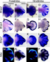
Bmp pathway gene expression in developing bat limbs. Analysis of Bmp signaling components in Carollia forelimbs and hindlimbs is shown. (A–D) Bmp2 expression in forelimbs (A and B) and hindlimbs (C and D) at stage 16 (A and C) and stage 17 (B and D). Roman numerals in A and C indicate digit number. (E–H) Bmp4 expression in forelimbs (E and F) and hindlimbs (G and H) at stage 16 (E and G) and stage 17 (F and H). (I–L) Bmp7 expression in forelimbs (I and J) and hindlimbs (K and L) at stage 16 (I and K) and stage 17 (J and L). (M and O) Msx1/2 protein expression on longitudinal sections of stage-15 forelimbs (M) and hindlimbs (O). (N and P) Msx2 RNA expression in forelimbs (N) and hindlimbs (P) at stage 17. Anterior is up in all images. (Scale bars, 1 mm. The scale bar in B also applies to F, J, and N. The scale bar in A also applies to C_–_E, G_–_I, K_–_M, O, and P.)
Fig. 3.
Fgf signaling and Gremlin expression in bat limbs. (A–D) Gremlin expression in forelimbs (A and B) and hindlimbs (C and D) at stage 16 (A and C) and stage 17 (B and D). Roman numerals in A and C indicate digit number. (E–H) Fgf8 expression in forelimbs (E and F) and hindlimbs (G and H) at stage 16 (E and G) and stage 17 (F and H). Fgf8 is expressed throughout the hindlimb AER, in the forelimb AER between digits I-III, and in the interdigits of the forelimb. Interdigital expression persists in the forelimb, but AER expression is restricted to the tips of digits II and III, and expression in the hindlimbs is found in remnants of the AER at the tips of all digits. (I–L) Spry2 expression in bat forelimbs (I and J) and hindlimbs (K and L) at stage 16 (I and K) and stage 17 (J and L). Spry2 expression correlates with the domains of Fgf8 expression. Anterior is up in all images. (Scale bars, 1 mm. The scale bar in B also applies to F and J. The scale bar in A also applies to C–E, G–I, K, and L.)
Fig. 4.
Functional analysis of Fgf and Bmp signaling in bat limbs. (A–D) Control (A and C) and Bmp- and SU5402-treated (B and D) cultured bat limbs (stage 16 late) after a 23-h incubation. (C and D) Active caspase-3 immunofluorescence on longitudinal bat forelimb sections shows interdigit region IV–V (boxed regions in A and B). Yellow circles are sections through beads, and asterisks mark the position of beads that are not in the plane of section or that fell out after sectioning of the limbs. (E) Schematic of the differences in gene expression in free-toed mouse limbs and webbed duck and bat limbs. Mouse forelimbs show proximally restricted Gremlin expression (red) and high levels of Bmp signaling (yellow) throughout the interdigit, which results in extensive cell death of interdigit tissue and free digits. Duck hindlimbs have strong proximal expression of Gremlin, which blocks Bmp-induced gene expression and apoptosis. Bat forelimbs exhibit Bmp signaling, but cell death is blocked, likely because of the widespread expression of Gremlin and the unique domain of Fgf8 signaling (blue) in forelimb interdigit regions.
Similar articles
- A second wave of Sonic hedgehog expression during the development of the bat limb.
Hockman D, Cretekos CJ, Mason MK, Behringer RR, Jacobs DS, Illing N. Hockman D, et al. Proc Natl Acad Sci U S A. 2008 Nov 4;105(44):16982-7. doi: 10.1073/pnas.0805308105. Epub 2008 Oct 28. Proc Natl Acad Sci U S A. 2008. PMID: 18957550 Free PMC article. - Development of bat flight: morphologic and molecular evolution of bat wing digits.
Sears KE, Behringer RR, Rasweiler JJ 4th, Niswander LA. Sears KE, et al. Proc Natl Acad Sci U S A. 2006 Apr 25;103(17):6581-6. doi: 10.1073/pnas.0509716103. Epub 2006 Apr 17. Proc Natl Acad Sci U S A. 2006. PMID: 16618938 Free PMC article. - Unique expression patterns of multiple key genes associated with the evolution of mammalian flight.
Wang Z, Dai M, Wang Y, Cooper KL, Zhu T, Dong D, Zhang J, Zhang S. Wang Z, et al. Proc Biol Sci. 2014 Apr 2;281(1783):20133133. doi: 10.1098/rspb.2013.3133. Print 2014 May 22. Proc Biol Sci. 2014. PMID: 24695426 Free PMC article. - The evolution and development of mammalian flight.
Cooper LN, Cretekos CJ, Sears KE. Cooper LN, et al. Wiley Interdiscip Rev Dev Biol. 2012 Sep-Oct;1(5):773-9. doi: 10.1002/wdev.50. Epub 2012 Apr 4. Wiley Interdiscip Rev Dev Biol. 2012. PMID: 23799572 Review. - Molecular determinants of bat wing development.
Sears KE. Sears KE. Cells Tissues Organs. 2008;187(1):6-12. doi: 10.1159/000109959. Epub 2007 Dec 11. Cells Tissues Organs. 2008. PMID: 18160799 Review.
Cited by
- HOXA13 and HOXD13 expression during development of the syndactylous digits in the marsupial Macropus eugenii.
Chew KY, Yu H, Pask AJ, Shaw G, Renfree MB. Chew KY, et al. BMC Dev Biol. 2012 Jan 11;12:2. doi: 10.1186/1471-213X-12-2. BMC Dev Biol. 2012. PMID: 22235805 Free PMC article. - A Review of the Genetics and Pathogenesis of Syndactyly in Humans and Experimental Animals: A 3-Step Pathway of Pathogenesis.
Al-Qattan MM. Al-Qattan MM. Biomed Res Int. 2019 Sep 15;2019:9652649. doi: 10.1155/2019/9652649. eCollection 2019. Biomed Res Int. 2019. PMID: 31637260 Free PMC article. Review. - Developmental processes underlying the evolution of a derived foot morphology in salamanders.
Jaekel M, Wake DB. Jaekel M, et al. Proc Natl Acad Sci U S A. 2007 Dec 18;104(51):20437-42. doi: 10.1073/pnas.0710216105. Epub 2007 Dec 10. Proc Natl Acad Sci U S A. 2007. PMID: 18077320 Free PMC article. - Embryonic staging system for the Black Mastiff Bat, Molossus rufus (Molossidae), correlated with structure-function relationships in the adult.
Nolte MJ, Hockman D, Cretekos CJ, Behringer RR, Rasweiler JJ 4th. Nolte MJ, et al. Anat Rec (Hoboken). 2009 Feb;292(2):155-68, spc 1. doi: 10.1002/ar.20835. Anat Rec (Hoboken). 2009. PMID: 19089888 Free PMC article. - BMPs are direct triggers of interdigital programmed cell death.
Kaltcheva MM, Anderson MJ, Harfe BD, Lewandoski M. Kaltcheva MM, et al. Dev Biol. 2016 Mar 15;411(2):266-276. doi: 10.1016/j.ydbio.2015.12.016. Epub 2016 Jan 27. Dev Biol. 2016. PMID: 26826495 Free PMC article.
References
- Ganan Y, Macias D, Basco RD, Merino R, Hurle JM. Dev Biol. 1998;196:33–41. - PubMed
- Yokouchi Y, Sakiyama J, Kameda T, Iba H, Suzuki A, Ueno N, Kuroiwa A. Development (Cambridge, UK) 1996;122:3725–3734. - PubMed
- Zou H, Niswander L. Science. 1996;272:738–741. - PubMed
- Macias D, Ganan Y, Sampath TK, Piedra ME, Ros MA, Hurle JM. Development (Cambridge, UK) 1997;124:1109–1117. - PubMed
- Guha U, Gomes WA, Kobayashi T, Pestell RG, Kessler JA. Dev Biol. 2002;249:108–120. - PubMed
Publication types
MeSH terms
Substances
LinkOut - more resources
Full Text Sources
Other Literature Sources


