Six2 is required for suppression of nephrogenesis and progenitor renewal in the developing kidney - PubMed (original) (raw)
Six2 is required for suppression of nephrogenesis and progenitor renewal in the developing kidney
Michelle Self et al. EMBO J. 2006.
Abstract
During kidney development and in response to inductive signals, the metanephric mesenchyme aggregates, becomes polarized, and generates much of the epithelia of the nephron. As such, the metanephric mesenchyme is a renal progenitor cell population that must be replenished as epithelial derivatives are continuously generated. The molecular mechanisms that maintain the undifferentiated state of the metanephric mesenchymal precursor cells have not yet been identified. In this paper, we report that functional inactivation of the homeobox gene Six2 results in premature and ectopic differentiation of mesenchymal cells into epithelia and depletion of the progenitor cell population within the metanephric mesenchyme. Failure to renew the mesenchymal cells results in severe renal hypoplasia. Gain of Six2 function in cortical metanephric mesenchymal cells was sufficient to prevent their epithelial differentiation in an organ culture assay. We propose that in the developing kidney, Six2 activity is required for maintaining the mesenchymal progenitor population in an undifferentiated state by opposing the inductive signals emanating from the ureteric bud.
Figures
Figure 1
Six2 expression during renal development. (A) At E10.5, Six2 (blue) was expressed in the metanephric blastema, which signals the UB (asterisk) to evaginate from the Wolffian duct. (B) Six2 is expressed at high levels in the dorsal MM (d) at E11.5 and is downregulated where pretubular aggregates will form (arrows) on the ventral side (v) of the UB. (C) At E14.5, Six2 expression persists in the peripheral mesenchyme of the renal cortex. (D) Six2 protein (brown; arrowhead) is localized in the nephrogenic zone but is absent from the epithelial derivatives of the MM, which express Cadherin-6 (gray; arrow). Scale bar, 100 μm.
Figure 2
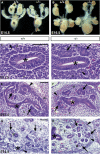
Six2 is crucial for kidney development. Analysis of urogenital tracts dissected at E14.5 (A) and E16.5 (B) revealed that _Six2_-null kidneys (k) were approximately 50% smaller than those of wild-type (+/+) littermates at E14.5 and 65% smaller at E16.5. The adrenal glands (a) and bladder (b) appeared normal. (C–H) Hematoxylin and eosin staining showed that at E11.5 in the wild-type kidney (C) the UB (asterisk) has induced the MM (arrowhead) to condense, but no pretubular aggregates or epithelia were present at this stage. (D) In _Six2_-null littermates, the MM has formed ectopic and premature epithelial vesicles (arrows) on the dorsal (d) side of the UB. (E) At E12.5, the wild-type MM on the ventral side of the UB tips has begun to transform into the epithelia of the renal vesicle (arrows). Condensing mesenchyme (arrowhead) was also detected on the dorsal side of the UB tips. (F) In _Six2_−/− littermates, the MM on the ventral and dorsal sides of the UB transitioned from mesenchyme to epithelia and formed precocious and ectopic renal vesicles (arrows). Note the lack of condensing MM (arrowhead) at the bud tips. (G) At E14.5, the wild-type kidney exhibited its typical uninduced and condensing mesenchyme (arrowhead) and growing branch tips (asterisk) in the cortex, maturing glomeruli (g; arrow) in the medulla, and interstitial stromal cells (s) dispersed throughout. (H) The _Six2_-null kidney revealed an absence of condensing mesenchyme in the cortex (arrowheads), unorganized epithelial structures (arrows) throughout the kidney including a few glomerular structures (g), and a normal distribution of stromal cells (s). Scale bar, 100 μm.
Figure 3
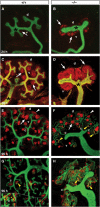
_Six2_-null kidneys exhibit precocious nephrogenesis. (A, B) Wild-type and _Six2_-null kidney explants maintained in culture and costained with E-cadherin to label the UB (green; asterisk) and Cadherin-6 to label developing nephrons (red; arrows). After 24 h culture, few cells expressed Cadherin-6 in the wild-type renal vesicles (A); instead, more advanced epithelial structures (arrows) on the dorsal and ventral sides of the UB tips were seen in the Six2 −/− explant (B). (C, D) After 48 h, immunohistochemistry was performed using anti-pan-cytokeratin (green) to label the UB (asterisk) and anti-laminin-A (red) to label epithelial structures. (C) Normal developing comma and S-shaped bodies (arrows) were seen on the ventral sides (v) of the bud tips of the wild-type kidney. (D) Numerous ectopic renal epithelial structures (arrows) and decreased branching of the UB were identified in the _Six2_-null explant. (E, F) Explants cultured for 96 h were labeled with Wt1 (red), Cadherin-6 (red), and E-cadherin (green). (E) A normal reserve of mesenchymal progenitors (arrowhead) at the tips of the UB (green) and normal developing glomeruli (arrow) throughout the kidney were observed in the wild-type explant. (F) Six2 −/− explants lacked MM in the periphery (white arrowhead) but formed glomeruli (arrow). (G, H) Explants cultured for 96 h were labeled with only Cadherin-6 (red) and E-cadherin (green) to identify any overlap in their expression at the boundary of the proximal and distal tubules (yellow arrowheads). The _Six2_-null explant (H) displayed abnormally extensive coexpression of these markers in mispatterned masses of developing tubules and rare connections of the tubules to the UB (c).
Figure 4
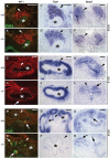
Molecular characterization of the _Six2_-null kidney. At E10.5, no obvious differences in the levels of expression of Wt1 (A, B; red), Eya1 (C, D), or Bmp7 (E, F) were detected in the MM (arrowheads) localized at the tip of the UB (asterisk) in _Six2_-null kidneys. No changes in their expression levels were also detected at E11.5 (G–L), although the size of the MM (arrowheads) surrounding the UB (asterisk and outlined) was reduced and premature and ectopic renal vesicles were already present (arrows) in Six2 −/−. At E12.5, epithelial vesicles (arrows) with MM progenitors residing in the cortex (arrowheads) are seen in control kidneys (M, O, Q). _Six2_-null kidney (N) displayed Wt1 (red)- and Cadherin-6 (green)-expressing ectopic renal vesicles (arrows) on the dorsal and ventral sides of the UB and an absence of MM in the cortex (arrowhead). (P) In agreement with the depletion of the MM surrounding the UB (asterisk; outlined with dashes), expression of Eya1 was lost in E12.5 _Six2_-null kidneys. (R) Bmp7 was expressed at normal levels in the ectopic renal vesicles and UB at this stage. Scale bar, 100 μm.
Figure 5
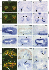
Expression of members of the Wnt pathway is ectopically and prematurely detected in _Six2_-null kidneys. (A, B) Pax2 (red) and Sall1 (green) were used to identify the E10.5 MM. At this stage, expression of both these markers was normal in Six2 −/− (B). (C) Wnt4 expression is localized in the ventral-most mesenchyme (arrow) of an E10.5 wild-type kidney; instead, it was ectopically expanded into the dorsal-most mesenchyme (arrowheads) of the _Six2_-null littermate (D). (E) At this stage, normal low levels of Sfrp2 were present in the wild-type mesenchyme surrounding the Wolffian duct (arrow). Sfrp2 expression was also ectopically expanded into the dorsal side of the UB (arrowheads) and its expression was highly upregulated in the mutant littermate (F). (G, H) At E11.5, Pax2 expression in the UB and MM of the _Six2_-null kidney was similar to that of the wild type, although the size of the MM was reduced. (I) At this stage, the expression of Six2 (brown) and that of Wnt4 (blue) in the MM were complementary, that is, Six2 was expressed predominantly on the dorsal side of the UB and Wnt4 (arrow) only on the ventral side. (J) In the _Six2_−/− kidney, Wnt4 expression ectopically expanded to the dorsal MM (arrowhead). (K) At E11.5, Sfrp2 expression in the wild-type MM was similar to that of Wnt4; dotted line indicates UB epithelium. (L) In the _Six2_−/− kidney, the level of Sfrp2 was upregulated and ectopically expanded to the dorsal side (arrowhead). (M) At E12.5, Pax2 and Sall1 expression remained in the MM (white arrowhead) and was also detected in renal vesicles (arrows). Sall1 was also expressed by the stromal population (green arrowhead). (N) At E12.5, Pax2 expression in the _Six2_−/− kidney highlighted the presence of ectopic supernumerary renal vesicles (arrows) surrounding the UB. Pax2 expression was normal in the UB and renal vesicles, but mesenchymal cells (white arrowhead) expressing Pax2 were absent. However, Sall1-expressing stromal cells (green arrowhead) were still present in the _Six2_-null kidney at this stage. (P, R) Wnt4 and Sfrp2 were expressed in the ectopic renal vesicles (arrows) of the E12.5 _Six2_−/− kidney at levels comparable to those of wild-type (O, Q) kidney. Scale bar, 100 μm.
Figure 6
Reciprocal inductive interactions are lost in _Six2_-null kidney. Wnt11 (A, B), Ret (C, D), and Gdnf (E, F) expression was normal in Six2 −/− kidney at E10.5, indicating that the initial inductive mechanism of the UB was unaffected. (G, H) At E11.5, Wnt11 expression was downregulated in the UB tips of the _Six2_−/− kidney as compared to wild-type littermates but that of Ret (I, J) and Gdnf (K, L) was normal. (M, N) At E12.5, reciprocal inductive interactions were lost in the _Six2_−/− kidney as indicated by the lack of Wnt11 expression in the UB (dashed outline). (O, P) Ret expression remained at normal levels in the _Six2_-null UB at E12.5. (Q, R) Gdnf expression confirmed the abnormal reduction in the size of the MM population and the presence of ectopic developing nephrons (arrows). Scale bar, 100 μm.
Figure 7
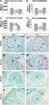
_Six2_−/− kidneys exhibit increased apoptosis at E11.5. (A, B) No differences in the mean total number of cells (A) or in the mean percentage of proliferating cells (B) were measured in the E10.5 _Six2_-null MM. (C, D) At E11.5, _Six2_−/− kidneys were significantly smaller than wild type (C; _P_=0.006); however, the number of proliferating cells was comparable. (E, F) Similar to its wild-type littermate, no apoptotic cells were detected at E10.5 in _Six2_-mutant kidneys. The UB (asterisk) and the area of the metanephric blastema (dashed line) are indicated. (G, H) An abnormal increase in the number of apoptotic cells was identified in the _Six2_-null MM (arrows) surrounding the UB at this stage. (I, J) At E12.5, there was no significance difference in the number of apoptotic cells between wild-type and _Six2_−/− kidneys. (K, L) An increase in the number of apoptotic cells was again detected at E17.5 in the mutant kidney (arrow) as compared to wild-type controls. Scale bar, 100 μm.
Figure 8
Overexpression of Six2 in wild-type kidney organ cultures. Forty-eight hours after microinjection and electroporation of EGFP or FLAG-Six2 expression plasmids, sections of E12.5 kidney organ cultures were labeled with antibodies specific for Pax2 (red; A–D), laminin (red; E–H), EGFP (green), or FLAG-Six2 (green). (A, B) EGFP and Pax2 were coexpressed in epithelial structures (arrows), and EGFP was also expressed in peripheral mesenchyme (arrowhead). (C, D) Cells expressing FLAG-Six2 were almost exclusively found in peripheral and interstitial mesenchyme (arrowheads), separated from Pax2-positive cells. (E, F) EGFP-positive cells were located within the developing tubules (arrows), as demarcated by laminin-containing basement membranes, and in the peripheral mesenchyme (arrowheads). (G, H) FLAG-Six2-expressing cells (arrowheads) were not surrounded by laminin-containing basement membranes and exhibited a mesenchymal phenotype. (I, J) In situ hybridization for Foxd1 followed by immunohistochemistry using anti-GFP (I) or anti-FLAG (J) antibodies indicated that the cells expressing FLAG-Six2 resided mainly in the interstitial stroma, whereas cells expressing the EGFP control vector resided in all cell populations. Scale bar, 100 μm.
Similar articles
- Osr1 acts downstream of and interacts synergistically with Six2 to maintain nephron progenitor cells during kidney organogenesis.
Xu J, Liu H, Park JS, Lan Y, Jiang R. Xu J, et al. Development. 2014 Apr;141(7):1442-52. doi: 10.1242/dev.103283. Epub 2014 Mar 5. Development. 2014. PMID: 24598167 Free PMC article. - Osr1 expression demarcates a multi-potent population of intermediate mesoderm that undergoes progressive restriction to an Osr1-dependent nephron progenitor compartment within the mammalian kidney.
Mugford JW, Sipilä P, McMahon JA, McMahon AP. Mugford JW, et al. Dev Biol. 2008 Dec 1;324(1):88-98. doi: 10.1016/j.ydbio.2008.09.010. Epub 2008 Sep 19. Dev Biol. 2008. PMID: 18835385 Free PMC article. - Six2 defines and regulates a multipotent self-renewing nephron progenitor population throughout mammalian kidney development.
Kobayashi A, Valerius MT, Mugford JW, Carroll TJ, Self M, Oliver G, McMahon AP. Kobayashi A, et al. Cell Stem Cell. 2008 Aug 7;3(2):169-81. doi: 10.1016/j.stem.2008.05.020. Cell Stem Cell. 2008. PMID: 18682239 Free PMC article. - Stem cells in the embryonic kidney.
Nishinakamura R. Nishinakamura R. Kidney Int. 2008 Apr;73(8):913-7. doi: 10.1038/sj.ki.5002784. Epub 2008 Jan 16. Kidney Int. 2008. PMID: 18200005 Review. - Nephron progenitors in the metanephric mesenchyme.
Nishinakamura R, Uchiyama Y, Sakaguchi M, Fujimura S. Nishinakamura R, et al. Pediatr Nephrol. 2011 Sep;26(9):1463-7. doi: 10.1007/s00467-011-1806-0. Epub 2011 Feb 19. Pediatr Nephrol. 2011. PMID: 21336811 Review.
Cited by
- Six2 and Wnt regulate self-renewal and commitment of nephron progenitors through shared gene regulatory networks.
Park JS, Ma W, O'Brien LL, Chung E, Guo JJ, Cheng JG, Valerius MT, McMahon JA, Wong WH, McMahon AP. Park JS, et al. Dev Cell. 2012 Sep 11;23(3):637-51. doi: 10.1016/j.devcel.2012.07.008. Epub 2012 Aug 16. Dev Cell. 2012. PMID: 22902740 Free PMC article. - Genetic approaches to human renal agenesis/hypoplasia and dysplasia.
Sanna-Cherchi S, Caridi G, Weng PL, Scolari F, Perfumo F, Gharavi AG, Ghiggeri GM. Sanna-Cherchi S, et al. Pediatr Nephrol. 2007 Oct;22(10):1675-84. doi: 10.1007/s00467-007-0479-1. Epub 2007 Apr 17. Pediatr Nephrol. 2007. PMID: 17437132 Free PMC article. Review. - Effect of Hypoxia on Branching Characteristics and Cell Subpopulations during Kidney Organ Culture.
Hamon M, Cheng HM, Johnson M, Yanagawa N, Hauser PV. Hamon M, et al. Bioengineering (Basel). 2022 Dec 14;9(12):801. doi: 10.3390/bioengineering9120801. Bioengineering (Basel). 2022. PMID: 36551007 Free PMC article. - Single-Cell Chromatin and Gene-Regulatory Dynamics of Mouse Nephron Progenitors.
Hilliard S, Tortelote G, Liu H, Chen CH, El-Dahr SS. Hilliard S, et al. J Am Soc Nephrol. 2022 Jul;33(7):1308-1322. doi: 10.1681/ASN.2021091213. Epub 2022 Apr 5. J Am Soc Nephrol. 2022. PMID: 35383123 Free PMC article. - Six1 promotes skeletal muscle thyroid hormone response through regulation of the MCT10 transporter.
Girgis J, Yang D, Chakroun I, Liu Y, Blais A. Girgis J, et al. Skelet Muscle. 2021 Nov 19;11(1):26. doi: 10.1186/s13395-021-00281-6. Skelet Muscle. 2021. PMID: 34809717 Free PMC article.
References
- Armstrong JF, Pritchard-Jones K, Bickmore WA, Hastie ND, Bard JB (1993) The expression of the Wilms' tumour gene, WT1, in the developing mammalian embryo. Mech Dev 40: 85–97 - PubMed
- Carroll TJ, Park JS, Hayashi S, Majumdar A, McMahon AP (2005) Wnt9b plays a central role in the regulation of mesenchymal to epithelial transitions underlying organogenesis of the mammalian urogenital system. Dev Cell 9: 283–292 - PubMed
- Cho EA, Patterson LT, Brookhiser WT, Mah S, Kintner C, Dressler GR (1998) Differential expression and function of cadherin-6 during renal epithelium development. Development 125: 803–812 - PubMed
Publication types
MeSH terms
Substances
Grants and funding
- R01 DK054740/DK/NIDDK NIH HHS/United States
- R25 CA023944/CA/NCI NIH HHS/United States
- R21DK068560/DK/NIDDK NIH HHS/United States
- R01 DK062914/DK/NIDDK NIH HHS/United States
- DK062914/DK/NIDDK NIH HHS/United States
- CA-21765/CA/NCI NIH HHS/United States
- P30 CA021765/CA/NCI NIH HHS/United States
- R21 DK068560/DK/NIDDK NIH HHS/United States
- DK054740/DK/NIDDK NIH HHS/United States
LinkOut - more resources
Full Text Sources
Other Literature Sources
Molecular Biology Databases
Research Materials

