Structural basis for recognition of the nonclassical MHC molecule HLA-G by the leukocyte Ig-like receptor B2 (LILRB2/LIR2/ILT4/CD85d) - PubMed (original) (raw)
Structural basis for recognition of the nonclassical MHC molecule HLA-G by the leukocyte Ig-like receptor B2 (LILRB2/LIR2/ILT4/CD85d)
Mitsunori Shiroishi et al. Proc Natl Acad Sci U S A. 2006.
Abstract
HLA-G is a nonclassical MHC class I (MHCI) molecule that can suppress a wide range of immune responses in the maternal-fetal interface. The human inhibitory immune receptors leukocyte Ig-like receptor (LILR) B1 [also called LIR1, Ig-like transcript 2 (ILT2), or CD85j] and LILRB2 (LIR2/ILT4/CD85d) preferentially recognize HLA-G. HLA-G inherently exhibits various forms, including beta(2)-microglobulin (beta(2)m)-free and disulfide-linked dimer forms. Notably, LILRB1 cannot recognize the beta(2)m-free form of HLA-G or HLA-B27, but LILRB2 can recognize the beta(2)m-free form of HLA-B27. To date, the structural basis for HLA-G/LILR recognition remains to be examined. Here, we report the 2.5-A resolution crystal structure of the LILRB2/HLA-G complex. LILRB2 exhibits an overlapping but distinct MHCI recognition mode compared with LILRB1 and dominantly recognizes the hydrophobic site of the HLA-G alpha3 domain. NMR binding studies also confirmed these LILR recognition differences on both conformed (heavy chain/peptide/beta(2)m) and free forms of beta(2)m. Binding studies using beta(2)m-free MHCIs revealed differential beta(2)m-dependent LILR-binding specificities. These results suggest that subtle structural differences between LILRB family members cause the distinct binding specificities to various forms of HLA-G and other MHCIs, which may in turn regulate immune suppression.
Conflict of interest statement
The authors declare no conflict of interest.
Figures
Fig. 1.
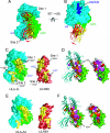
Overall structure of the LILRB2 complex of HLA-G. (A) Ribbon diagram of the LILRB2/HLA-G1 complex. Red, LILRB2; cyan, HLA-G heavy chain; green, β2m; blue, nonapeptide. LILRB2 uses two binding interfaces: D2-β2m (site 1) and D1-α3 (site 2) domain. The stick models of the amino acids involved in the complex formation are shown as follows. Site 1: orange, D2 domain of LILRB2 (Trp-67, Asp-177, Asn-179, and Val-183); sky blue, β2m (Lys-6 and Lys-91). Site 2: magenta, D1 domain of LILRB2 (Arg-36, Tyr-38, Lys-42, Ile-47, and Thr-48); blue, α3 domain (Phe-195, Tyr-197, and Glu-229). The descriptions of site 1 and site 2 apply to Figs. 1–4. (B) 2_F_o − _F_c map (green mesh) contoured at 1σ onto the stick model of the LILRB2/HLA-G complex (around Phe-195 and Tyr-197 of HLA-G).
Fig. 2.
Structural comparison of the LILRB2/HLA-G and LILRB1/HLA-A2 complexes. (A) The LILRB2/HLA-G complex is superimposed onto the LILRB1/HLA-A2 complex around the MHCI regions. The molecular surface of HLA-G is shown. Cyan, HLA-G heavy chain; green, β2m; red, LILRB2; yellow, LILRB1. The D1 domain of LILRB2 has more binding interface on the α3 domain (site 2). The different interactions are observed in D2–β2m interfaces (site 1). (B) This complex image is produced by rotating the image in A around the horizontal axis ≈90°. (C and E) The buried surface areas of LILRB2/HLA-G (C) and LILRB1/HLA-A2 (E). The buried surface areas were calculated by using the program SURFACE (CCP4 suite) with a 1.4-Å probe radius. Cyan, HLA heavy chain; green, β2m; red, LILRB1 or LILRB2. The buried surfaces are shown in yellow. (D and F) NMR analysis of LILRB2–HLA-Cw7 and LILRB1–HLA-Cw7 interactions. The model orientation is similar to that of C and E. Mapping of amino acids whose 1H-15N heteronuclear sequential quantum correlation peaks were perturbed upon complex formation is shown (orange, >0.09 ppm chemical shift changed; magenta, >50% intensity reduced; red, disappeared; green, <0.09 ppm chemical shift changed; white, unassigned). (D) Interaction of conformed (heavy chain/peptide/β2m) (Left) and free (Right) forms of β2m with LILRB2. (F) The same binding analysis as in D with LILRB1.
Fig. 3.
LILR binding interfaces (site 2) of the α3 domain of the LILRB2/HLA-G and LILRB1/HLA-A2 complexes. (A and C) LILRB2/HLA-G complex. (B and D) LILRB1/HLA-A2 complex. Cyan, HLA-G heavy chain; light blue, HLA-A2 heavy chain; green and light green, β2m; magenta, LILRB2; yellow, LILRB1. (A and B) The binding interface around the 195–197 loop of HLA-G. (C and D) The binding interface around the cleft between the first 310 helix and the C strand of LILRBs.
Fig. 4.
SPR analyses. Binding of LILRB2 (Left) and LILRB1 (Right) to HLA-G heterotrimer (red lines) and β2m-free HLA-G heavy chain (green lines). Heterotrimers and β2m-free forms of MHCIs were immobilized on the sensor chip at ≈2,000 response units (RU). Black lines show the responses to the control protein (BSA).
Similar articles
- Human inhibitory receptors Ig-like transcript 2 (ILT2) and ILT4 compete with CD8 for MHC class I binding and bind preferentially to HLA-G.
Shiroishi M, Tsumoto K, Amano K, Shirakihara Y, Colonna M, Braud VM, Allan DS, Makadzange A, Rowland-Jones S, Willcox B, Jones EY, van der Merwe PA, Kumagai I, Maenaka K. Shiroishi M, et al. Proc Natl Acad Sci U S A. 2003 Jul 22;100(15):8856-61. doi: 10.1073/pnas.1431057100. Epub 2003 Jul 9. Proc Natl Acad Sci U S A. 2003. PMID: 12853576 Free PMC article. - HLA-G molecule.
Kamishikiryo J, Maenaka K. Kamishikiryo J, et al. Curr Pharm Des. 2009;15(28):3318-24. doi: 10.2174/138161209789105153. Curr Pharm Des. 2009. PMID: 19860681 - LILRA3 binds both classical and non-classical HLA class I molecules but with reduced affinities compared to LILRB1/LILRB2: structural evidence.
Ryu M, Chen Y, Qi J, Liu J, Fan Z, Nam G, Shi Y, Cheng H, Gao GF. Ryu M, et al. PLoS One. 2011 Apr 29;6(4):e19245. doi: 10.1371/journal.pone.0019245. PLoS One. 2011. PMID: 21559424 Free PMC article. - Immune modulation of HLA-G dimer in maternal-fetal interface.
Kuroki K, Maenaka K. Kuroki K, et al. Eur J Immunol. 2007 Jul;37(7):1727-9. doi: 10.1002/eji.200737515. Eur J Immunol. 2007. PMID: 17587197 Free PMC article. Review. - Recognition of classical and heavy chain forms of HLA-B27 by leukocyte receptors.
Allen RL, Trowsdale J. Allen RL, et al. Curr Mol Med. 2004 Feb;4(1):59-65. doi: 10.2174/1566524043479329. Curr Mol Med. 2004. PMID: 15011960 Review.
Cited by
- Weighted gene co-expression network-based identification of genetic effect of mRNA vaccination and previous infection on SARS-CoV-2 infection.
He Y, Sun M, Xu Y, Hu C, Wang Y, Zhang Y, Fang J, Jin L. He Y, et al. Cell Immunol. 2023 Mar;385:104689. doi: 10.1016/j.cellimm.2023.104689. Epub 2023 Feb 10. Cell Immunol. 2023. PMID: 36780771 Free PMC article. - Biology of HLA-G in cancer: a candidate molecule for therapeutic intervention?
Amiot L, Ferrone S, Grosse-Wilde H, Seliger B. Amiot L, et al. Cell Mol Life Sci. 2011 Feb;68(3):417-31. doi: 10.1007/s00018-010-0583-4. Epub 2010 Nov 10. Cell Mol Life Sci. 2011. PMID: 21063893 Free PMC article. Review. - HLA-G and humanized mouse models as a novel therapeutic approach in transplantation.
Ajith A, Portik-Dobos V, Horuzsko DD, Kapoor R, Mulloy LL, Horuzsko A. Ajith A, et al. Hum Immunol. 2020 Apr;81(4):178-185. doi: 10.1016/j.humimm.2020.02.006. Epub 2020 Feb 21. Hum Immunol. 2020. PMID: 32093884 Free PMC article. Review. - HLA-dependent tumour development: a role for tumour associate macrophages?
Marchesi M, Andersson E, Villabona L, Seliger B, Lundqvist A, Kiessling R, Masucci GV. Marchesi M, et al. J Transl Med. 2013 Oct 6;11:247. doi: 10.1186/1479-5876-11-247. J Transl Med. 2013. PMID: 24093459 Free PMC article. Review. - Cell Surface B2m-Free Human Leukocyte Antigen (HLA) Monomers and Dimers: Are They Neo-HLA Class and Proto-HLA?
Ravindranath MH, Ravindranath NM, Selvan SR, Hilali FE, Amato-Menker CJ, Filippone EJ. Ravindranath MH, et al. Biomolecules. 2023 Jul 28;13(8):1178. doi: 10.3390/biom13081178. Biomolecules. 2023. PMID: 37627243 Free PMC article. Review.
References
- LeMaoult J, Le Discorde M, Rouas-Freiss N, Moreau P, Menier C, McCluskey J, Carosella ED. Tissue Antigens. 2003;62:273–284. - PubMed
- Gonen-Gross T, Achdout H, Gazit R, Hanna J, Mizrahi S, Markel G, Goldman-Wohl D, Yagel S, Horejsi V, Levy O, et al. J Immunol. 2003;171:1343–1351. - PubMed
- Shiroishi M, Kuroki K, Ose T, Rasubala L, Shiratori I, Arase H, Tsumoto K, Kumagai I, Kohda D, Maenaka K. J Biol Chem. 2006;281:10439–10447. - PubMed
Publication types
MeSH terms
Substances
LinkOut - more resources
Full Text Sources
Other Literature Sources
Molecular Biology Databases
Research Materials


