Mutations in the Yersinia pseudotuberculosis type III secretion system needle protein, YscF, that specifically abrogate effector translocation into host cells - PubMed (original) (raw)
Mutations in the Yersinia pseudotuberculosis type III secretion system needle protein, YscF, that specifically abrogate effector translocation into host cells
Alison J Davis et al. J Bacteriol. 2007 Jan.
Abstract
The trafficking of effectors, termed Yops, from Yersinia spp. into host cells is a multistep process that requires the type III secretion system (TTSS). The TTSS has three main structural parts: a base, a needle, and a translocon, which work together to ensure the polarized movement of Yops directly from the bacterial cytosol into the host cell cytosol. To understand the interactions that take place at the interface between the tip of the TTSS needle and the translocon, we developed a screen to identify mutations in the needle protein YscF that separated its function in secretion from its role in translocation. We identified 25 translocation-defective (TD) yscF mutants, which fall into five phenotypic classes. Some classes exhibit aberrant needle structure and/or reduced levels of Yop secretion, consistent with known functions for YscF. Strikingly, two yscF TD classes formed needles and secreted Yops normally but displayed distinct translocation defects. Class I yscF TD mutants showed diminished pore formation, suggesting incomplete pore insertion and/or assembly. Class II yscF TD mutants formed pores but showed nonpolar translocation, suggesting unstable needle-translocon interactions. These results indicate that YscF functions in Yop secretion and translocation can be genetically separated. Furthermore, the identification of YscF residues that are required for the assembly of the translocon and/or productive interactions with the translocon has allowed us to initiate the mapping of the needle-translocon interface.
Figures
FIG. 1.
yscF secretion-positive mutants are defective for cell rounding during infection. HEp-2 cells were infected at an MOI of 10:1 with various Yersinia strains (_ΔyscF+_pTRC99A-yscF, _ΔyscF+_pTRC99A, ΔyopB+pTRC99A, ΔyopN+pTRC99A, ΔyscF+pD250, ΔyscF+pD319, ΔyscF+pD409, ΔyscF+pE40, and ΔyscF+pE354) grown in secretion media with IPTG to induce yscF production. Pictures were taken 1 hour postinfection.
FIG. 2.
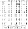
Compilation of yscF TD mutants and their phenotypes in various assays. All yscF TD mutants and control strains were tested for levels of secreted and translocated YopE (Fig. 3), YscF polymers (Fig. 4A), cellular and secreted YscF protein (Fig. 4B), YopD and YopE leaked during infection (Fig. 5A), and SRBC hemolysis (Fig. 6). (+) or (−) indicates detection of YscF polymers after overproduction of YscF protein (Fig. 4C). (nd), not done.
FIG. 3.
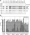
Levels of secreted and translocated YopE from yscF TD mutants. Yersinia strains containing pTRC99A vector alone (+p) or pTRC99A expressing various yscF TD mutants were grown in secretion media for 2 h at 26°C, induced with IPTG, and shifted to 37°C for 2 h. (A) Bacteria were sedimented, and culture supernatants containing secreted proteins from equal numbers of cells were precipitated with TCA. Secreted proteins were separated by SDS-PAGE and stained with Coomassie blue. Molecular mass standards are shown on the left. Secreted Yops are indicated on the right. (B) Bacterial pellets were used to infect HEp-2 cells at an MOI of 50:1 for 1 hour. HEp-2 cells were gently washed in PBS, and plasma membranes were lysed with 0.1% NP-40. HEp-2 cytosol was collected, centrifuged to remove bacteria, and solubilized in sample buffer. Proteins in HEp-2 cytosol and bacterial culture supernatants were separated by SDS-PAGE, transferred to PVDF membranes, and probed with antisera to YopE. Chemiluminescent signals for YopE were quantified, and the amount secreted or translocated in the complemented ΔyscF+pTRC99A-yscF strain was set to 100% (Δ_yscF_+F). All strains were tested a minimum of three times, and the averages for secreted YopE (black bars) and translocated YopE (gray bars) are shown as percentages of the WT signal. Error bars indicate the standard deviations from the means.
FIG. 4.
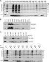
yscF TD mutants form external YscF polymers. Yersinia strains were grown in secretion media as in described in the legend to Fig. 3. (A) Chemical cross-linking. The chemical cross-linker BS3 or water was added to the bacteria and subsequently quenched with Tris-HCl. Bacteria were collected and pellets solubilized in sample buffer. Proteins were separated by SDS-PAGE, transferred to PVDF, and probed with antisera to YscF. Molecular mass standards are shown on the right in kDa. The positions of the YscF monomer (∼7 kDa) and YscF cross-linked multimers are shown on the left. A number of YscF antiserum cross-reactive background bands are visible in every lane (see ΔF+p, with [+] or without [−] BS3; +p is the Δ_yscF_ strain containing vector pTRC99A alone) and are not cross-linker dependent. (B) YscF protein levels. Culture supernatants and bacterial pellets were collected from equal numbers of cells, and proteins were separated by SDS-PAGE. Cell-associated (top panel) and secreted (bottom panel) proteins were transferred to PVDF and probed with antisera to the YscF protein. (C) YscF protein levels after overexpression with increased amounts of IPTG. yscF mutants were induced with 25 μM IPTG and compared to the control strains induced with 10 μM IPTG. Culture supernatants and bacterial pellets were collected and processed as described for panel B. (D) Chemical cross-linking of overproduced yscF mutants. Strains were induced as described for panel C, cross-linked, and processed as described for panel B.
FIG. 5.
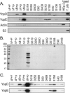
Most yscF TD mutants maintain tight needle-translocon connections during infection. Yersinia strains containing pTRC99A or pTRC99A expressing various yscF TD mutants were grown in 2× YT media with 5 mM calcium for 2 h at 26°C, induced with IPTG, and shifted to 37°C for 2 h. (A) Bacteria were added to monolayers of HEp-2 cells at an MOI of 50:1 and centrifuged to initiate cell contact and subsequent Yops translocation. After 1 hour of infection, tissue culture supernatants were collected and centrifuged to remove bacteria. Proteins were precipitated with TCA, separated by SDS-PAGE, and analyzed by Western blotting with antisera to the YopE and YopD proteins. Controls for disrupted HEp-2 cells and bacteria included uninfected cells lysed with Triton X-100 (lysed HEp-2) and bacteria solubilized in SDS-containing sample buffer (lysed Y. pseudotuberculosis [Y. ptb]). Antisera to actin were used to detect leakage of HEp-2 cytosol during infection, and antisera to the ribosomal subunit S2 were used to detect lysis of bacteria. (B) Secreted proteins in the presence of calcium. Y. pseudotuberculosis bacteria were centrifuged, and culture supernatants were collected. Secreted proteins were precipitated with TCA, separated by SDS-PAGE, and stained with Coomassie blue. Molecular mass standards are shown on the left. The asterisk points to the E158 mutant with barely detectable secreted Yops. (C) Western blot of secreted proteins from panel B, probed with antiserum to YopE and YopD.
FIG. 6.
Hemolysis of sheep red blood cells by yscF TD strains. Yersinia bacteria were grown in secretion media for 2 h at 26°C, induced with IPTG, and shifted to 37°C for 2 h. Bacteria were mixed 1:1 with sheep red blood cells and gently centrifuged to initiate cell contact. Infections were allowed to occur for 3 h at 37°C, and hemoglobin released into the culture supernatant was detected by reading absorbance values at 545 nm. The amount of RBC hemolysis conferred by the ΔyscF+pTRC99A-yscF strain was set to 100% (ΔF+F), and hemolysis levels in all other strains were normalized to WT levels. Experiments were performed in triplicate at least twice, and results for one representative experiment are shown. Error bars show the standard deviations from the means for the triplicate samples.
FIG. 7.
Models of proposed mechanisms for translocation-defective yscF mutations. Needle, LcrV, and translocon components are shown before and after cell contact. (WT) The needle, LcrV tip, and translocon coordinate for Yop translocation. (A) Model A. Disrupted LcrV tip assembly leads to poor translocon formation. (B) Model B. Disrupted interactions between the LcrV tip and/or the translocon leads to leakage of Yops and inefficient translocation. (C) Model C. Unstable YscF-YscF interactions lead to needle breakage before or after cell contact. (D) Model D. LcrV binding to the needle is abolished and no pores are formed. See Discussion for details.
FIG. 8.
Mapping of YscF residues A27, D28, A30, N31, and N47 onto the surface of a T3SS needle. (Left) Surface representation of the end-on view of the Shigella T3SS needle, with each subunit colored differently. The equivalent residues (T23, Q24, L26, Q27, and N43) of the Shigella subunit protein MxiH have been highlighted in gray. (Right) Cutaway surface representation of the side-on view of the Shigella T3SS, with the equivalent Yersinia YscF residues that were mutated highlighted in gray. Class I mutation equivalents A27, A30, and N31 are colored light gray, and class II mutation equivalents D28 and N47 are colored dark gray. This figure was prepared using the PyMOL program (11).
Similar articles
- A dominant-negative needle mutant blocks type III secretion of early but not late substrates in Yersinia.
Davis AJ, Díaz DA, Mecsas J. Davis AJ, et al. Mol Microbiol. 2010 Apr;76(1):236-59. doi: 10.1111/j.1365-2958.2010.07096.x. Epub 2010 Feb 28. Mol Microbiol. 2010. PMID: 20199604 Free PMC article. - Genetic analysis of the formation of the Ysc-Yop translocation pore in macrophages by Yersinia enterocolitica: role of LcrV, YscF and YopN.
Marenne MN, Journet L, Mota LJ, Cornelis GR. Marenne MN, et al. Microb Pathog. 2003 Dec;35(6):243-58. doi: 10.1016/s0882-4010(03)00154-2. Microb Pathog. 2003. PMID: 14580388 - The Yersinia pestis type III secretion system: expression, assembly and role in the evasion of host defenses.
Plano GV, Schesser K. Plano GV, et al. Immunol Res. 2013 Dec;57(1-3):237-45. doi: 10.1007/s12026-013-8454-3. Immunol Res. 2013. PMID: 24198067 Review. - Regulation of the Yersinia type III secretion system: traffic control.
Dewoody RS, Merritt PM, Marketon MM. Dewoody RS, et al. Front Cell Infect Microbiol. 2013 Feb 6;3:4. doi: 10.3389/fcimb.2013.00004. eCollection 2013. Front Cell Infect Microbiol. 2013. PMID: 23390616 Free PMC article. Review.
Cited by
- The pathogenesis, detection, and prevention of Vibrio parahaemolyticus.
Wang R, Zhong Y, Gu X, Yuan J, Saeed AF, Wang S. Wang R, et al. Front Microbiol. 2015 Mar 5;6:144. doi: 10.3389/fmicb.2015.00144. eCollection 2015. Front Microbiol. 2015. PMID: 25798132 Free PMC article. - Yersinia pseudotuberculosis virulence determinants invasin, YopE, and YopT modulate RhoG activity and localization.
Mohammadi S, Isberg RR. Mohammadi S, et al. Infect Immun. 2009 Nov;77(11):4771-82. doi: 10.1128/IAI.00850-09. Epub 2009 Aug 31. Infect Immun. 2009. PMID: 19720752 Free PMC article. - Innate immune recognition of Yersinia pseudotuberculosis type III secretion.
Auerbuch V, Golenbock DT, Isberg RR. Auerbuch V, et al. PLoS Pathog. 2009 Dec;5(12):e1000686. doi: 10.1371/journal.ppat.1000686. Epub 2009 Dec 4. PLoS Pathog. 2009. PMID: 19997504 Free PMC article. - Modified needle-tip PcrV proteins reveal distinct phenotypes relevant to the control of type III secretion and intoxication by Pseudomonas aeruginosa.
Sato H, Hunt ML, Weiner JJ, Hansen AT, Frank DW. Sato H, et al. PLoS One. 2011 Mar 29;6(3):e18356. doi: 10.1371/journal.pone.0018356. PLoS One. 2011. PMID: 21479247 Free PMC article. - YscP and YscU switch the substrate specificity of the Yersinia type III secretion system by regulating export of the inner rod protein YscI.
Wood SE, Jin J, Lloyd SA. Wood SE, et al. J Bacteriol. 2008 Jun;190(12):4252-62. doi: 10.1128/JB.00328-08. Epub 2008 Apr 18. J Bacteriol. 2008. PMID: 18424518 Free PMC article.
References
- Amann, E., B. Ochs, and K. J. Abel. 1988. Tightly regulated tac promoter vectors useful for the expression of unfused and fused proteins in Escherichia coli. Gene 68:301-315. - PubMed
- Blocker, A., N. Jouihri, E. Larquet, P. Gounon, F. Ebel, C. Parsot, P. Sansonetti, and A. Allaoui. 2001. Structure and composition of the Shigella flexneri “needle complex”, a part of its type III secreton. Mol. Microbiol. 39:652-663. - PubMed
Publication types
MeSH terms
Substances
Grants and funding
- R21 NS053740/NS/NINDS NIH HHS/United States
- P3034928/PHS HHS/United States
- F32 GM067517/GM/NIGMS NIH HHS/United States
- GM67517/GM/NIGMS NIH HHS/United States
- R01 AI056068/AI/NIAID NIH HHS/United States
LinkOut - more resources
Full Text Sources
Other Literature Sources
Molecular Biology Databases
Miscellaneous


