Human visfatin expression: relationship to insulin sensitivity, intramyocellular lipids, and inflammation - PubMed (original) (raw)
doi: 10.1210/jc.2006-1303. Epub 2006 Nov 7.
Aiwei Yao-Borengasser, Neda Rasouli, Angela M Bodles, Bounleut Phanavanh, Mi-Jeong Lee, Tasha Starks, Leslie M Kern, Horace J Spencer 3rd, Robert E McGehee Jr, Susan K Fried, Philip A Kern
Affiliations
- PMID: 17090638
- PMCID: PMC2893416
- DOI: 10.1210/jc.2006-1303
Human visfatin expression: relationship to insulin sensitivity, intramyocellular lipids, and inflammation
Vijayalakshmi Varma et al. J Clin Endocrinol Metab. 2007 Feb.
Abstract
Context: Visfatin (VF) is a recently described adipokine preferentially secreted by visceral adipose tissue (VAT) with insulin mimetic properties.
Objective: The aim of this study was to examine the association of VF with insulin sensitivity, intramyocellular lipids (IMCL), and inflammation in humans.
Design and patients: VF mRNA was examined in paired samples of VAT and abdominal sc adipose tissue (SAT) obtained from subjects undergoing surgery. Plasma VF and VF mRNA was also examined in SAT and muscle tissue, obtained by biopsy from well-characterized subjects with normal or impaired glucose tolerance, with a wide range in body mass index (BMI) and insulin sensitivity (S(I)).
Setting: The study was conducted at a University Hospital and General Clinical Research Center.
Intervention: S(I) was measured, and fat and muscle biopsies were performed. In impaired glucose tolerance subjects, these procedures were performed before and after treatment with pioglitazone or metformin.
Main outcome measures: We measured the relationship between VF and obesity, S(I), adipose tissue inflammation, IMCL, and response to insulin sensitizers.
Results: No significant difference in VF mRNA was seen between SAT and VAT depots. VAT VF mRNA associated positively with BMI, whereas SAT VF mRNA decreased with BMI. SAT VF correlated positively with S(I), and the association of SAT VF mRNA with S(I) was independent of BMI. IMCL and markers of inflammation (adipose CD68 and plasma TNFalpha) were negatively associated with SAT VF. Impaired glucose tolerance subjects treated with pioglitazone showed no change in SAT VF mRNA despite a significant increase in S(I). Plasma VF and muscle VF mRNA did not correlate with BMI or S(I) or IMCL, and there was no change in muscle VF with either pioglitazone or metformin treatments.
Conclusion: SAT VF is highly expressed in lean, more insulin-sensitive subjects and is attenuated in subjects with high IMCL, low S(I), and high levels of inflammatory markers. VAT VF and SAT VF are regulated oppositely with BMI.
Figures
FIG. 1
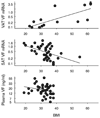
Relationship between VF mRNA expression and BMI. Top, VAT VF mRNA expression and BMI (n = 15). Middle, SAT VF mRNA expression and BMI (n = 39). VF mRNA was determined by real time RT-PCR analysis and was expressed in relation to endogenous 18S RNA. Bottom, Relationship between plasma VF and BMI (n = 29). Plasma VF was measured by ELISA as described in Subjects and Methods. The natural log of VAT VF mRNA, SAT VF mRNA, and plasma VF, respectively, was plotted against BMI.
FIG. 2
Relationship between SAT VF expression and SI (n = 32). VF mRNA level was determined by real-time RT-PCR analysis and was expressed in relation to endogenous 18S RNA. SI value was determined by the method of frequently sampled iv glucose tolerance. The natural log of VF was plotted against the natural log of SI.
FIG. 3
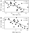
Relationship between SATVF expression and IMCL (n = 31). A, Type 1 muscle fiber IMCL. B, Type 2 muscle fiber IMCL. The natural log of SAT VF mRNA was plotted against the natural log of IMCL type I and type II, respectively.
FIG. 4
Effects of pioglitazone (n = 17) and metformin (n = 19) treatment on VF expression in adipose tissue. VF mRNA levels were determined as described in Subjects and Methods.
Similar articles
- Retinol binding protein 4 expression in humans: relationship to insulin resistance, inflammation, and response to pioglitazone.
Yao-Borengasser A, Varma V, Bodles AM, Rasouli N, Phanavanh B, Lee MJ, Starks T, Kern LM, Spencer HJ 3rd, Rashidi AA, McGehee RE Jr, Fried SK, Kern PA. Yao-Borengasser A, et al. J Clin Endocrinol Metab. 2007 Jul;92(7):2590-7. doi: 10.1210/jc.2006-0816. Epub 2007 Jun 26. J Clin Endocrinol Metab. 2007. PMID: 17595259 Free PMC article. - Reduced plasma visfatin/pre-B cell colony-enhancing factor in obesity is not related to insulin resistance in humans.
Pagano C, Pilon C, Olivieri M, Mason P, Fabris R, Serra R, Milan G, Rossato M, Federspil G, Vettor R. Pagano C, et al. J Clin Endocrinol Metab. 2006 Aug;91(8):3165-70. doi: 10.1210/jc.2006-0361. Epub 2006 May 23. J Clin Endocrinol Metab. 2006. PMID: 16720654 - Association between volume and glucose metabolism of abdominal adipose tissue in healthy population.
Kwon HW, Lee SM, Lee JW, Oh JE, Lee SW, Kim SY. Kwon HW, et al. Obes Res Clin Pract. 2017 Sep-Oct;11(5 Suppl 1):133-143. doi: 10.1016/j.orcp.2016.12.007. Epub 2017 Jan 7. Obes Res Clin Pract. 2017. PMID: 28073639 Review. - An update on visfatin/pre-B cell colony-enhancing factor, an ubiquitously expressed, illusive cytokine that is regulated in obesity.
Stephens JM, Vidal-Puig AJ. Stephens JM, et al. Curr Opin Lipidol. 2006 Apr;17(2):128-31. doi: 10.1097/01.mol.0000217893.77746.4b. Curr Opin Lipidol. 2006. PMID: 16531748 Review.
Cited by
- NAMPT-Mediated NAD(+) Biosynthesis in Adipocytes Regulates Adipose Tissue Function and Multi-organ Insulin Sensitivity in Mice.
Stromsdorfer KL, Yamaguchi S, Yoon MJ, Moseley AC, Franczyk MP, Kelly SC, Qi N, Imai S, Yoshino J. Stromsdorfer KL, et al. Cell Rep. 2016 Aug 16;16(7):1851-60. doi: 10.1016/j.celrep.2016.07.027. Epub 2016 Aug 4. Cell Rep. 2016. PMID: 27498863 Free PMC article. - Adipokines: Deciphering the cardiovascular signature of adipose tissue.
Galley JC, Singh S, Awata WMC, Alves JV, Bruder-Nascimento T. Galley JC, et al. Biochem Pharmacol. 2022 Dec;206:115324. doi: 10.1016/j.bcp.2022.115324. Epub 2022 Oct 27. Biochem Pharmacol. 2022. PMID: 36309078 Free PMC article. Review. - The role of the adipocytokines vaspin and visfatin in vascular endothelial function and insulin resistance in obese children.
Yin C, Hu W, Wang M, Xiao Y. Yin C, et al. BMC Endocr Disord. 2019 Nov 26;19(1):127. doi: 10.1186/s12902-019-0452-6. BMC Endocr Disord. 2019. PMID: 31771561 Free PMC article. - Novel Biomolecules in the Pathogenesis of Gestational Diabetes Mellitus.
Ruszała M, Niebrzydowska M, Pilszyk A, Kimber-Trojnar Ż, Trojnar M, Leszczyńska-Gorzelak B. Ruszała M, et al. Int J Mol Sci. 2021 Oct 27;22(21):11578. doi: 10.3390/ijms222111578. Int J Mol Sci. 2021. PMID: 34769010 Free PMC article. Review. - Suppressive effects of berberine on atherosclerosis via downregulating visfatin expression and attenuating visfatin-induced endothelial dysfunction.
Wan Q, Liu Z, Yang Y, Cui X. Wan Q, et al. Int J Mol Med. 2018 Apr;41(4):1939-1948. doi: 10.3892/ijmm.2018.3440. Epub 2018 Jan 30. Int J Mol Med. 2018. PMID: 29393413 Free PMC article.
References
- Cummings DE, Schwartz MW. Genetics and pathophysiology of human obesity. Annu Rev Med. 2003;54:453–471. - PubMed
- Wajchenberg BL. Subcutaneous and visceral adipose tissue: their relation to the metabolic syndrome. Endocr Rev. 2000;21:697–738. - PubMed
- Fukuhara A, Matsuda M, Nishizawa M, Segawa K, Tanaka M, Kishimoto K, Matsuki Y, Murakami M, Ichisaka T, Murakami H, Watanabe E, Takagi T, Akiyoshi M, Ohtsubo T, Kihara S, Yamashita S, Makishima M, Funahashi T, Yamanaka S, Hiramatsu R, Matsuzawa Y, Shimomura I. Visfatin: a protein secreted by visceral fat that mimics the effects of insulin. Science. 2005;307:426–430. - PubMed
- Kitani T, Okuno S, Fujisawa H. Growth phase-dependent changes in the subcellular localization of pre-B-cell colony-enhancing factor. FEBS Lett. 2003;544:74–78. - PubMed
Publication types
MeSH terms
Substances
Grants and funding
- R37 DK039176/DK/NIDDK NIH HHS/United States
- DK 71346/DK/NIDDK NIH HHS/United States
- DK 71277/DK/NIDDK NIH HHS/United States
- R01 DK071277/DK/NIDDK NIH HHS/United States
- R01 DK071346-03/DK/NIDDK NIH HHS/United States
- UL1 TR001998/TR/NCATS NIH HHS/United States
- M01 RR014288/RR/NCRR NIH HHS/United States
- M01RR14288/RR/NCRR NIH HHS/United States
- R01 HD034522-04/HD/NICHD NIH HHS/United States
- R01 DK039176/DK/NIDDK NIH HHS/United States
- R01 DK071346-01/DK/NIDDK NIH HHS/United States
- R01 DK071346-02/DK/NIDDK NIH HHS/United States
- R01 DK071346/DK/NIDDK NIH HHS/United States
- R01 DK052398/DK/NIDDK NIH HHS/United States
- R25 GM083247/GM/NIGMS NIH HHS/United States
- R01 DK071346-04/DK/NIDDK NIH HHS/United States
- DK 52398/DK/NIDDK NIH HHS/United States
- DK 39176/DK/NIDDK NIH HHS/United States
LinkOut - more resources
Full Text Sources
Medical
Research Materials


