Deregulated Syk inhibits differentiation and induces growth factor-independent proliferation of pre-B cells - PubMed (original) (raw)
Deregulated Syk inhibits differentiation and induces growth factor-independent proliferation of pre-B cells
Thomas Wossning et al. J Exp Med. 2006.
Abstract
The nonreceptor protein spleen tyrosine kinase (Syk) is a key mediator of signal transduction in a variety of cell types, including B lymphocytes. We show that deregulated Syk activity allows growth factor-independent proliferation and transforms bone marrow-derived pre-B cells that are then able to induce leukemia in mice. Syk-transformed pre-B cells show a characteristic pattern of tyrosine phosphorylation, increased c-Myc expression, and defective differentiation. Treatment of Syk-transformed pre-B cells with a novel Syk-specific inhibitor (R406) reduces tyrosine phosphorylation and c-Myc expression. In addition, R406 treatment removes the developmental block and allows the differentiation of the Syk-transformed pre-B cells into immature B cells. Because R406 treatment also prevents the proliferation of c-Myc-transformed pre-B cells, our data indicate that endogenous Syk kinase activity may be required for the survival of pre-B cells transformed by other oncogenes. Collectively, our data suggest that Syk is a protooncogene involved in the transformation of lymphocytes, thus making Syk a potential target for the treatment of leukemia.
Figures
Figure 1.
Syk enables growth factor–independent proliferation. (A) SLP65−/− pre–B cells were retrovirally transduced with IRES-GFP vectors encoding either GFP alone, SLP-65, or Syk. (left) Immunoblot analysis for the expression of Syk and SLP-65 in a WT pre–B cell line and transduced SLP-65−/− pre–B cells sorted for GFP expression. (right) Enrichment of Syk-expressing cells. The proportion of GFP+ cells in transduced cultures was determined by FACS at 1 d and 1 wk after transduction. Data are representative of three independent experiments. (B) Syk allows proliferation in the absence of IL-7. FACS profiles showing SLP-65−/− pre–B cells transduced with either GFP or Syk and cultured in the absence of IL-7 for 3 wk. Data are representative of five independent experiments. Numbers represent the percentage of cells in the indicated quadrant. FSC, forward scatter.
Figure 2.
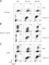
TEL-Syk transforms pre–B cells in vitro. (A) WT pre–B cell lines were retrovirally transduced with IRES-GFP vectors encoding either GFP alone, TEL-Syk, or BCR-Abl. Cells were cultured in the absence of IL-7 and analyzed by FACS at days 1 and 4 after transduction. (B) Transformation of SLP65−/− pre–B cell lines. FACS analysis as described in (A) but using SLP65−/− pre–B cells. (C) Transformation of freshly isolated WT BM-derived pre–B cells. FACS analysis as described in (A) but using freshly isolated BM-derived pre–B cells. Data are representative of five independent experiments. Numbers represent the percentage of cells in the indicated quadrant. FSC, forward scatter.
Figure 3.
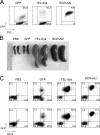
TEL-Syk–expressing cells cause leukemia upon injection in mice. (A) Freshly isolated WT BM-derived pre–B cells (shown in Fig. 2 C) were retrovirally transduced with IRES-GFP vectors for the expression of either GFP alone, TEL-Syk, or BCR-Abl and injected into the tail vein of RAG-2/γC−/− mice. (B) Spleens of the mice 3 wk after injection. (C) FACS profiles showing the expression of GFP and CD19 in cells from the indicated spleens of duplicate mice. Data are representative of five independent experiments. Numbers represent the percentage of cells in the indicated quadrant. FSC, forward scatter.
Figure 4.
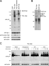
TEL-Syk induces phosphorylation of specific substrates. (A) Immunoblot analysis of pre–B cells transduced with IRES-GFP vectors for the expression of either GFP, TEL-Syk, the kinase-negative TEL-SykK402A, or BCR-Abl. Data are representative of three independent experiments. (B) Silver staining of tyrosine-phosphorylated proteins from TEL-Syk–expressing cells. Total cellular lysates of indicated cells were subjected to immunoprecipitation with antiphosphotyrosine antibodies (4G10). After SDS-PAGE and silver staining, bands marked with an asterisk were cut out and analyzed by mass spectrometry. Proteins identified are indicated next to the respective bands. Data are representative of two independent experiments. (C) The specific Syk inhibitor R406 blocks substrate phosphorylation in TEL-Syk–transformed cells. Shown is an immunoblot analysis of TEL-Syk–transformed cells treated with the Syk inhibitors R406 (2 μM) or piceatannol (50 μM), with the BCR-Abl inhibitor STI-571 (2 μM), or with the Src family kinase inhibitor PP2 (5 μM) for the indicated times. Data are representative of five independent experiments.
Figure 5.
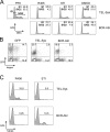
The Syk inhibitor R406 blocks proliferation and induces differentiation of TEL-Syk–transformed cells. (A) Syk inhibition blocks proliferation. Histograms showing the DNA content of TEL-Syk– or BCR-Abl–transformed cells treated for 36 h with DMSO, 2 μM of the Syk inhibitor R406, or 2 μM of the Abl inhibitor STI-571. TEL-Syk– but not BCR-Abl–transformed cells were also treated with 5 μM of the Src family kinase inhibitor PP2. Data are representative of more than five independent experiments. (B) TEL-Syk blocks pre–B cell differentiation. Pre–B cells expressing GFP, TEL-Syk, or BCR-Abl were cultured for 3 d in the absence of IL-7 and analyzed for the expression of κ L chain by FACS. Data are representative of three independent experiments. (C) Inhibition of TEL-Syk with R406 induces differentiation. FACS analysis of κ L chain expression on TEL-Syk– or BCR-Abl–transformed cells treated for 3 d with either 2 μM R406 or 2 μM STI-571. Data are representative of two independent experiments. Numbers represent the percentage of cells in the indicated quadrant or gate.
Figure 6.
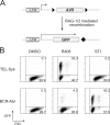
A GFP-based system to measure Syk kinase inhibition. (A) Structure of the recombination reporter plasmid. Induction of RAG-1/2 expression by differentiation-inducing stimuli leads to recombination of the two RSS (black triangles) flanking the inverted GFP cDNA. Flipping the GFP cassette brings the start codon close to the LTR, thereby enabling GFP expression. (B) FACS analysis showing GFP expression in pre–B cells transformed by TEL-Syk or BCR-Abl and containing the recombination reporter plasmid. Cells were treated for 3 d with 2 μM R406 or 2 μM STI-571. Data are representative of three independent experiments. Numbers represent the percentage of cells in the indicated quadrant. FSC, forward scatter; LTR, long terminal repeat.
Figure 7.
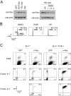
Transformation by Syk induces c-Myc up-regulation in pre–B cells. (A, left) Syk-transformed pre–B cells show augmented c-Myc expression. Immunoblot analysis of pre–B cells transduced with IRES-GFP vectors for the expression of GFP, Syk, TEL-Syk, or BCR-Abl. Data are representative of four independent experiments. (right) Inhibition of Syk decreases c-Myc expression in TEL-Syk–transformed cells. Immunoblot analysis of pre–B cells transformed by TEL-Syk treated with DMSO, 2 μM STI-571 for 2 d, or 2 μM R406 for 1, 1.5, and 2 d, as indicated. Data are representative of two independent experiments. (B) Proliferation of c-Myc–transformed pre–B cells depends on Syk activity. Histograms showing DNA content of c-Myc–transformed cells that were treated for 36 h with DMSO, 2 μM R406, or 2 μM STI-571. Data are representative of more than five independent experiments. (C) Pre-BCR expression is required for the transformation of Ig-α−/− pro–B cells by c-Myc. FACS profiles show Ig-α−/− pro–B cells (left) or Ig-α−/− cells reconstituted with Ig-α (right). Cells were retrovirally transduced with IRES-GFP vectors expressing either GFP alone or c-Myc, cultured in the absence of IL-7, and analyzed at days 1, 6, and 9 after transduction. Numbers represent the percentage of cells in the indicated quadrant or gate. FSC, forward scatter.
Similar articles
- Specific inhibition of spleen tyrosine kinase suppresses leukocyte immune function and inflammation in animal models of rheumatoid arthritis.
Coffey G, DeGuzman F, Inagaki M, Pak Y, Delaney SM, Ives D, Betz A, Jia ZJ, Pandey A, Baker D, Hollenbach SJ, Phillips DR, Sinha U. Coffey G, et al. J Pharmacol Exp Ther. 2012 Feb;340(2):350-9. doi: 10.1124/jpet.111.188441. Epub 2011 Oct 31. J Pharmacol Exp Ther. 2012. PMID: 22040680 - Inhibition of constitutive and BCR-induced Syk activation downregulates Mcl-1 and induces apoptosis in chronic lymphocytic leukemia B cells.
Gobessi S, Laurenti L, Longo PG, Carsetti L, Berno V, Sica S, Leone G, Efremov DG. Gobessi S, et al. Leukemia. 2009 Apr;23(4):686-97. doi: 10.1038/leu.2008.346. Epub 2008 Dec 18. Leukemia. 2009. PMID: 19092849 - Spleen tyrosine kinase inhibition prevents chemokine- and integrin-mediated stromal protective effects in chronic lymphocytic leukemia.
Buchner M, Baer C, Prinz G, Dierks C, Burger M, Zenz T, Stilgenbauer S, Jumaa H, Veelken H, Zirlik K. Buchner M, et al. Blood. 2010 Jun 3;115(22):4497-506. doi: 10.1182/blood-2009-07-233692. Epub 2010 Mar 24. Blood. 2010. PMID: 20335218 - Therapeutic prospect of Syk inhibitors.
Ruzza P, Biondi B, Calderan A. Ruzza P, et al. Expert Opin Ther Pat. 2009 Oct;19(10):1361-76. doi: 10.1517/13543770903207039. Expert Opin Ther Pat. 2009. PMID: 19670961 Review. - Autoinhibition and adapter function of Syk.
Kulathu Y, Grothe G, Reth M. Kulathu Y, et al. Immunol Rev. 2009 Nov;232(1):286-99. doi: 10.1111/j.1600-065X.2009.00837.x. Immunol Rev. 2009. PMID: 19909371 Review.
Cited by
- B-cell antigen receptor signaling enhances chronic lymphocytic leukemia cell migration and survival: specific targeting with a novel spleen tyrosine kinase inhibitor, R406.
Quiroga MP, Balakrishnan K, Kurtova AV, Sivina M, Keating MJ, Wierda WG, Gandhi V, Burger JA. Quiroga MP, et al. Blood. 2009 Jul 30;114(5):1029-37. doi: 10.1182/blood-2009-03-212837. Epub 2009 Jun 2. Blood. 2009. PMID: 19491390 Free PMC article. - Human pre-B cell receptor signal transduction: evidence for distinct roles of PI3kinase and MAP-kinase signalling pathways.
Anbazhagan K, Rabbind Singh A, Isabelle P, Stella I, Céline AD, Bissac E, Bertrand B, Rémy N, Naomi T, Vincent F, Rochette J, Lassoued K. Anbazhagan K, et al. Immun Inflamm Dis. 2013 Oct;1(1):26-36. doi: 10.1002/iid3.4. Epub 2013 Oct 30. Immun Inflamm Dis. 2013. PMID: 25400915 Free PMC article. - At the intersection of DNA damage and immune responses.
Bednarski JJ, Sleckman BP. Bednarski JJ, et al. Nat Rev Immunol. 2019 Apr;19(4):231-242. doi: 10.1038/s41577-019-0135-6. Nat Rev Immunol. 2019. PMID: 30778174 Free PMC article. Review. - GM-CSF receptor/SYK/JNK/FOXO1/CD11c signaling promotes atherosclerosis.
Tsukui D, Kimura Y, Kono H. Tsukui D, et al. iScience. 2023 Jul 11;26(8):107293. doi: 10.1016/j.isci.2023.107293. eCollection 2023 Aug 18. iScience. 2023. PMID: 37520709 Free PMC article. - Is there a role for differentiating therapy in non-APL AML?
Koeffler HP. Koeffler HP. Best Pract Res Clin Haematol. 2010 Dec;23(4):503-8. doi: 10.1016/j.beha.2010.09.014. Epub 2010 Nov 23. Best Pract Res Clin Haematol. 2010. PMID: 21130414 Free PMC article. Review.
References
- Rolink, A.G., E. ten Boekel, T. Yamagami, R. Ceredig, J. Andersson, and F. Melchers. 1999. B cell development in the mouse from early progenitors to mature B cells. Immunol. Lett. 68:89–93. - PubMed
- Karasuyama, H., A. Rolink, Y. Shinkai, F. Young, F.W. Alt, and F. Melchers. 1994. The expression of Vpre-B/lambda 5 surrogate light chain in early bone marrow precursor B cells of normal and B cell-deficient mutant mice. Cell. 77:133–143. - PubMed
Publication types
MeSH terms
Substances
LinkOut - more resources
Full Text Sources
Other Literature Sources
Miscellaneous

