Targeting the expression of platelet-derived growth factor receptor by reactive stroma inhibits growth and metastasis of human colon carcinoma - PubMed (original) (raw)
Targeting the expression of platelet-derived growth factor receptor by reactive stroma inhibits growth and metastasis of human colon carcinoma
Yasuhiko Kitadai et al. Am J Pathol. 2006 Dec.
Abstract
The stromal cells within colon carcinoma express high levels of the platelet-derived growth factor receptor (PDGF-R), whereas colon cancer cells do not. Here, we examined whether blocking PDGF-R could inhibit colon cancer growth in vivo. KM12SM human colon cancer cells were injected subcutaneously (ectopic implantation) into the cecal wall (orthotopic implantation) or into the spleen (experimental liver metastasis) of nude mice. In the colon and liver, the tumors induced active stromal reaction, whereas in the subcutis, the stromal reaction was minimal. Groups of mice (n=10) received saline (control), the tyrosine kinase inhibitor imatinib, irinotecan, or a combination of imatinib and irinotecan. Four weeks of treatment with imatinib and irinotecan significantly inhibited tumor growth (relative to control or single-agent therapy) in the cecum and liver but not in the subcutis. The combination therapy completely inhibited lymph node metastasis. Imatinib alone or in combination with irinotecan inhibited phosphorylation of PDGF-Rbeta of tumor-associated stromal cells and pericytes. Combination therapy also significantly decreased stromal reaction, tumor cell proliferation, and pericyte coverage of tumor microvessels and increased apoptosis of tumor cells and tumor-associated stromal cells. These data demonstrate that blockade of PDGF-R signaling pathways in tumor-associated stromal cells and pericytes inhibits the progressive growth and metastasis of colon cancer cells.
Figures
Figure 1
Growth of KM12SM cells implanted in the subcutis (ectopic site) of nude mice. KM12SM cells (5 × 10⁵) were implanted into the subcutis of nude mice. Mean tumor volumes were determined as described in Materials and Methods. The tumor volume in mice treated with irinotecan was smaller than that in control group, but the difference was not statistically significant. Irinotecan and imatinib did not affect tumor growth at the subcutaneous site. Mean ± SE values.
Figure 2
Experimental liver metastasis. Liver tumor colonies were produced by the intrasplenic injection of KM12SM cells, followed by splenectomy. The mice were treated for 4 weeks with saline (control), imatinib alone, irinotecan alone, or the combination of imatinib and irinotecan. The livers were fixed in Bouin’s fixative.
Figure 3
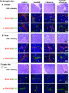
Double immunofluorescence staining for CD31 and PDGF-Rβ or pPDGF-Rβ in KM12SM cecal (A), liver (B), and subcutaneous (C) tumors. Tumor sections were stained with H&E, anti-CD31 antibody (green), and anti-PDGF-Rβ or pPDGF-Rβ (in red fluorescence) as described in Materials and Methods. A and B: Expression of PDGF-Rβ by tumor-associated stromal cells was found in cecal and liver tumors from all treatment groups. Phosphorylation of PDGF-Rβ of tumor-associated stromal cells was inhibited by treatment with imatinib and imatinib and irinotecan. C: In subcutaneous tumors, the expression of PDGF-Rβ by perivascular stroma was low, and the PDGF-Rβ was not phosphorylated. T, tumor nest; S, stroma.
Figure 4
Fluorescence double-labeled immunohistochemistry of KM12SM human colon cancer cells growing in the cecum or subcutis of nude mice. Representative images show immunohistochemistry for CD31 (endothelial marker), desmin (pericyte marker), and α-SMA (marker for myofibroblast and pericyte) in green and PDGF-Rβ in red. Orthotopic KM12SM tumors (A and B) had abundant stroma in which α-SMA+ and desmin+ stromal cells (myofibroblasts and pericytes) expressed PDGF-Rβ (yellow), whereas ectopic tumors did not (C).
Figure 5
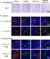
Analysis of cell proliferation (Ki-67) (A), apoptosis (TUNEL/PDGF-Rβ and TUNEL/CD31) (B), and pericyte coverage of microvessels (desmin/CD31) (C). Mice with cecal KM12SM tumors were treated with control, imatinib, irinotecan, or imatinib and irinotecan. A: KM12SM tumors were resected and processed for immunohistochemical evaluation of Ki-67 as described in Materials and Methods. B: Double immunofluorescence staining of TUNEL and PDGF-Rβ or CD31. Stromal cells (PDGF-Rβ+) stained with red fluorescence, and apoptotic cells (TUNEL+) stained with green fluorescence. Colocalization of stromal cells undergoing apoptosis yielded yellow images. Peri-endothelial cells (pericytes and stromal fibroblasts) undergoing apoptosis. C: Pericyte coverage on tumor-associated endothelial cells in the KM12SM cecal tumors. Tumor sections were stained with anti-CD31 antibody in green fluorescence and anti-desmin antibody (pericyte marker) in red fluorescence. T, tumor nest; S, stroma.
Similar articles
- Anti-stromal therapy with imatinib inhibits growth and metastasis of gastric carcinoma in an orthotopic nude mouse model.
Sumida T, Kitadai Y, Shinagawa K, Tanaka M, Kodama M, Ohnishi M, Ohara E, Tanaka S, Yasui W, Chayama K. Sumida T, et al. Int J Cancer. 2011 May 1;128(9):2050-62. doi: 10.1002/ijc.25812. Int J Cancer. 2011. PMID: 21387285 - Stroma-directed imatinib therapy impairs the tumor-promoting effect of bone marrow-derived mesenchymal stem cells in an orthotopic transplantation model of colon cancer.
Shinagawa K, Kitadai Y, Tanaka M, Sumida T, Onoyama M, Ohnishi M, Ohara E, Higashi Y, Tanaka S, Yasui W, Chayama K. Shinagawa K, et al. Int J Cancer. 2013 Feb 15;132(4):813-23. doi: 10.1002/ijc.27735. Epub 2012 Aug 6. Int J Cancer. 2013. PMID: 22821812 - Expression of activated platelet-derived growth factor receptor in stromal cells of human colon carcinomas is associated with metastatic potential.
Kitadai Y, Sasaki T, Kuwai T, Nakamura T, Bucana CD, Hamilton SR, Fidler IJ. Kitadai Y, et al. Int J Cancer. 2006 Dec 1;119(11):2567-74. doi: 10.1002/ijc.22229. Int J Cancer. 2006. PMID: 16988946 - [Molecular targets in colon cancer].
Borner MM. Borner MM. Ther Umsch. 2006 Apr;63(4):243-8. doi: 10.1024/0040-5930.63.4.243. Ther Umsch. 2006. PMID: 16689454 Review. German. - PDGF receptors as targets in tumor treatment.
Ostman A, Heldin CH. Ostman A, et al. Adv Cancer Res. 2007;97:247-74. doi: 10.1016/S0065-230X(06)97011-0. Adv Cancer Res. 2007. PMID: 17419949 Review.
Cited by
- Microenvironment Cell Contribution to Lymphoma Immunity.
Kumar D, Xu ML. Kumar D, et al. Front Oncol. 2018 Jul 27;8:288. doi: 10.3389/fonc.2018.00288. eCollection 2018. Front Oncol. 2018. PMID: 30101129 Free PMC article. Review. - Autologous bone marrow stromal cells genetically engineered to secrete an igf-I receptor decoy prevent the growth of liver metastases.
Wang N, Fallavollita L, Nguyen L, Burnier J, Rafei M, Galipeau J, Yakar S, Brodt P. Wang N, et al. Mol Ther. 2009 Jul;17(7):1241-9. doi: 10.1038/mt.2009.82. Epub 2009 Apr 14. Mol Ther. 2009. PMID: 19367255 Free PMC article. - Imatinib treatment of poor prognosis mesenchymal-type primary colon cancer: a proof-of-concept study in the preoperative window period (ImPACCT).
Ubink I, Bloemendal HJ, Elias SG, Brink MA, Schwartz MP, Holierhoek YCW, Verheijen PM, Boerman AW, Mathijssen RHJ, de Leng WWJ, de Weger RA, van Grevenstein WMU, Koopman M, Lolkema MP, Kranenburg O, Borel Rinkes IHM. Ubink I, et al. BMC Cancer. 2017 Apr 19;17(1):282. doi: 10.1186/s12885-017-3264-y. BMC Cancer. 2017. PMID: 28424071 Free PMC article. - Dasatinib reverses cancer-associated fibroblasts (CAFs) from primary lung carcinomas to a phenotype comparable to that of normal fibroblasts.
Haubeiss S, Schmid JO, Mürdter TE, Sonnenberg M, Friedel G, van der Kuip H, Aulitzky WE. Haubeiss S, et al. Mol Cancer. 2010 Jun 27;9:168. doi: 10.1186/1476-4598-9-168. Mol Cancer. 2010. PMID: 20579391 Free PMC article. - Role of cancer microenvironment in metastasis: focus on colon cancer.
Gout S, Huot J. Gout S, et al. Cancer Microenviron. 2008 Dec;1(1):69-83. doi: 10.1007/s12307-008-0007-2. Epub 2008 Mar 14. Cancer Microenviron. 2008. PMID: 19308686 Free PMC article.
References
- Lynch HT, de la Chapelle A. Hereditary colorectal cancer. N Engl J Med. 2003;348:919–932. - PubMed
- Bond JH. Colorectal cancer update: prevention, screening, treatment, and surveillance for high-risk groups. Med Clin North Am. 2000;84:1163–1182. - PubMed
- Ito M, Yoshida K, Kyo E, Ayhan A, Nakayama H, Yasui W, Ito H, Tahara E. Expression of several growth factors and their receptor genes in human colon carcinomas. Virchows Arch B Cell Pathol Incl Mol Pathol. 1990;59:173–178. - PubMed
- Heldin CH, Ostman A, Ronnstrand L. Signal transduction via platelet-derived growth factor receptors. Biochim Biophys Acta. 1998;1378:F79–F113. - PubMed
- Li X, Ponten A, Aase K, Karlsson L, Abramsson A, Uutela M, Backstrom G, Hellstrom M, Bostrom H, Li H, Soriano P, Betsholtz C, Heldin CH, Alitalo K, Ostman A, Eriksson U. PDGF-C is a new protease-activated ligand for the PDGF alpha-receptor. Nat Cell Biol. 2000;2:302–309. - PubMed
Publication types
MeSH terms
Substances
LinkOut - more resources
Full Text Sources
Other Literature Sources
Miscellaneous


