Orchiectomy for suspected microscopic tumor in patients with anti-Ma2-associated encephalitis - PubMed (original) (raw)
Orchiectomy for suspected microscopic tumor in patients with anti-Ma2-associated encephalitis
R M Mathew et al. Neurology. 2007.
Abstract
Objective: To report the presence of microscopic neoplasms of the testis in men with anti-Ma2-associated encephalitis (Ma2-encephalitis) and to discuss the clinical implications.
Methods: Orchiectomy specimens were examined using immunohistochemistry with Ma2 and Oct4 antibodies.
Results: Among 25 patients with Ma2-encephalitis younger than 50 years, 19 had germ-cell tumors, and 6 had no evidence of cancer. These 6 patients underwent orchiectomy because they fulfilled five criteria: 1) demonstration of anti-Ma2 antibodies in association with MRI or clinical features compatible with Ma2-encephalitis, 2) life-threatening or progressive neurologic deficits, 3) age < 50 years, 4) absence of other tumors, and 5) new testicular enlargement or risk factors for germ-cell tumors, mainly cryptorchidism or ultrasound evidence of testicular microcalcifications. All orchiectomy specimens showed intratubular-germ cell neoplasms unclassified type (IGCNU) and other abnormalities including microcalcifications, atrophy, fibrosis, inflammatory infiltrates, or hypospermatogenesis. Ma2 was expressed by neoplastic cells in three of three patients examined. Even though most patients had severe neurologic deficits at the time of orchiectomy (median progression of symptoms, 10 months), 4 had partial improvement and prolonged stabilization (8 to 84 months, median 22.5 months) and two did not improve after the procedure.
Conclusions: In young men with Ma2-encephalitis, 1) the disorder should be attributed to a germ-cell neoplasm of the testis unless another Ma2-expressing tumor is found, 2) negative tumor markers, ultrasound, body CT, or PET do not exclude an intratubular germ-cell neoplasm of the testis, and 3) if no tumor is found, the presence of the five indicated criteria should prompt consideration of orchiectomy.
Figures
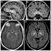
Figure 1
MRI of patients with anti-Ma2-associated encephalitis. The images demonstrate the fluid-attenuated inversion-recovery (FLAIR) MRI sequences of three patients. The MRI of Patient 1 (A and B) shows the areas more frequently involved in patients with anti-Ma2-associated encephalitis, including medial temporal lobes, hypothalamus, and upper brainstem. C corresponds to Patient 2 and shows involvement of the medial temporal lobes and hypothalamus. D corresponds to Patient 3 whose symptoms of severe hypokinesis correlated with bilateral involvement of the pallidum; this patient also had FLAIR hyperintensities involving the medial thalami, pulvinars, hippocampi, and upper brainstem (not shown here).
Figure 2
Demonstration of germ-cell tumors with Oct4 antibody. All panels correspond to paraffin embedded orchiectomy specimens immunolabeled with anti-Oct4 antibody (A, Patient 5; B, Patient 1; C and D, Patient 3). Note the intense immunolabeling of the neoplastic cells (brown staining) but lack of reactivity of the epithelium of normal tubules (arrows in B). C shows Oct4-expressing cells detectable at low magnification; there are several inflammatory infiltrates, one of them surrounds a tubule with neoplastic cells (arrow) that is shown amplified in D. All sections counterstained with hematoxylin. A, B, D, x200; C, x5.
Figure 3
Demonstration of small foci of neoplastic cells using PLAP and Oct4 antibodies. All panels correspond to paraffin embedded orchiectomy specimens from Patient 6. In A, the neoplastic cells are demonstrated with PLAP staining; the arrow indicates a focus of neoplastic cells that is shown at higher magnification in B. In C, another focus of neoplastic cells is demonstrated with Oct4 staining; the arrow points to the area amplified in D. Note that the Oct4 staining is more intense than PLAP and easier to detect at low magnification. All sections counterstained with hematoxylin. A, C, x5; B, D, x400.
Figure 4
Expression of Ma2 by intratubular germ-cell neoplasms. (A) Paraffin section of human hippocampus obtained from autopsy of a neurologically normal individual, immunolabeled with biotinylated IgG containing anti-Ma2 antibodies. Note that in normal brain Ma2 is expressed in the cytoplasm and nucleus of neurons with a pattern of dot-like nuclear reactivity resembling speckles (arrow). (B) Paraffin section of a seminiferous tubule of Patient 5 immunolabeled with biotinylated IgG containing anti-Ma2 antibodies. Ma2 protein is expressed as dot-like speckles by the neoplastic germ-cells (arrow heads), but not by normal epithelium (arrows). Confirmation of Ma2 expression by neoplastic cells was also obtained with a double labeling immunofluorescence method shown in C, D. (C, D) Sections of seminiferous tubules of Patients 3 (C) and 5 (D) immunolabeled with anti-Oct4 (green) and biotinylated IgG containing anti-Ma2 antibodies (red). The expression of Ma2 (red dots) by several neoplastic cells (green nuclei) is indicated with arrows; some cells also show mild diffuse coexpression of Ma2 (yellow). A, B, avidin-biotin-peroxidase method; counterstained with hematoxylin, x400. C, D, double labeling immunofluorescence method, x400.
Comment in
- Can antibodies in serum predict the presence of microscopic tumors?
Voltz R. Voltz R. Neurology. 2007 Mar 20;68(12):887-8. doi: 10.1212/01.wnl.0000259612.19249.4d. Neurology. 2007. PMID: 17372122 No abstract available. - Orchiectomy for suspected microscopic tumor in patients with anti-Ma2-associated encephalitis.
Prüss H, Zschenderlein R, Prass K. Prüss H, et al. Neurology. 2007 Aug 14;69(7):709; author reply 709-10. doi: 10.1212/01.wnl.0000285429.16252.32. Neurology. 2007. PMID: 17698799 No abstract available.
Similar articles
- Severe hypokinesis caused by paraneoplastic anti-Ma2 encephalitis associated with bilateral intratubular germ-cell neoplasm of the testes.
Matsumoto L, Yamamoto T, Higashihara M, Sugimoto I, Kowa H, Shibahara J, Nakamura K, Shimizu J, Ugawa Y, Goto J, Dalmau J, Tsuji S. Matsumoto L, et al. Mov Disord. 2007 Apr 15;22(5):728-31. doi: 10.1002/mds.21314. Mov Disord. 2007. PMID: 17269131 Free PMC article. - [Anti-Ma2-associated encephalitis and paraneoplastic limbic encephalitis].
Yamamoto T, Tsuji S. Yamamoto T, et al. Brain Nerve. 2010 Aug;62(8):838-51. Brain Nerve. 2010. PMID: 20714032 Review. Japanese. - Clinical analysis of anti-Ma2-associated encephalitis.
Dalmau J, Graus F, Villarejo A, Posner JB, Blumenthal D, Thiessen B, Saiz A, Meneses P, Rosenfeld MR. Dalmau J, et al. Brain. 2004 Aug;127(Pt 8):1831-44. doi: 10.1093/brain/awh203. Epub 2004 Jun 23. Brain. 2004. PMID: 15215214 - Anti-Ma2-associated encephalitis in a patient with testis carcinoma.
Suwijn SR, Klieverik LP, Odekerken VJJ. Suwijn SR, et al. Neurology. 2016 Apr 12;86(15):1461. doi: 10.1212/WNL.0000000000002574. Neurology. 2016. PMID: 27163661 No abstract available. - [Limbic encephalitis with antibodies against intracellular antigens].
Morita A, Kamei S. Morita A, et al. Brain Nerve. 2010 Apr;62(4):347-55. Brain Nerve. 2010. PMID: 20420174 Review. Japanese.
Cited by
- Autoimmune-mediated encephalitis.
Demaerel P, Van Dessel W, Van Paesschen W, Vandenberghe R, Van Laere K, Linn J. Demaerel P, et al. Neuroradiology. 2011 Nov;53(11):837-51. doi: 10.1007/s00234-010-0832-0. Epub 2011 Jan 27. Neuroradiology. 2011. PMID: 21271243 Review. - Autoimmune Encephalitis: Pathophysiology and Imaging Review of an Overlooked Diagnosis.
Kelley BP, Patel SC, Marin HL, Corrigan JJ, Mitsias PD, Griffith B. Kelley BP, et al. AJNR Am J Neuroradiol. 2017 Jun;38(6):1070-1078. doi: 10.3174/ajnr.A5086. Epub 2017 Feb 9. AJNR Am J Neuroradiol. 2017. PMID: 28183838 Free PMC article. Review. - Treatment of anti-Ma2/Ta paraneoplastic syndrome.
Kraker J. Kraker J. Curr Treat Options Neurol. 2009 Jan;11(1):46-51. doi: 10.1007/s11940-009-0007-7. Curr Treat Options Neurol. 2009. PMID: 19094836 - Movement disorders in paraneoplastic and autoimmune disease.
Panzer J, Dalmau J. Panzer J, et al. Curr Opin Neurol. 2011 Aug;24(4):346-53. doi: 10.1097/WCO.0b013e328347b307. Curr Opin Neurol. 2011. PMID: 21577108 Free PMC article. Review. - Limbic encephalitis and related cortical syndromes.
Rubio-Agusti I, Salavert M, Bataller L. Rubio-Agusti I, et al. Curr Treat Options Neurol. 2013 Apr;15(2):169-84. doi: 10.1007/s11940-012-0212-7. Curr Treat Options Neurol. 2013. PMID: 23250843
References
- Graus F, Dalmau J, Rene R, et al. Anti-Hu antibodies in patients with small-cell lung cancer: association with complete response to therapy and improved survival. J Clin Oncol. 1997;15:2866–2872. - PubMed
- Darnell RB, Posner JB. Paraneoplastic syndromes involving the nervous system. N Engl J Med. 2003;349:1543–1554. - PubMed
- Younes-Mhenni S, Janier MF, Cinotti L, et al. FDG-PET improves tumour detection in patients with paraneoplastic neurological syndromes. Brain. 2004;127:2331–2338. - PubMed
- Dalmau J, Graus F, Villarejo A, et al. Clinical analysis of anti-Ma2-associated encephalitis. Brain. 2004;127:1831–1844. - PubMed
- Montironi R. Intratubular germ cell neoplasia of the testis: testicular intraepithelial neoplasia. Eur Urol. 2002;41:651–654. - PubMed
Publication types
MeSH terms
Substances
LinkOut - more resources
Full Text Sources
Other Literature Sources
Medical


